


21 August 2026
: Case report 
Left Ventricular Summit Ventricular Tachycardia Identified by Electrocardiographic Pattern Recognition and Managed With Radiofrequency Ablation
Challenging differential diagnosis, Management of emergency care, Rare disease
Mariana Goes Moreira ABCDEF 1, Carina Abigail Hardy ABD 1, Leandro Menezes Alves da Costa ACD 1, Ana Carolina Menezes Borsoi ACD 1, Rodrigo Goldenstein Schainberg ACD 1, Anna Beatriz Gori Montes ACD 1, Rafael Amorim Belo Nunes ACD 1, Thiago Midlej Brito ACD 1, Daniel Castanho Genta Pereira ACD 1, Roger Pereira de Oliveira ACD 1, Thiago Luis Scudeler ABCDEF 1*
DOI: 10.12659/AJCR.953321
Am J Case Rep 2026; 27:e953321


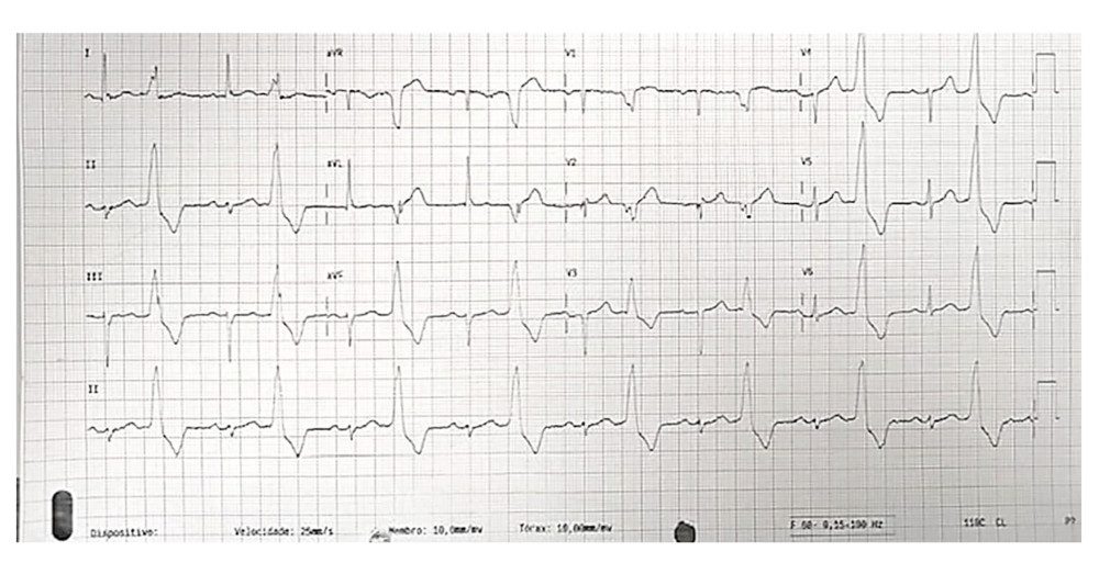
Figure 1 Emergency department 12-lead electrocardiogram (ECG). The 12-lead ECG shows sinus rhythm with frequent premature ventricular complexes (PVCs). The PVCs have left bundle branch block-like morphology, inferior axis, and early precordial transition, with an R/S ratio greater than 1 in lead V2.