


17 August 2026
: Case report 
A Case Report of a 64-Year-Old Man With Urothelial Carcinoma With Brain and Leptomeningeal Metastases Treated With Intrathecal Methotrexate as Part of Multimodal Management
Unusual or unexpected effect of treatment, Rare disease
Leland F. Damron ABCDEF 1, Leigh Kinney ADEF 1, Nikhita Kathuria-Prakash ADEF 2, Marilena Iliopoulos EF 3, Dimitrios Stefanoudakis EF 4, Lidia P. Lopez BDEF 2, Marah Sakkal BE 5, Negar Khanlou BCD 6, Steven D. Hart BCD 6, Neda A. Moatamed BCD 6, Nicolaos J. Palaskas AEF 2, Alexandra Drakaki ACDEG 2,4*
DOI: 10.12659/AJCR.951572
Am J Case Rep 2026; 27:e951572


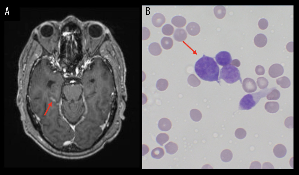
Figure 5 Radiologic and cytologic evidence of leptomeningeal spread. (A) T1-enhanced magnetic resonance imaging (MRI) showing more prominent sulcal enhancement in the right temporal lobe compared with prior images, suggestive of leptomeningeal disease. (B) May-Grünwald Giemsa (MGG) stain of the patient’s cerebrospinal fluid (CSF) shows scattered malignant cells with high nuclear to cytoplasmic ratios, dark chromatin, and irregular nuclear membranes (60× objective).