


17 August 2026: Articles 
A Case Report of a 64-Year-Old Man With Urothelial Carcinoma With Brain and Leptomeningeal Metastases Treated With Intrathecal Methotrexate as Part of Multimodal Management
Unusual or unexpected effect of treatment, Rare disease
Leland F. Damron ABCDEF 1, Leigh Kinney ADEF 1, Nikhita Kathuria-Prakash ADEF 2, Marilena Iliopoulos EF 3, Dimitrios Stefanoudakis EF 4, Lidia P. Lopez BDEF 2, Marah Sakkal BE 5, Negar Khanlou BCD 6, Steven D. Hart BCD 6, Neda A. Moatamed BCD 6, Nicolaos J. Palaskas AEF 2, Alexandra Drakaki ACDEG 2,4*
DOI: 10.12659/AJCR.951572
Am J Case Rep 2026; 27:e951572


Abstract
BACKGROUND: The central nervous system (CNS) is a rare site for upper tract urothelial carcinoma (UTUC) metastasis and typically carries a poor prognosis. Little is known about effective treatments. This report describes a 64-year-old man with UTUC with brain and leptomeningeal metastases. He was treated with multiple lines of chemotherapy, immunotherapy, surgery, radiation, and intrathecal methotrexate as part of multimodal therapy delivered through multidisciplinary care.
CASE REPORT: A 64-year-old man with multiple sclerosis was diagnosed with UTUC. Initial management included a nephroureterectomy followed by platinum-based chemotherapy. Despite this treatment, his malignancy recurred with pulmonary metastases, treated with enfortumab vedotin and local ablation. After this treatment, he achieved systemic disease control. However, he later presented with neurologic symptoms, and imaging showed brain metastases. He underwent resection followed by stereotactic radiation therapy and immunotherapy. Even with this multimodal treatment, he developed leptomeningeal spread, evidenced by cytology-positive cerebrospinal fluid (CSF). Following progression on multiple systemic therapies, he received intrathecal methotrexate with temporary clearance of tumor cells from his CSF before disease progression. This treatment course, including chemotherapy, immunotherapy, and intrathecal methotrexate, prolonged the patient’s life before he unfortunately died from his disease.
CONCLUSIONS: This case report outlines a treatment approach for a rare complication of UTUC. This regimen includes both standard-of-care therapy and inventive strategies extrapolated from data on other solid tumors. Additionally, it contributes to the limited data on the management of CNS metastasis in rare solid tumor malignancies, which could inform future clinical decision-making.
Keywords: Brain Diseases, Case Reports, Immunotherapy, Methotrexate, Neoplasms
Introduction
Upper tract urothelial carcinoma (UTUC) is a rare malignancy with a poor prognosis, accounting for 5–10% of urothelial carcinoma cases and occurring in the renal pelvis, renal calyx, or ureter [1,2]. Thirty percent of patients have metastatic disease at the time of diagnosis, most commonly to the lungs, bone, and liver [2,3]. While rare, urothelial carcinoma can metastasize to the brain and leptomeninges, and this development is associated with a poor prognosis [4]. Prognostic signs include the status of muscle invasion, the presence of hydronephrosis, tumor stage and grade, and certain molecular markers [5].
Historically, platinum-based chemotherapy has been the standard of care for metastatic urothelial carcinoma; however, systemic treatment for advanced disease has evolved in recent years [5]. Current first-line therapy for locally advanced or metastatic urothelial carcinoma is pembrolizumab and enfortumab vedotin [6]. Other options include platinum-based chemotherapy or dose-dense methotrexate, vinblastine, doxorubicin, and cisplatin, with different maintenance therapy strategies [1]. Second-line options include single-agent immune checkpoint inhibitors, single-agent enfortumab vedotin, erdafitinib, or fam-trastuzumab deruxtecan-nxki [1,7–9]. Sacituzumab govitecan-hziy, an antibody-drug conjugate targeting trop2, was granted accelerated approval by the Food and Drug Administration but was later withdrawn [10,11]. This followed a phase 3 confirmatory trial that failed to meet its primary endpoint of improved overall survival and showed increased toxicity compared with chemotherapy [10,11]. Despite this progress, participation in clinical trials is always encouraged. The role of radical nephroureterectomy in patients with metastatic UTUC has been explored in several observational studies, which primarily involve patients with a single metastatic site [12,13].
While UTUC is highly metastatic, central nervous system (CNS) involvement is infrequent, and evidence to guide management is limited. Some systemic therapies are available, but many show limited efficacy, with poor CNS penetration across the blood-brain barrier and pose a challenge in treating CNS metastases from solid tumors [14]. As a result, direct intrathecal therapy may play an important role. Additional treatment modalities include stereotactic radiosurgery, whole-brain radiotherapy (WBRT), and surgical resection [15]. Optimal management strategies for CNS metastases in rare malignancies such as UTUC remain inadequately defined.
Only a small number of case reports have described leptomeningeal spread from UTUC. Each case report has noted the aggressive clinical course and limited therapeutic options in these rare cases [16–18]. Reported cases include a case of carcinomatous meningitis from bladder cancer, a case of leptomeningeal spread managed with WBRT, and lastly, a case of isolated leptomeningeal spread from urothelial carcinoma [16–18]. This report describes a 64-year-old male patient with brain and leptomeningeal metastases from UTUC treated with intrathecal methotrexate as part of multimodal therapy and multidisciplinary care. This case illustrates the therapeutic challenges of CNS-confined metastatic UTUC and describes potential CNS-directed interventions that could be extrapolated to other rare cases with limited evidence.
Case Report
A 64-year-old African American man with multiple sclerosis (MS) on peginterferon beta 1a, sickle cell trait, stage 3 chronic kidney disease, and hypertension was diagnosed with high-grade urothelial carcinoma of the right upper urinary tract. This malignancy was identified incidentally on a routine MRI of the thoracic spine for MS surveillance. Further scans demonstrated a distended ureter with thickening and stricture (Figure 1). Cystoscopy and ureteroscopy showed a mid-ureteral tumor, and biopsy confirmed high-grade papillary urothelial carcinoma. He underwent robot-assisted nephroureterectomy, with pathology showing pT3Nx invasive high-grade urothelial carcinoma with focal micropapillary features and negative margins.
The patient received 4 cycles of adjuvant gemcitabine (1000 mg/m2, intravenous) with split-dose cisplatin (35 mg/m2, intravenous). After this treatment, he had no evidence of active disease based on imaging and circulating cell-free DNA according to the Guardant360 platform. However, 2 months later, his malignancy relapsed, with imaging suggestive of pulmonary spread (Figure 2A). In addition, a biopsy confirmed metastasis (Figure 3). He received enfortumab vedotin by intravenous infusion, starting at 1.0 mg/kg and reduced to 0.75 mg/kg for a total of 14 cycles over 10 months. This was discontinued due to worsening neuropathy. At this point, the lung was the only remaining site of metastatic disease, so he underwent microwave radiofrequency ablation with complete resolution of the cancer.
After stopping enfortumab vedotin, imaging showed stable disease for about 8 months. The patient then developed confusion, memory impairment, and gait instability. A brain MRI identified multifocal lesions in the right parieto-occipital and right temporal lobes (Figure 2B). No active disease was seen elsewhere. At this time, his neurological exam revealed disorientation to person, place, and time, as well as blurry vision in the left eye and the medial field of the right eye. He underwent craniotomy for resection of the right parietal tumor and stereotactic radiosurgery, with tumor pathology consistent with CNS spread (Figure 4). Following this surgery, his mental status improved, but he was noted to have some expressive aphasia. Although clinical trial enrollment would have been appropriate, his history of MS made him ineligible. Limited safety data also exist for immune checkpoint inhibitors in patients with pre-existing autoimmune conditions such as MS.
Both immune checkpoint inhibitors and enfortumab vedotin have demonstrated activity in patients with brain metastases [19,20]. Therefore, he was started on pembrolizumab (200 mg or 400 mg, intravenous) and rechallenged with enfortumab vedotin (0.75 mg/kg, intravenous). He received 10 cycles before disease progression (Table 1). At the time of progression on this regimen, he had no new neurologic symptoms, but he continued to have expressive aphasia.
The patient was subsequently treated with a combination of nivolumab (3 mg/kg intravenously) and ipilimumab (1 mg/kg intravenously). The decision to use this combination therapy for his CNS-confined disease was based on evidence from studies showing intracranial efficacy in metastatic clear cell carcinoma and melanoma [21,22]. Due to the limited response duration, he received only 4 cycles. For his new CNS metastases, he underwent stereotactic radiotherapy followed by sacituzumab govitecan (10 mg/kg or 7.5 mg/kg, intravenous). The decision to give sacituzumab govitecan was derived from preliminary data showing good drug penetration across the blood-brain barrier and efficacy in treating CNS metastases in breast cancer [23]. This treatment, extrapolated from breast cancer research, was pursued because of the limited data on the management of UTUC with brain metastases and the patient’s excellent performance status despite rapid intracranial disease progression. After 2 cycles, MRI revealed further disease progression and leptomeningeal spread (Figure 5A), confirmed by lumbar puncture (Figure 5B). No new neurologic symptoms were present at the time of leptomeningeal spread, and his expressive aphasia had improved. The patient continued MS therapy throughout treatment without exacerbation.
Following multidisciplinary evaluation and in the context of CNS-confined disease with preserved performance status, he received methotrexate (12 mg, intrathecal). Before the first dose of intrathecal methotrexate, his Karnofsky Performance Status score was 80. The first dose was via lumbar injection and later via Ommaya reservoir (3 doses, 42 and 28 days apart). Cerebrospinal fluid (CSF) was analyzed before each treatment to assess disease status and response to therapy. Impressively, the patient cleared his CSF and had negative cytology after the first dose of intrathecal methotrexate. CSF cytology remained negative at several time points; however, 2 months after initiation of intrathecal therapy, progression was noted on clinical, radiographic, and CSF analyses (Table 1). At this point, the patient developed worsening aphasia and memory impairment as well as slurred speech. After 3 doses of intrathecal methotrexate, his Karnofsky Performance Status score was 50.
Due to marked clinical deterioration, the patient received palliative whole-brain radiation therapy. Within several weeks, he developed seizures and hydrocephalus, necessitating intubation and intensive care unit (ICU) admission. Under ICU care, he died from his malignancy.
Discussion
This case report describes the possible benefits of multimodal therapy and multidisciplinary care in CNS-confined UTUC and highlights the role of intrathecal methotrexate and other CNS-directed interventions when evidence-based guidelines are lacking. Aggressive systemic disease was controlled through surgical resection, platinum-based chemotherapy, multiple lines of immunotherapy, antibody-drug conjugates, and local ablation. Overall survival exceeded expectations from historical data [24]. However, this outcome cannot be attributed to intrathecal methotrexate alone (Table 1). This treatment course deviates from the current standard of care. At the initiation of his treatment, platinum-based chemotherapy was the recommended first-line treatment for UTUC. Further divergence from guidelines occurred because of enfortumab-induced neuropathy, limited clinical trial options due to MS, and an unusual metastatic pattern with CNS-isolated disease.
The decision to pursue multiple lines of CNS-targeted and systemic therapies reflects limited evidence for this clinical scenario, consistently preserved patient performance status, and the patient’s desire for aggressive treatment. At the time of leptomeningeal spread, the disease was confined to the CNS, and the patient had an acceptable neurologic status, supporting the use of intrathecal methotrexate despite a lack of evidence in UTUC. This case functions as a reminder that, in rare metastatic presentations, such as CNS-confined metastatic UTUC, treatment may rely on extrapolation from more common malignancies, shared decision-making, and clinician judgment in the absence of established treatment evidence.
While brain metastasis is a common complication of many solid tumor malignancies, it is rare for urothelial carcinoma, and especially UTUC [24]. Leptomeningeal spread is even less common and is associated with an exceptionally low median overall survival of 2–3 months [25]. A recent review from the Society for Neuro-Oncology and the American Society of Clinical Oncology reiterated this therapeutic gap and stated the importance of multimodal therapy and continued research into this area [26]. Published case reports on UTUC with CNS spread extrapolate data from CNS metastases of other solid tumor malignancies to guide treatment. This most commonly involves surgical resection followed by adjuvant radiation therapy, but not often intrathecal treatment. More robust data on CNS management in other solid tumors, such as breast and kidney cancer, present off-label therapeutic options to explore [22,23]. For patients with good performance status, social support, and willingness to pursue treatments extrapolated from other malignancies, these options may help prolong survival and manage symptoms associated with CNS metastases.
While the literature is limited, several published case reports have similarly described the clinical course of patients with CNS and leptomeningeal metastases from genitourinary malignancies and suggested treatment approaches [16–18]. Tomioka et al and Lim et al described patients with genitourinary cancers who had a rapid neurologic decline after the identification of leptomeningeal disease [16,17]. The primary intervention in both cases was WBRT, and compared with the patient described in this case report, they had rapid neurologic progression and worse functional status [16,17]. Tadepalli et al described a case of carcinomatous meningitis in a patient with a known bladder primary [18]. This patient’s chief complaint was a headache and cranial nerve III palsy [18]. After identification of leptomeningeal disease, he was treated with biweekly intrathecal methotrexate for 6 weeks followed by WBRT [18]. The patient was still alive when the case report was published [18]. Compared with these case reports, the patient described in this case had a relatively prolonged clinical course and maintained better functional status throughout his treatment [16–18]. Importantly, the case presented by Tadepalli et al also included intrathecal methotrexate and WBRT, and, while overall survival for this patient is unknown, they, too, experienced at least transient benefit from multimodal therapy [18].
While this case report provides an innovative treatment strategy that ultimately extended this patient’s survival, several limitations ought to be considered. First, this is an anecdotal case report based on a single patient with a rare metastatic pattern. In addition, this patient maintained excellent performance status for the vast majority of his treatment course and desired aggressive treatment, which may not be true for other patients. For all of these reasons, the generalizability of this case report is limited. Another limitation is the inability to study each intervention in isolation due to serial treatments; as a result, it is challenging to attribute intrathecal methotrexate alone to the temporary clearance of leptomeningeal disease, given that the patient had previously received many cycles of chemotherapy, immunotherapy, and radiation. Lastly, there are biological differences among malignancies, so extrapolation of this data to other solid tumor malignancies should be done with caution. Regardless of these inherent constraints, this case report provides helpful perspectives for clinicians who encounter rare malignancies with limited treatment evidence.
Conclusions
This case report adds to the limited literature on UTUC with CNS metastasis and includes treatment strategies extrapolated from other malignancies, given the lack of evidence-based applicable data. This multimodal treatment course with multidisciplinary team-based management was associated with prolonged survival compared with historical outcomes, without substantially affecting functional status and patient quality of life for most of his therapy. It encourages “outside of the box” thinking, as a randomized trial cannot be conducted because of the rarity of this disease and patient characteristics. This report may function as a valuable source for future treatment decisions for patients with CNS involvement in uncommon malignancies such as UTUC.
Figures
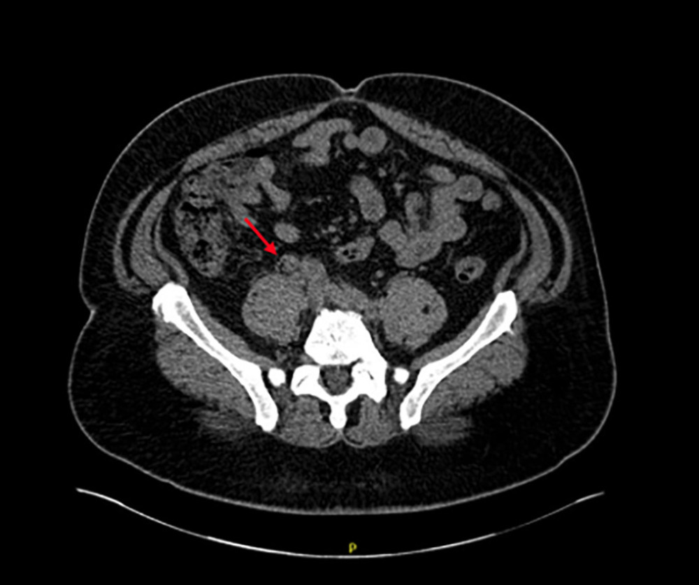 Figure 1. Computed tomography (CT) of the kidneys, ureters, and bladder with findings suggestive of primary upper tract urothelial carcinoma including distended proximal ureter. Severe right hydronephrosis was present but not fully visualized in this image.
 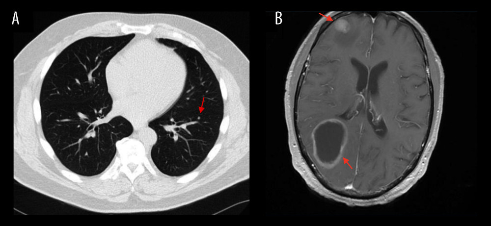 Figure 2. Radiologic progression of metastatic disease. (A) Computed tomography (CT) of the chest without contrast showing a newly enlarged 4-mm solid pulmonary nodule in the left lower lobe, suspicious for metastatic disease. (B) Contrast-enhanced magnetic resonance imaging (MRI) of the brain (axial T1-weighted post-contrast sequence) demonstrating a rim-enhancing cystic lesion in the right parietal lobe (posterior lesion), measuring 52×36 mm with no significant surrounding edema, with associated mass effect of the right ventricular trigone. In addition, a solid enhancing mass was visible in the right anterior frontal lobe measuring 13×15 mm, surrounded by vasogenic edema.
 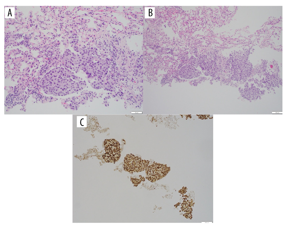 Figure 3. Histopathologic confirmation of pulmonary metastasis from urothelial carcinoma. (A) A high power (200×) hematoxylin and eosin (H&E)-stained section showing tumor cells with round-to-oval central nuclei and moderate amounts of eosinophilic cytoplasm, consistent with urothelial origin. (B) A low power (100×) H&E-stained section showing lung parenchyma with sheets of neoplastic cells within alveolar spaces. (C) GATA3 positive nuclear staining supportive of urothelial origin (GATA3 IPOX 100×).
 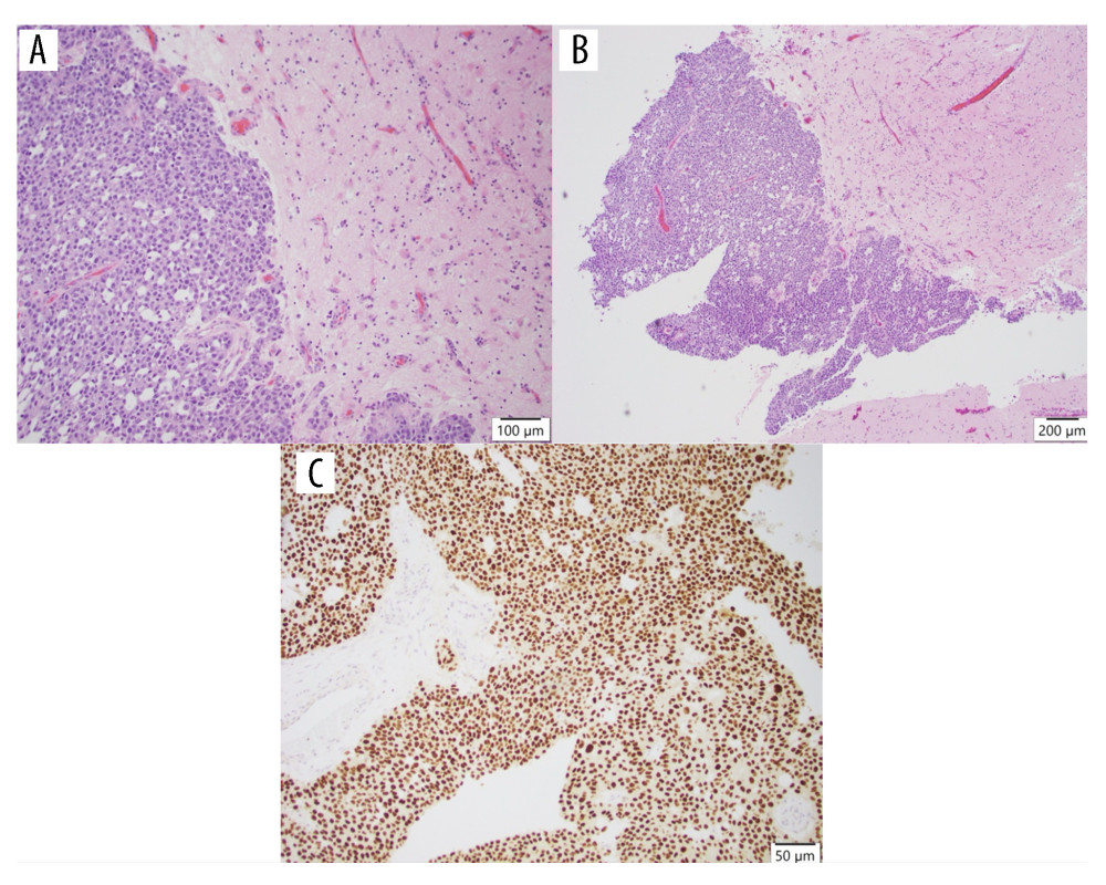 Figure 4. Histopathologic findings of brain metastasis. (A, B) Hematoxylin and eosin (H&E) histopathology at 100× and 200× power showing sheets of neoplastic epithelioid cells with moderately pleomorphic, enlarged nuclei and prominent nucleoli. Mitotic figures are present. Reactive gliosis is present in the surrounding brain tissue. (C) Nuclear immunoreactivity with antibody to GATA3 is consistent with the known urothelial origin.
 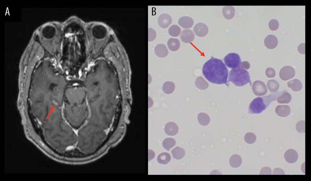 Figure 5. Radiologic and cytologic evidence of leptomeningeal spread. (A) T1-enhanced magnetic resonance imaging (MRI) showing more prominent sulcal enhancement in the right temporal lobe compared with prior images, suggestive of leptomeningeal disease. (B) May-Grünwald Giemsa (MGG) stain of the patient’s cerebrospinal fluid (CSF) shows scattered malignant cells with high nuclear to cytoplasmic ratios, dark chromatin, and irregular nuclear membranes (60× objective).
 References
1. Escobar D, Wang C, Suboc N, Diagnosis and management of upper tract urothelial carcinoma: A review: Cancers (Basel), 2025; 17(15); 2467
2. Kolawa A, D’Souza A, Tulpule V, Overview, diagnosis, and perioperative systemic therapy of upper tract urothelial carcinoma: Cancers (Basel), 2023; 15(19); 4813
3. Tufano A, Cordua N, Nardone V, Prognostic significance of organ-specific metastases in patients with metastatic upper tract urothelial carcinoma: J Clin Med, 2022; 11(18); 5310
4. Brenneman RJ, Gay HA, Christodouleas JP, Review: Brain metastases in bladder cancer: Bladder Cancer, 2020; 6(3); 237-48
5. Masson-Lecomte A, Birtle A, Pradere B, European Association of Urology Guidelines on upper urinary tract urothelial carcinoma: Summary of the 2025 update: Eur Urol, 2025; 87(6); 697-716
6. Powles T, Valderrama BP, Gupta S, Enfortumab vedotin and pembrolizumab in untreated advanced urothelial cancer: N Engl J Med, 2024; 390(10); 875-88
7. Powles T, Park SH, Caserta C, Avelumab first-line maintenance for advanced urothelial carcinoma: Results from the JAVELIN bladder 100 trial after ≥2 years of follow-up: J Clin Oncol, 2023; 41(19); 3486-92
8. Rizzo A, Buti S, Giannatempo P, Pembrolizumab in patients with advanced upper tract urothelial carcinoma: A real-world study from ARON-2 project: Clin Exp Metastasis, 2024; 41(5); 655-65
9. Bajorin DF, Witjes JA, Gschwend JE, Adjuvant nivolumab versus placebo in muscle-invasive urothelial carcinoma: N Engl J Med, 2021; 384(22); 2102-14
10. Powles T, Tagawa S, Vulsteke C, Sacituzumab govitecan in advanced urothelial carcinoma: TROPiCS-04, a phase III randomized trial: Ann Oncol, 2025; 36(5); 561-71
11. Tagawa ST, Balar AV, Petrylak DP, TROPHY-U-01: A phase II open-label study of sacituzumab govitecan in patients with metastatic urothelial carcinoma progressing after platinum-based chemotherapy and checkpoint inhibitors: J Clin Oncol, 2021; 39(22); 2474-85
12. Nazzani S, Preisser F, Mazzone E, Survival effect of nephroureterectomy in metastatic upper urinary tract urothelial carcinoma: Clin Genitourin Cancer, 2019; 17(3); e602-e11
13. Moschini M, Xylinas E, Zamboni S, Efficacy of surgery in the primary tumor site for metastatic urothelial cancer: Analysis of an international, multicenter, multidisciplinary database: Eur Urol Oncol, 2020; 3(1); 94-101
14. Lowery FJ, Yu D, Brain metastasis: Unique challenges and open opportunities: Biochim Biophys Acta, 2017; 1867(1); 49-57
15. Diamantopoulos LN, Khaki AR, Sonpavde GP, Central nervous system metastasis in urothelial carcinoma: Institutional experience and a comprehensive review of the literature: Clin Genitourin Cancer, 2020; 18(3); e266-e76
16. Tomioka M, Kawase M, Kato D, Leptomeningeal carcinomatosis in urothelial carcinoma of the urinary bladder: A report of a patient with a fulminant course who died of cancer after definitive therapies: Case Rep Urol, 2021; 2021; 5543939
17. Lim C, Winquist E, Joseph M, Nathan P, Isolated leptomeningeal carcinomatosis in urothelial carcinoma: A case report of positive CSF cytology with negative imaging: Case Rep Oncol Med, 2025; 2025; 8889550
18. Tadepalli S, Coleman T, Hacket LA, Liles GB, Carcinomatous meningitis: The natural history of successfully treated metastatic bladder cancer: Case Rep Oncol, 2011; 4(2); 406-12
19. Niedersuess-Beke D, Mayrhofer K, Krauter J, Real-world evidence for enfortumab vedotin in patients with metastatic urothelial cancer: An Austrian multicentre study: Clin Genitourin Cancer, 2025; 23(1); 102278
20. Brastianos PK, Kim AE, Giobbie-Hurder A, Phase II trial of pembrolizumab in patients with brain metastases: J Clinical Oncol, 2023; 41(16 Suppl); 2006
21. Tawbi HA, Forsyth PA, Algazi A, Combined nivolumab and ipilimumab in melanoma metastatic to the brain: N Engl J Med, 2018; 379(8); 722-30
22. Emamekhoo H, Olsen MR, Carthon BC, Safety and efficacy of nivolumab plus ipilimumab in patients with advanced renal cell carcinoma with brain metastases: CheckMate 920: Cancer, 2022; 128(5); 966-74
23. Diéras V, Weaver R, Tolaney SM, Abstract PD13-07: Subgroup analysis of patients with brain metastases from the phase 3 ASCENT study of sacituzumab govitecan versus chemotherapy in metastatic triple-negative breast cancer: Cancer Res, 2021; 81(4 Suppl); PD13-07
24. Di Bello F, de Angelis M, Siech C, Survival of metastatic urothelial carcinoma of urinary bladder according to number and location of visceral metastases: Clin Genitourin Cancer, 2024; 22(5); 102139
25. Le Rhun E, Taillibert S, Chamberlain MC, Carcinomatous meningitis: Leptomeningeal metastases in solid tumors: Surg Neurol Int, 2013; 4(Suppl 4); S265-88
26. Wilcox JA, Chukwueke UN, Ahn MJ, Leptomeningeal metastases from solid tumors: A Society for Neuro-Oncology and American Society of Clinical Oncology consensus review on clinical management and future directions: Neuro Oncol, 2024; 26(10); 1781-804
Figures
Figure 1. Computed tomography (CT) of the kidneys, ureters, and bladder with findings suggestive of primary upper tract urothelial carcinoma including distended proximal ureter. Severe right hydronephrosis was present but not fully visualized in this image.Figure 2. Radiologic progression of metastatic disease. (A) Computed tomography (CT) of the chest without contrast showing a newly enlarged 4-mm solid pulmonary nodule in the left lower lobe, suspicious for metastatic disease. (B) Contrast-enhanced magnetic resonance imaging (MRI) of the brain (axial T1-weighted post-contrast sequence) demonstrating a rim-enhancing cystic lesion in the right parietal lobe (posterior lesion), measuring 52×36 mm with no significant surrounding edema, with associated mass effect of the right ventricular trigone. In addition, a solid enhancing mass was visible in the right anterior frontal lobe measuring 13×15 mm, surrounded by vasogenic edema.Figure 3. Histopathologic confirmation of pulmonary metastasis from urothelial carcinoma. (A) A high power (200×) hematoxylin and eosin (H&E)-stained section showing tumor cells with round-to-oval central nuclei and moderate amounts of eosinophilic cytoplasm, consistent with urothelial origin. (B) A low power (100×) H&E-stained section showing lung parenchyma with sheets of neoplastic cells within alveolar spaces. (C) GATA3 positive nuclear staining supportive of urothelial origin (GATA3 IPOX 100×).Figure 4. Histopathologic findings of brain metastasis. (A, B) Hematoxylin and eosin (H&E) histopathology at 100× and 200× power showing sheets of neoplastic epithelioid cells with moderately pleomorphic, enlarged nuclei and prominent nucleoli. Mitotic figures are present. Reactive gliosis is present in the surrounding brain tissue. (C) Nuclear immunoreactivity with antibody to GATA3 is consistent with the known urothelial origin.Figure 5. Radiologic and cytologic evidence of leptomeningeal spread. (A) T1-enhanced magnetic resonance imaging (MRI) showing more prominent sulcal enhancement in the right temporal lobe compared with prior images, suggestive of leptomeningeal disease. (B) May-Grünwald Giemsa (MGG) stain of the patient’s cerebrospinal fluid (CSF) shows scattered malignant cells with high nuclear to cytoplasmic ratios, dark chromatin, and irregular nuclear membranes (60× objective). Tables
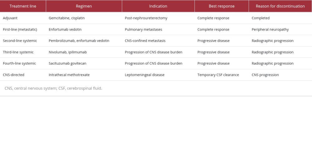 Table 1. Summary of systemic and CNS-directed therapies administered during the patient’s disease course. The table outlines the sequence of treatment lines, indications for therapy, best response, and reason for treatment discontinuation in this patient with metastatic upper tract urothelial carcinoma.Table 1. Summary of systemic and CNS-directed therapies administered during the patient’s disease course. The table outlines the sequence of treatment lines, indications for therapy, best response, and reason for treatment discontinuation in this patient with metastatic upper tract urothelial carcinoma.
 In Press
Case report 
Am J Case Rep In Press; DOI: 10.12659/AJCR.953543
Case report 
Am J Case Rep In Press; DOI: 10.12659/AJCR.954216
Case report 
Am J Case Rep In Press; DOI: 10.12659/AJCR.954932
Case report
Am J Case Rep In Press; DOI: 10.12659/AJCR.954146
Most Viewed Current Articles
07 Dec 2021 : Case report
28,712,695
DOI :10.12659/AJCR.934347
Am J Case Rep 2021; 22:e934347
06 Dec 2021 : Case report  180,721
DOI :10.12659/AJCR.934406
Am J Case Rep 2021; 22:e934406
21 Jun 2024 : Case report
124,419
DOI :10.12659/AJCR.944371
Am J Case Rep 2024; 25:e944371
08 May 2026 : Case report
95,462
DOI :10.12659/AJCR.952158
Am J Case Rep 2026; 27:e952158