


16 September 2026
: Case report 
Late Diagnosis of Noonan Syndrome Presenting With Infective Endocarditis: A Multidisciplinary Challenge in an Underserved Population
Challenging differential diagnosis, Diagnostic / therapeutic accidents, Management of emergency care, Rare disease, Congenital defects / diseases, Educational Purpose (only if useful for a systematic review or synthesis)
Nur Farhanah BDEF 1*, Jessica Novia Hadiyanto BEF 2, Sefri Noventi Sofia BD 3, Tania Tedjo Minuljo BD 4, Tri Indah Winarni DF 5
DOI: 10.12659/AJCR.952213
Am J Case Rep 2026; 27:e952213


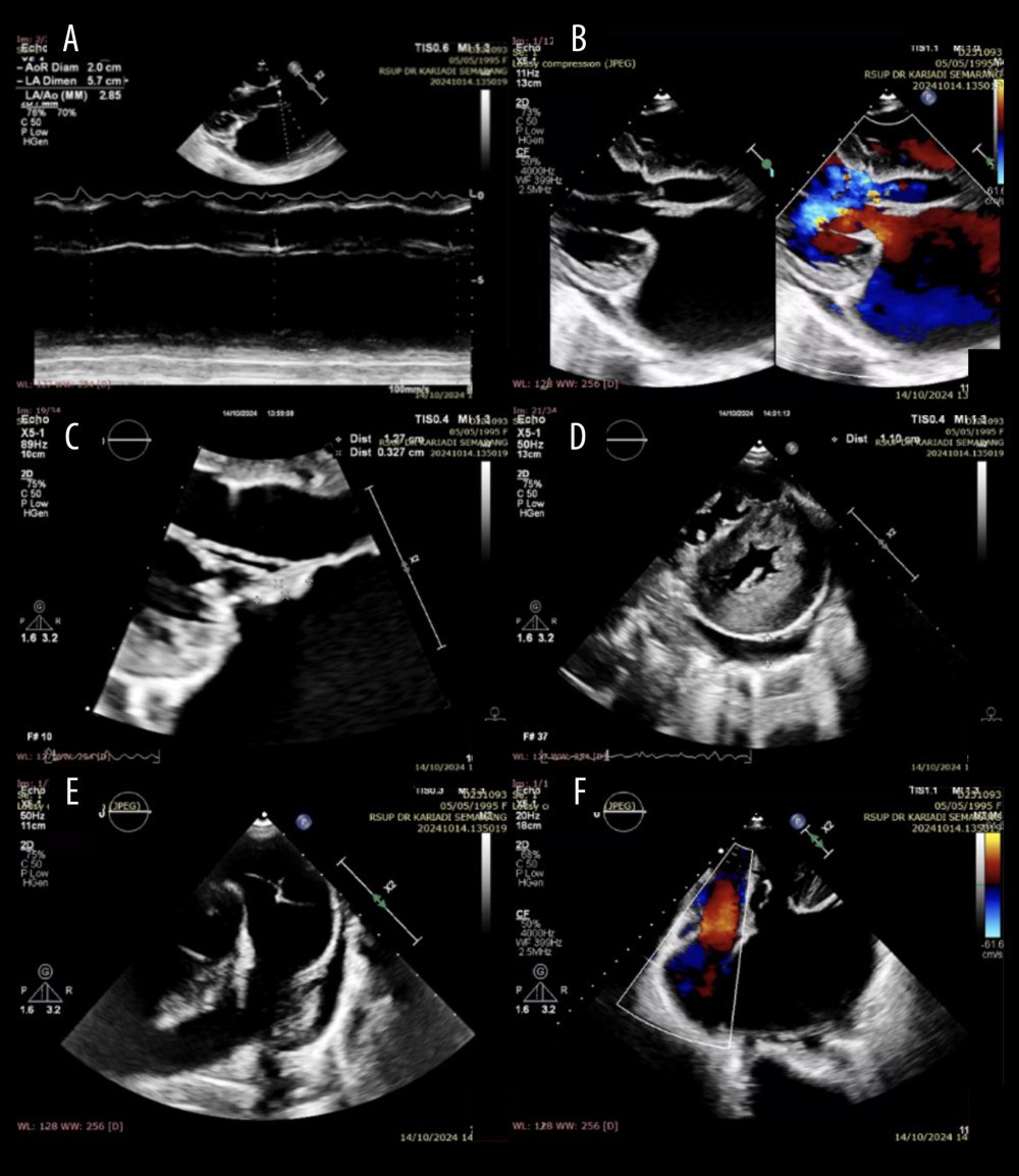
Figure 4 Echocardiogram showing mitral regurgitation with vegetations and pericardial effusion. (A) Dilated left atrium and eccentric left ventricular hypertrophy with a left ventricular ejection fraction (LVEF) of 69.9%. (B) Severe mitral regurgitation due to a flail anterior mitral leaflet (AML) with an oscillating mass on the AML. (C) Oscillating mass (+) on the AML measuring 1.2 × 0.3 cm. (D) Left ventricle (LV) showing a D-shape during systole and diastole. The LV was globally normokinetic. Loculated pericardial effusion was visible posterior to the LV. (E) A mass was present in the right ventricular outflow tract, suspected to be vegetation, pedunculated, measuring 1.3 × 0.6 cm. The main pulmonary artery was dilated. (F) Moderate tricuspid regurgitation (TR), TR vena contracta 0.3 cm, TR Vmax 2.4 m/s, MaxPG 23 mm Hg.