


16 September 2026: Articles 
Late Diagnosis of Noonan Syndrome Presenting With Infective Endocarditis: A Multidisciplinary Challenge in an Underserved Population
Challenging differential diagnosis, Diagnostic / therapeutic accidents, Management of emergency care, Rare disease, Congenital defects / diseases, Educational Purpose (only if useful for a systematic review or synthesis)
Nur Farhanah BDEF 1*, Jessica Novia Hadiyanto BEF 2, Sefri Noventi Sofia BD 3, Tania Tedjo Minuljo BD 4, Tri Indah Winarni DF 5
DOI: 10.12659/AJCR.952213
Am J Case Rep 2026; 27:e952213


Abstract
BACKGROUND: Noonan syndrome is a clinically diagnosed genetic disorder frequently associated with congenital and structural cardiac abnormalities such as valvular dysplasia and cardiomyopathy, which may predispose patients to infective endocarditis. Delayed recognition, particularly in underserved populations, can lead to advanced cardiac complications and complex presentations.
CASE REPORT: A 29-year-old woman from a rural area with no previous medical history presented with severe respiratory distress, weight loss, and newly detected cardiac murmurs. Physical examination revealed dysmorphic features suggestive of Noonan syndrome, including low-set ears, hypertelorism, short stature, and broad thorax, leading to a clinical diagnosis. Echocardiography demonstrated severe mitral regurgitation with mobile vegetations, a pedunculated mass in the right ventricular outflow tract, left atrial enlargement, reduced right ventricular systolic function (Tricuspid Annular Plane Systolic Excursion, 14 mm), and mild pericardial effusion. Blood cultures remained negative after prolonged incubation, consistent with blood culture-negative infective endocarditis. Concomitant hyperthyroidism contributed to heart failure decompensation. A multidisciplinary team initiated empirical antibiotics, heart failure therapy, thyroid control, and supportive care. The patient improved clinically and was discharged with a plan for elective valve surgery after infection control.
CONCLUSIONS: This case highlights infective endocarditis as a serious complication in late-diagnosed Noonan syndrome due to underlying structural cardiac abnormalities. Early recognition of Noonan syndrome and multidisciplinary management are essential to improve outcomes and ensure timely referral for definitive cardiac intervention.
Keywords: Case Reports, Genetics, Heart Defects, Congenital, infective endocarditis, Multidisciplinary Care, Noonan Syndrome, Case Reports, Noonan Syndrome, Endocarditis
Introduction
Noonan syndrome is an autosomal dominant genetic disorder caused by mutations in genes of the Ras-MAPK signaling pathway, crucial for cell proliferation and differentiation. The condition is characterized by distinct craniofacial features, short stature, and congenital heart defects, most commonly pulmonic stenosis and hypertrophic cardiomyopathy [1,2].
Infective endocarditis is a serious complication in patients with congenital heart defects, including those associated with Noonan syndrome. It poses diagnostic and therapeutic challenges, especially in the presence of negative blood cultures or delayed presentation [3,4]. Furthermore, infective endocarditis can exacerbate pre-existing cardiac dysfunction, leading to embolic events, severe valvular disease, or heart failure, requiring a multidisciplinary management approach [4,5].
This report discusses a case of a 29-year-old woman with Noonan syndrome and late-onset infective endocarditis, highlighting the challenges in diagnosis and management of such complex conditions. The case underscores the importance of timely diagnosis, advanced imaging modalities, and coordinated multidisciplinary care in optimizing patient outcomes.
Case Report
PHYSICAL EXAMINATION:
On admission, the patient appeared acutely ill and febrile (temperature 38.5 °C). Vital signs revealed a heart rate of 110 beats/min, blood pressure 100/60 mm Hg, respiratory rate 22 breaths/min, and oxygen saturation 92% on room air. Cardiac auscultation demonstrated a grade 3/6 holosystolic murmur at the apex radiating to the axilla, consistent with mitral regurgitation. Jugular venous pressure was elevated, and bilateral lower extremity pitting edema was present.
No peripheral stigmata of infective endocarditis (Osler nodes, Janeway lesions, Roth spots, or splinter hemorrhages) were identified.
The patient exhibited multiple dysmorphic features suggestive of Noonan syndrome, including hypertelorism, down-slanting palpebral fissures, low-set ears, short stature (< third percentile), and a broad thorax with widely spaced nipples. Cardiac findings included severe mitral regurgitation and mild pulmonary stenosis, fulfilling major diagnostic criteria, along with minor features including suspected lymphatic dysplasia. Based on the presence of multiple major and minor features, a clinical diagnosis of Noonan syndrome was established according to the van der Burgt criteria. Molecular genetic confirmation was not obtained because such testing was not available and was not readily accessible to the patient. Therefore, the diagnosis should be interpreted as a clinically established rather than genetically confirmed diagnosis (Figures 1, 2).
LABORATORY INVESTIGATIONS:
Initial laboratory evaluation demonstrated systemic inflammation with a white blood cell count of 15 000/mm3 and erythrocyte sedimentation rate (ESR) of 55 mm/h. Three sets of blood cultures obtained from separate venipuncture sites prior to antibiotic administration remained negative after standard incubation and extended monitoring up to 14 days, consistent with blood culture-negative infective endocarditis (BCNE).
Serologic testing for fastidious organisms commonly associated with BCNE (including
Additional laboratory findings included: HbA1c: 5.1%; thyroid stimulating hormone (TSH): 6.65 μIU/mL (elevated); free T4: 24.74 pmol/L (elevated). These findings were consistent with thyroid dysfunction, which was considered a contributing factor to hemodynamic instability but not central to the pathogenesis of endocarditis.
CARDIAC IMAGING:
Chest radiography demonstrated cardiomegaly with bilateral pleural effusion and pulmonary congestion, consistent with acute decompensated heart failure (Figure 3). Transthoracic echocardiography revealed severe mitral regurgitation with a mobile echodensity attached to the anterior mitral leaflet, suggestive of vegetation, as well as a pedunculated mass within the right ventricular outflow tract without evidence of obstruction. Additional findings included left atrial enlargement, preserved left ventricular systolic function, with a left ventricular ejection fraction (LVEF) of 77%, reduced right ventricular systolic function (Tricuspid Annular Plane Systolic Excursion, 14 mm), and circumferential pericardial effusion measuring 9 mm (Figure 4).
DIAGNOSIS OF INFECTIVE ENDOCARDITIS:
Based on the modified Duke criteria, the diagnosis of infective endocarditis was established based on the following: (A) 1 major criterion: echocardiographic evidence of oscillating intracardiac mass consistent with vegetation; (B) 3 minor criteria: fever > 38 °C, predisposing structural heart disease, and laboratory evidence of inflammation.
MANAGEMENT AND CLINICAL COURSE:
The patient was managed by a multidisciplinary team that included expertise in cardiology, infectious disease, endocrinology, and genetics. Empirical intravenous antimicrobial therapy was initiated for suspected BCNE. Concurrent management included heart failure therapy and thyroid dysfunction control. Clinical improvement was defined by resolution of fever, normalization of inflammatory markers, improvement in dyspnea (New York Heart Association class IV to II), reduction of peripheral edema, and hemodynamic stabilization. Follow-up echocardiography demonstrated stable valvular findings without progression of vegetation size or new complications.
FOLLOW-UP AND SURGICAL PLANNING:
Given the presence of severe mitral regurgitation secondary to structural valve disease and prior infective involvement, the patient was scheduled for elective mitral valve surgery following completion of antimicrobial therapy and confirmation of infection control. The patient subsequently underwent definitive mitral valve surgery but developed acute cardiogenic shock and died in the immediate postoperative period despite intensive resuscitative efforts.
Discussion
NOONAN SYNDROME, STRUCTURAL HEART DISEASE, AND SUSCEPTIBILITY TO INFECTIVE ENDOCARDITIS:
Noonan syndrome is commonly associated with congenital and structural heart abnormalities, including valvular dysplasia, myxomatous degeneration, pulmonic stenosis, and hypertrophic cardiomyopathy, all of which may alter intracardiac flow patterns and endothelial integrity. Although the precise incidence of infective endocarditis in patients with Noonan syndrome is not well defined in the literature, congenital heart disease itself is a recognized predisposing factor for infective endocarditis.
Although infective endocarditis has rarely been reported in patients with Noonan syndrome, several published cases support the plausibility of this association in the presence of structural cardiac abnormalities. Hatemi et al reported a 9-year-old girl with Noonan syndrome, dysplastic pulmonary valve stenosis, and pulmonary valve/pulmonary artery vegetations requiring surgery. More recently, another case described a middle-aged man with Noonan syndrome and severe pulmonary stenosis who developed isolated pulmonary valve endocarditis with pulmonary regurgitation and annular abscess, ultimately requiring surgical intervention. In contrast, our patient was an adult woman with late-recognized Noonan syndrome who presented predominantly with severe mitral regurgitation and mitral valve vegetation rather than isolated right-sided valvular disease. This difference suggests that, although pulmonary valve lesions are more commonly emphasized in Noonan syndrome, infective endocarditis in patients with Noonan syndrome may involve different valves depending on the underlying structural substrate.
Abnormal valve morphology such as thickened or dysplastic leaflets combined with chronic turbulent flow and severe regurgitation, may promote endothelial injury and facilitate bacterial adhesion, thereby increasing susceptibility to infective endocarditis. In the present case, it remains uncertain whether the severe mitral regurgitation observed represented a pre-existing structural abnormality related to Noonan syndrome or was secondary to valvular damage caused by infective endocarditis [6,7]. The presence of significant regurgitation and turbulent flow could have contributed to endothelial injury, thereby increasing susceptibility to bacterial adhesion and infection. However, it is also possible that the observed mitral regurgitation developed as a complication of the infectious process itself. Furthermore, the coexistence of Noonan syndrome and infective endocarditis in this patient may be contributory, but a coincidental occurrence cannot be entirely excluded. This highlights the complexity of establishing a direct causal relationship and underscores the importance of careful interpretation when syndromic cardiac abnormalities and infective processes overlap.
IMPACT OF DELAYED DIAGNOSIS IN UNDERSERVED POPULATIONS:
Delayed recognition of Noonan syndrome in adults remains a significant clinical challenge, particularly in underserved or resource-limited settings. Subtle phenotypic features may be overlooked in childhood, allowing progressive cardiac remodeling and valvular deterioration to remain unrecognized until advanced stages. In our patient, the absence of early diagnosis and routine cardiac surveillance likely contributed to the development of severe valvular disease complicated by infective endocarditis.
This case underscores that late diagnosis, rather than genetic predisposition alone, may play a critical role in the emergence of life-threatening complications in Noonan syndrome, emphasizing the need for heightened clinical awareness and early referral when syndromic features coexist with cardiac findings [8,9].
BLOOD CULTURE-NEGATIVE ENDOCARDITIS AND DIAGNOSTIC CHALLENGES:
The diagnosis of BCNE further complicated clinical decision-making in this case. BCNE is frequently associated with prior antibiotic exposure or infection with fastidious organisms such as
Empirical antimicrobial therapy was therefore selected to provide broad coverage for common BCNE-associated pathogens, reflecting a pragmatic approach in resource-limited settings. This highlights the importance of integrating guideline-based diagnostic frameworks with clinical judgment when definitive microbiological confirmation is unavailable [8,9].
THYROID DYSFUNCTION AS A CONTRIBUTING, NOT CAUSATIVE, FACTOR:
Thyroid function tests in this patient demonstrated an atypical pattern characterized by elevated TSH and elevated free T4 levels, which is not consistent with primary hyperthyroidism. This biochemical profile raises the possibility of alternative diagnoses such as secondary (TSH-mediated) hyperthyroidism or thyroid hormone resistance. However, further diagnostic evaluation, including pituitary imaging and dynamic endocrine testing, was not pursued given the acute clinical priorities and limited resource availability.
In the context of severe systemic illness, non-thyroidal factors, assay interference, or transient dysregulation of the hypothalamic-pituitary-thyroid axis may also contribute to such discordant findings. In this case, thyroid dysfunction was managed supportively and considered a contributing factor to hemodynamic instability rather than a primary driver of the patient’s presentation [9,10].
MULTIDISCIPLINARY MANAGEMENT AND CLINICAL IMPLICATIONS:
The patient’s preoperative management highlights the importance of multidisciplinary collaboration involving cardiology, infectious disease, endocrinology, and genetics. Coordinated management allowed simultaneous control of infection, heart failure, and metabolic derangements, while facilitating preparation for definitive surgical intervention. Despite comprehensive multidisciplinary care and successful preoperative optimization, the patient unfortunately died in the immediate postoperative period following mitral valve surgery [6,7,10].
PROGNOSIS AND FUTURE CARE:
Although the patient initially improved with multidisciplinary management and was considered suitable for mitral valve surgery, she unfortunately developed acute cardiogenic shock and died in the immediate postoperative period despite intensive resuscitative efforts. This outcome reflects the guarded prognosis of advanced infective endocarditis complicated by severe valvular destruction in patients with late-diagnosed Noonan syndrome. Consequently, long-term follow-up could not be obtained. Nevertheless, this case highlights the importance of early recognition, multidisciplinary management, and timely referral before irreversible cardiac complications develop [9,10].
Conclusions
This case demonstrates that infective endocarditis can represent a severe complication of late-diagnosed Noonan syndrome, primarily resulting from underlying structural cardiac abnormalities and delayed clinical recognition rather than confirmed molecular genetic mechanisms. The alteration of intracardiac hemodynamics by advanced valvular disease and limited access to early healthcare contributed to disease severity and diagnostic complexity. Early recognition of Noonan syndrome, systematic application of guideline-based diagnostic criteria for infective endocarditis, and timely multidisciplinary management remain essential to prevent irreversible cardiac complications, particularly in underserved populations. This case report has been reported in line with the Surgical CAse REport (SCARE) 2023 guidelines, and written informed consent was obtained from the patient for publication.
Figures
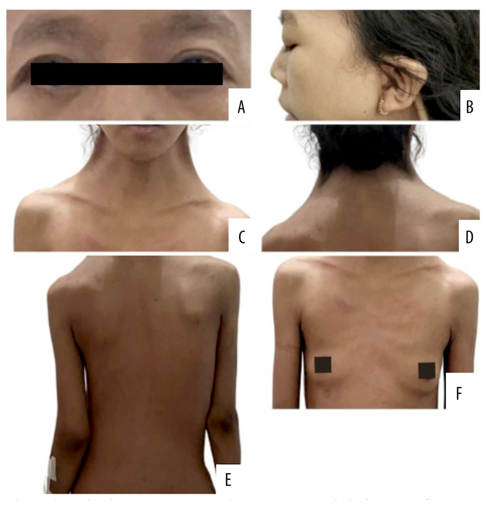 Figure 1. Patient’s clinical manifestation showing the characteristic features of Noonan syndrome. The images depict characteristic features including hypertelorism, down-slanting palpebral fissures (A), low-set ears (B), webbed neck (C), low posterior hairline (D), scoliosis (E), and a shield-shaped chest with widely spaced nipples (F).
 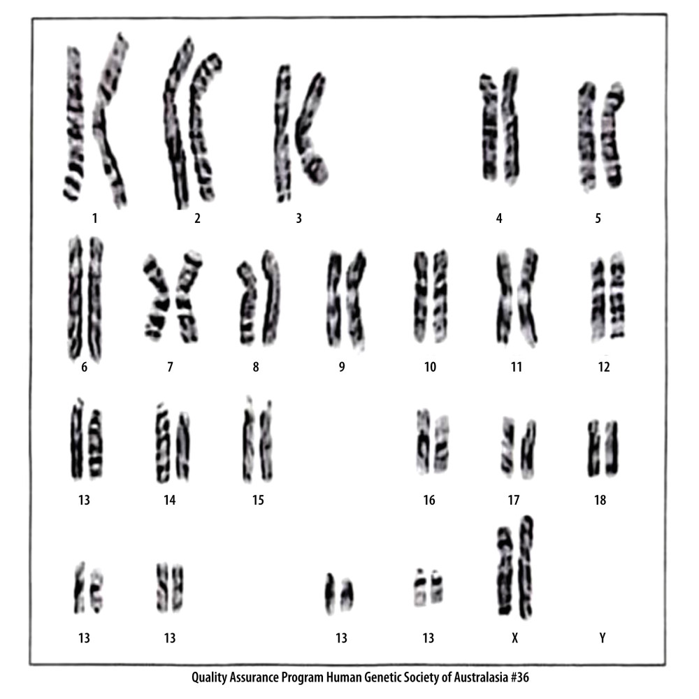 Figure 2. Karyotype analysis of the current patient showing a normal female karyotype (46, XX). Karyotyping was performed to exclude Turner syndrome, an important differential diagnosis in female patients with suspected Noonan syndrome. As expected for Noonan syndrome, no chromosomal abnormalities were identified. Molecular confirmation using a RASopathy gene panel (eg, PTPN11 and related genes) was not available.
 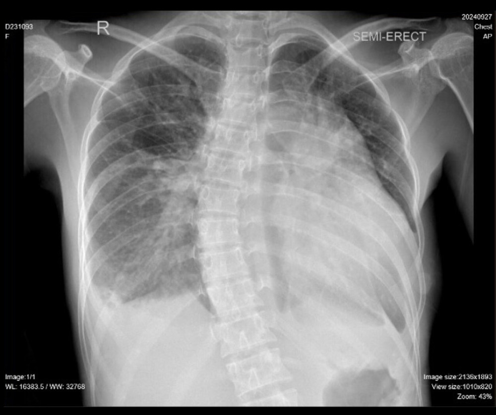 Figure 3. Chest Radiograph. Chest X-ray demonstrating cardiomegaly, pleural effusion, pulmonary edema, and vascular congestion.
 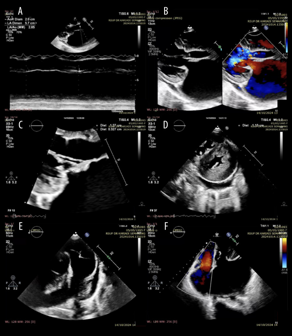 Figure 4. Echocardiogram showing mitral regurgitation with vegetations and pericardial effusion. (A) Dilated left atrium and eccentric left ventricular hypertrophy with a left ventricular ejection fraction (LVEF) of 69.9%. (B) Severe mitral regurgitation due to a flail anterior mitral leaflet (AML) with an oscillating mass on the AML. (C) Oscillating mass (+) on the AML measuring 1.2 × 0.3 cm. (D) Left ventricle (LV) showing a D-shape during systole and diastole. The LV was globally normokinetic. Loculated pericardial effusion was visible posterior to the LV. (E) A mass was present in the right ventricular outflow tract, suspected to be vegetation, pedunculated, measuring 1.3 × 0.6 cm. The main pulmonary artery was dilated. (F) Moderate tricuspid regurgitation (TR), TR vena contracta 0.3 cm, TR Vmax 2.4 m/s, MaxPG 23 mm Hg.
 References
1. Allanson JE, Roberts AE, Noonan syndrome: The evolving clinical spectrum: Eur J Pediatr, 2020; 179(5); 689-705
2. Bhattacharya D, Mathur S, Ozge A, Congenital heart defects in Noonan syndrome: Contemporary management and outcomes: Curr Cardiol Rep, 2022; 24(6); 729-41
3. Habib G, Lancellotti P, Antunes MJ, 2021 ESC Guidelines for the prevention, diagnosis, and management of infective endocarditis: Eur Heart J, 2021; 42(40); 4127-200
4. Niesert AC, Feuchtner GM, Krejci B, Infective endocarditis: Current perspectives and multidisciplinary approaches: World J Cardiol, 2021; 13(8); 251-71
5. Gopalakrishnan A, Shanmugasundaram M, Lingamaneni P, Advances in the management of congenital heart disease and endocarditis: Cardiovasc Diagn Ther, 2022; 12(2); 223-33
6. Cannon PJ, Silvestry SC, Moons P, Management of adult congenital heart disease: A focus on valvular pathology and heart failure: J Am Coll Cardiol, 2020; 75(15); 1934-48
7. Rodriguez-Gonzalez FJ, Manlhiot C, Kilburn L, Infective endocarditis in congenital heart disease: Diagnostic challenges and management strategies: Circulation, 2021; 144(8); 644-56
8. Habib G, Lancellotti P, Erba PA, 2023 ESC Guidelines for the management of infective endocarditis: Eur Heart J, 2023; 44(20); 1770-85
9. Yoon SY, Lee YJ, Jeong WJ, Coexistence of Graves’ disease and Noonan syndrome: A case report and review of the literature: Pediatr Int, 2020; 62(6); 779-82
10. Dhandapani M, Chokalingam A, Pillai A, Thyroid dysfunction and cardiac complications in genetic syndromes: A focus on Noonan Syndrome: J Clin Endocrinol Metab, 2021; 106(2); e646-e51
Figures
Figure 1. Patient’s clinical manifestation showing the characteristic features of Noonan syndrome. The images depict characteristic features including hypertelorism, down-slanting palpebral fissures (A), low-set ears (B), webbed neck (C), low posterior hairline (D), scoliosis (E), and a shield-shaped chest with widely spaced nipples (F).Figure 2. Karyotype analysis of the current patient showing a normal female karyotype (46, XX). Karyotyping was performed to exclude Turner syndrome, an important differential diagnosis in female patients with suspected Noonan syndrome. As expected for Noonan syndrome, no chromosomal abnormalities were identified. Molecular confirmation using a RASopathy gene panel (eg, PTPN11 and related genes) was not available.Figure 3. Chest Radiograph. Chest X-ray demonstrating cardiomegaly, pleural effusion, pulmonary edema, and vascular congestion.Figure 4. Echocardiogram showing mitral regurgitation with vegetations and pericardial effusion. (A) Dilated left atrium and eccentric left ventricular hypertrophy with a left ventricular ejection fraction (LVEF) of 69.9%. (B) Severe mitral regurgitation due to a flail anterior mitral leaflet (AML) with an oscillating mass on the AML. (C) Oscillating mass (+) on the AML measuring 1.2 × 0.3 cm. (D) Left ventricle (LV) showing a D-shape during systole and diastole. The LV was globally normokinetic. Loculated pericardial effusion was visible posterior to the LV. (E) A mass was present in the right ventricular outflow tract, suspected to be vegetation, pedunculated, measuring 1.3 × 0.6 cm. The main pulmonary artery was dilated. (F) Moderate tricuspid regurgitation (TR), TR vena contracta 0.3 cm, TR Vmax 2.4 m/s, MaxPG 23 mm Hg. In Press
Case report 
Am J Case Rep In Press; DOI: 10.12659/AJCR.954074
Case report 
Am J Case Rep In Press; DOI: 10.12659/AJCR.953592
Case report 
Am J Case Rep In Press; DOI: 10.12659/AJCR.954225
Case report 
Am J Case Rep In Press; DOI: 10.12659/AJCR.952870
Most Viewed Current Articles
07 Dec 2021 : Case report
28,712,481
DOI :10.12659/AJCR.934347
Am J Case Rep 2021; 22:e934347
06 Dec 2021 : Case report  180,580
DOI :10.12659/AJCR.934406
Am J Case Rep 2021; 22:e934406
21 Jun 2024 : Case report
124,319
DOI :10.12659/AJCR.944371
Am J Case Rep 2024; 25:e944371
08 May 2026 : Case report
95,142
DOI :10.12659/AJCR.952158
Am J Case Rep 2026; 27:e952158