


17 September 2026
: Case report 
Transient Endothelial Damage Following FS-LASIK Combined With Corneal Cross-Linking Surgery: Case Report
Unusual clinical course, Unusual or unexpected effect of treatment, Diagnostic / therapeutic accidents
Li Wang ACG 1, Yun Wang B 1, Xiaofeng Zhang AG 1*
DOI: 10.12659/AJCR.952499
Am J Case Rep 2026; 27:e952499


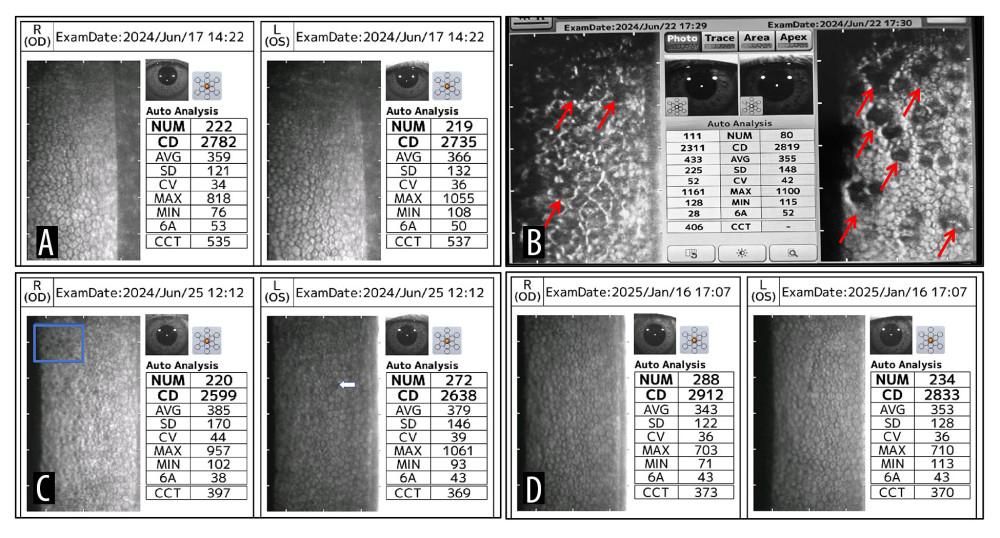
Figure 2 Images showing corneal endothelial microscopy. (A) Preoperative endothelial microscopy. (B) Corneal endothelial microscopy on postoperative day 3 showing hyporeflective zones on the endothelial surface in both eyes; the right eye (OD), affected by corneal edema, is not clearly visualized. Red arrows indicate the hyporeflective zones. (C) At 1 week postoperatively, the number and area of hyporeflective zones are significantly reduced; however, a few small hyporeflective zones remain visible (blue box in the right eye and white arrow in the left eye [OS]). (D) At 6 months postoperatively, endothelial cell density and percentage of hexagonal cells show further recovery, with no significant hyporeflective zones observed.