


17 September 2026: Articles 
Transient Endothelial Damage Following FS-LASIK Combined With Corneal Cross-Linking Surgery: Case Report
Unusual clinical course, Unusual or unexpected effect of treatment, Diagnostic / therapeutic accidents
Li Wang ACG 1, Yun Wang B 1, Xiaofeng Zhang AG 1*
DOI: 10.12659/AJCR.952499
Am J Case Rep 2026; 27:e952499


Abstract
BACKGROUND: Refractive surgery in high myopia with thinner corneas and borderline topography is challenging, with the most serious complication being regression and postoperative ectasia. We report early corneal endothelial damage in a patient with high myopia after undergoing femtosecond laser–assisted in situ keratomileusis (FS-LASIK) with corneal cross-linking.
CASE REPORT: This article reports an observational case of an 18-year-old man with high myopia who underwent FS-LASIK combined with corneal cross-linking. FS-LASIK was performed using a Zeiss femtosecond laser for flap creation (90 µm right eye, 95 µm left eye) and a EX500 excimer laser for corneal ablation (130 µm right eye, 131 µm left eye). Accelerated cross-linking was conducted with 0.23% riboflavin infiltration beneath the corneal stromal flap for 90 seconds, followed by UVA irradiation at 30 mW/cm² for 90 seconds (total energy dose, 2.7 J/cm²). On postoperative day 3, slit-lamp examination revealed corneal edema in the right eye and hyporeflective endothelial changes in both eyes. Endothelial microscopy showed significant reductions in endothelial cell density and hexagonal cell percentage compared with preoperative values. By 1 week postoperatively, the corneal edema and endothelial morphological irregularities had resolved, and the number and area of hyporeflective endothelial defects were markedly reduced. At 2 months postoperatively, visual acuity of both eyes was 20/20, hyporeflective zones on the endothelial surface disappeared, and corneal endothelial cell count, percentage of hexagonal cells, and coefficient of variation in cell size returned to preoperative levels.
CONCLUSIONS: This case highlights early, reversible endothelial damage following FS-LASIK with corneal cross-linking, underscoring the importance of careful patient selection, detailed preoperative counseling, and strong consideration of alternative treatments.
Keywords: Case Reports, Corneal Endothelial Cell Loss, Cross-Linking Reagents, Lasik, Myopia
Introduction
Due to its rapid and painless visual recovery, femtosecond laser–assisted in situ keratomileusis (FS-LASIK) has become a commonly performed laser vision correction procedure, with excellent visual outcomes and a strong safety profile. However, one of the most serious complications that can occur after laser vision correction is iatrogenic keratectasia. Postoperative corneal instability leads to the appearance of irregular astigmatism and impaired visual acuity [1]. FS-LASIK requires the creation of a corneal flap followed by excimer laser ablation of the corneal stroma, which inevitably alters corneal biomechanics, reducing postoperative corneal stability. Collagen cross-linking was first described in a clinical study in 2003 [2]. The aim of this procedure is to restore the stiffness of the cornea and thus halt the progression of a pathologic ectatic cornea, including keratoconus and iatrogenic post–laser vision correction ectasia. In the past, the use of cross-linking was combined with LASIK to reduce the risk of postoperative keratectasia by potentially strengthening the cornea weakened by laser vision correction surgery [3]. These procedures were appropriately named LASIK-Xtra [3,4].
However, with the application of riboflavin and the development of corneal cross-linking, corneal-related complications caused by the cross-linking occasionally occur. Reports of complications following cross-linking include stromal infiltrates [5], corneal haze and scarring [6], keratitis, and perforation, most of which are associated with cross-linking treatment for keratoconus. A matter of notable concern is that corneal cross-linking surgery can also include the risk of endothelial damage. Mazzotta et al [7] studied the morphological and clinical effects of topography-guided delivery of high-fluence pulsed-light UVA energy emission on the cornea and demonstrated that no endothelial damage was observed in terms of morphology and cell count. However, it cannot be ruled out that excessive ultraviolet illumination may cause transient damage to corneal endothelial cells. We present a rare case of transient endothelial injury following LASIK Xtra, emphasizing the importance of careful patient selection and risk communication. This report supports preoperative screening and surgical decision-making.
Case Report
An 18-year-old male patient presented for refractive surgery evaluation. His refractive error (−9.75 diopters sphere [DS] / −1.75 diopters cylinder [DC] × 45° corrected to 20/20 in the right eye, −10.00 DS / −1.00 DC × 160° corrected to 20/20 in the left eye) had remained stable for over a year, and his corrected visual acuity was 20/20. Corneal topography examination (Pentacam, Oculus, Germany) showed specific corneal parameters: corneal thickness measurements were 518 μm in right eye and 528 μm in left eye. Keratometry readings were 44.4/45.8 D in the right eye and 45.1/46.1 D in the left eye (Figure 1). The corneal data were measured by corneal topography (CEM-530, Nidek Co, Japan). The endothelial cell count (cells/mm2) in the right eye was 2782 ± 222 cells/mm2 and in the left eye was 2735 ± 219 cells/mm2 (Figure 2A, Table 1).
We discussed various surgical options and associated risks thoroughly with the patient, including collamer lens implantation, which was declined due to financial constraints and patient preference. Ultimately, FS-LASIK Xtra was performed using a Zeiss femtosecond laser for flap creation (90 μm right eye, 95 μm left eye) and an EX500 excimer laser for corneal ablation (130 μm right eye, 131 μm left eye). Residual stromal thickness exceeded 290 μm (298 μm right eye, 302 μm left eye). Accelerated cross-linking was conducted with 0.23% riboflavin (VibeX Rapid, Avedro, USA) for 90 seconds, followed by UVA irradiation at 30 mW/cm2 for 90 seconds (KXL system, Avedro Inc, USA), completing a total fluency dose of 2.7 J/cm2. Postoperatively, the patient was prescribed levofloxacin eye drops 4 times a day, fluorometholone eye drops 8 times a day, reduced by 1 drop every 4 days for a month, and sodium hyaluronate eye drops 4 times a day.
Three days postoperatively, uncorrected visual acuity in both eyes was 20/20. Slit lamp examination revealed mild corneal edema in the right eye, and hyporeflective endothelial changes were present in both eyes (Figure 3A, 3B, indicated by black arrows). The central corneal endothelial cell count was detected using a corneal endothelial microscope and showed a large number of hyporeflective zones on the endothelial surface of both eyes (Figure 2B, indicated by red arrows). Endothelial microscopy also revealed the endothelial cell count in the right eye was 2311 ± 111 cells/mm2, significantly lower after surgery compared with before surgery, and the percentage of hexagonal cells declined significantly from 53% before surgery to 28% after surgery (Table 1).
One week after surgery, slit lamp examination showed that corneal epithelial edema in the right eye had disappeared, hyporeflective endothelial changes had resolved, and endothelial microscopy (Figure 2C) showed a significant increase in endothelial cell count compared with 3 days postoperatively, with an increase in the percentage of hexagonal cells and a significant reduction in the number and area of hyporeflective zones on the endothelial surface, although a few small hyporeflective zones were still visible (Figure 2C, right eye, blue box; left eye, white arrow). Six months after surgery, uncorrected visual acuity was stable at 20/20, slit lamp examination showed a smooth endothelial surface, and corneal optical coherence tomography showed no significant abnormalities (Figure 3C, 3D). Endothelial parameters had also fully recovered to preoperative levels (Figure 2D). Specific values are shown in Table 1.
As the patient’s cornea did not show localized edema or posterior corneal endothelial keratic precipitates, we ruled out the diagnosis of UVA-induced corneal endotheliitis [8]. To date, this patient has been diagnosed with reversible endothelial damage following FS-LASIK combined with corneal cross-linking in both eyes.
Discussion
Based on the patient’s stable refractive history and careful risk assessment using the Randleman scoring system, the postoperative corneal ectasia risk score was 2, placing the patient in the low-risk category. Although LASIK surgery was feasible, the required high-diopter correction represented a borderline risk for ectasia, prompting the use of adjunctive cross-linking. The combination of FS-LASIK and accelerated corneal cross-linking (LASIK Xtra) has been proposed to strengthen the cornea and reduce postoperative ectasia risks. However, this approach is not without potential complications. Current reports on complications after corneal cross-linking surgery are usually related to corneal infections that occur during the postoperative epithelial healing period, most of which are concentrated on cross-linking treatment for keratoconus [9,10]. To our knowledge, this is the first reported case demonstrating complications of reversible endothelial damage after FS-LASIK combined with rapid cross-linking reinforcement surgery, reflecting the rarity of this case report.
In the standard cross-linking protocol, the surface UVA irradiance of 3 mW/cm2 has been shown to penetrate human keratocytes up to a depth of 300 μm. This irradiance is reduced to 0.15 mW/cm2 at the endothelial level, which is considered to be under the cytotoxic threshold value (0.36 mW/cm2) [11]. Meanwhile, the riboflavin shielding effect enables the absorption of excess ultraviolet light, thereby protecting endothelial cells. Mahmoud et al [12] studied the effect of LASIK with accelerated cross-linking on the corneal endothelium in patients with myopic diabetes and found that LASIK with accelerated cross-linking was safe for the corneal endothelium in this patient population, but note that a longer follow-up is needed. However, the patient in the present report developed transient corneal endothelial changes. Cingu et al [13] studied accelerated collagen cross-linking for the treatment of progressive keratoconus and reported transient endothelial changes. At early follow-up visits (1 week, 1 month, and 3 months), mean endothelial cell density was significantly lower in UVA/riboflavin cross-linking–treated eyes compared with preoperative values and also lower than that of untreated fellow eyes. All endothelial parameters recovered by 6 months in treated eyes, both compared with preoperative values and between treated and fellow eyes. These findings support the results of our present case report. This phenomenon may be explained by cellular migration, changes in reflectivity due to altered pump function [14], or reorganization of the cytoskeleton without actual apoptosis. In addition, it may also be due to cell rearrangement, hiding the actual damage to endothelial cells [15]. These corneal endothelial changes mean that the endothelium might be damaged but might not be totally lost or dead. Therefore, the recovery occurred when the undamaged endothelium slides in and replaces the function in the area of the damaged endothelium [16].
In contrast to transepithelial and epi-off cross-linking, the method we have termed under-flap cross-linking involves soaking the stromal bed with riboflavin, repositioning the flap, and then applying UV light to the corneal surface. This method allows riboflavin to rapidly and abundantly penetrate the corneal stroma, leading to increased saturation. The combination of higher riboflavin concentration and stronger UVA irradiation, resulting in greater energy delivery, may contribute to the reactive changes observed in the corneal endothelium. Meanwhile, various mechanisms may have a potential effect on the corneal endothelium during laser refractive procedures. Risks generated by an excimer laser include mechanical trauma from shock waves, oxidation, and thermal effects from UV light [17] that may harm endothelial cells. Accumulated energy delivered in procedures involving both the excimer lasers and interface-related inflammation [18] after FS-LASIK may contribute to even greater corneal endothelial cell damage [19]. Moreover, a possible interaction between excimer laser ablation and subsequent UVA exposure may further heighten endothelial vulnerability. The structural and biochemical changes induced by excimer laser treatment could alter the corneal microenvironment, potentially amplifying oxidative stress and photothermal effects when followed by cross-linking.
Current studies suggest that excessive corneal thinness (preoperative corneal thickness < 400 μm) is also a critical contributing factor to endothelial cell damage following cross-linking procedures. Nevertheless, Abbondanza et al [20] studied corneal cross-linking in corneas with thinnest pachymetry well below 400 μm using 9.0 mW/cm2 UVA irradiation for 10 minutes and found no adverse events and no loss of endothelial cell density. In another report, Gokhale [21] presented a case of severe corneal endothelial damage after the cross-linking with progressive keratoconus with a corneal thickness of more than 400 μm. The patient had presented with massive corneal edema 1 month after the cross-linking procedure, and, despite intense medical treatment, a ring-shaped corneal scar persisted, resulting in deterioration of visual acuity. These findings suggest that conventional safety parameters may not fully account for individualized corneal susceptibility, particularly in young patients with thin corneas and a need for high refractive correction. This observation may suggest increased endothelial susceptibility in younger patients; however, this hypothesis requires further validation.
Conclusions
This case underscores the critical importance of individualized patient selection and optimization of procedural parameters when performing LASIK Xtra for high myopia. Clinicians must therefore rigorously evaluate not only the static parameters (corneal thickness, Randleman score, ablation depth) but also consider dynamic risk factors such as oxidative load, riboflavin distribution, and cumulative energy exposure, while also assessing the changes in intraocular pressure in patients with high myopia [22]. For patients with endothelial damage, the energy dosage of cross-linking may be appropriately reduced based on clinical judgment. Although our patient refused to undergo implantable collamer lens surgery in this report, for highly myopic patients, implantable collamer lens surgery offers a safer alternative that preserves corneal biomechanics, avoids corneal tissue ablation, and can substantially mitigate the risk of postoperative corneal ectasia associated with corneal refractive surgery for high myopia. Implantable collamer lens surgery can avoid the need for corneal cross-linking surgery and eliminates the risk of photo-oxidative endothelial insult. Moving forward, biochemical and biomechanical modeling of riboflavin diffusion and UVA absorption in LASIK-flap–modified corneas may provide predictive metrics for endothelial safety [23,24]. Establishing individualized UVA dosage thresholds and optimizing riboflavin concentration gradients could help mitigate transient endothelial vulnerability. This report also had limitations, and longer-term clinical observation is still warranted to evaluate the endothelial cell changes observed in this patient.
Figures
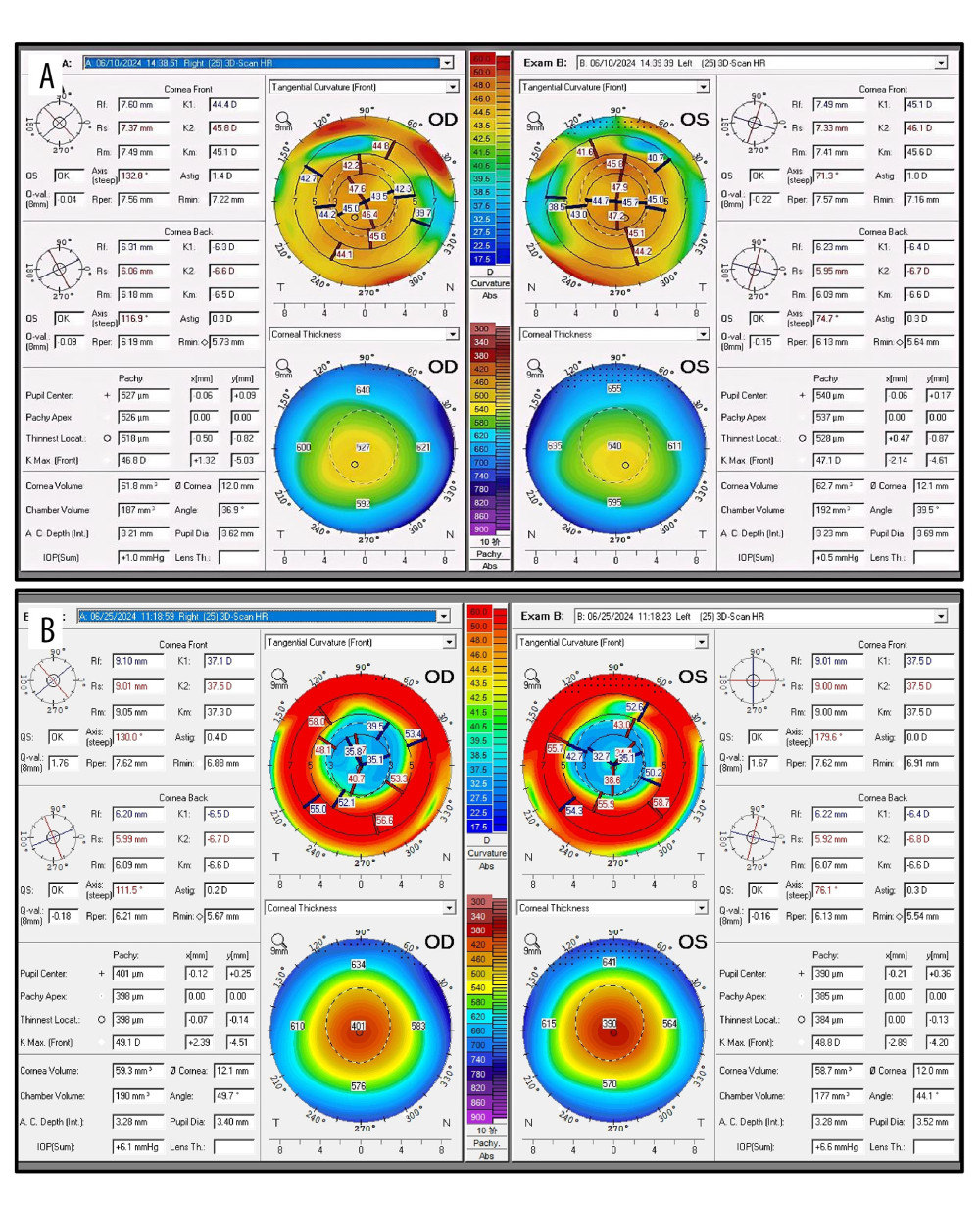 Figure 1. Images showing corneal topography. (A) Preoperative; (B) 2 days after surgery. Abbreviations: OD, right eye; OS, left eye.
 ![Images showing corneal endothelial microscopy. (A) Preoperative endothelial microscopy. (B) Corneal endothelial microscopy on postoperative day 3 showing hyporeflective zones on the endothelial surface in both eyes; the right eye (OD), affected by corneal edema, is not clearly visualized. Red arrows indicate the hyporeflective zones. (C) At 1 week postoperatively, the number and area of hyporeflective zones are significantly reduced; however, a few small hyporeflective zones remain visible (blue box in the right eye and white arrow in the left eye [OS]). (D) At 6 months postoperatively, endothelial cell density and percentage of hexagonal cells show further recovery, with no significant hyporeflective zones observed.](https://jours.isi-science.com/imageXml.php?i=amjcaserep-27-e952499-g002.jpg&idArt=952499&w=1000) Figure 2. Images showing corneal endothelial microscopy. (A) Preoperative endothelial microscopy. (B) Corneal endothelial microscopy on postoperative day 3 showing hyporeflective zones on the endothelial surface in both eyes; the right eye (OD), affected by corneal edema, is not clearly visualized. Red arrows indicate the hyporeflective zones. (C) At 1 week postoperatively, the number and area of hyporeflective zones are significantly reduced; however, a few small hyporeflective zones remain visible (blue box in the right eye and white arrow in the left eye [OS]). (D) At 6 months postoperatively, endothelial cell density and percentage of hexagonal cells show further recovery, with no significant hyporeflective zones observed.
Figure 2. Images showing corneal endothelial microscopy. (A) Preoperative endothelial microscopy. (B) Corneal endothelial microscopy on postoperative day 3 showing hyporeflective zones on the endothelial surface in both eyes; the right eye (OD), affected by corneal edema, is not clearly visualized. Red arrows indicate the hyporeflective zones. (C) At 1 week postoperatively, the number and area of hyporeflective zones are significantly reduced; however, a few small hyporeflective zones remain visible (blue box in the right eye and white arrow in the left eye [OS]). (D) At 6 months postoperatively, endothelial cell density and percentage of hexagonal cells show further recovery, with no significant hyporeflective zones observed. 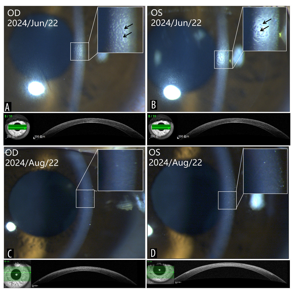 Figure 3. Images showing slit lamp examination and anterior segment optical coherence tomography (AS-OCT; Anterion, Heidelberg, Germany). (A) right (OD) and (B) left (OS) hyporeflective endothelial changes (indicated by the black arrows) visible on the endothelial surface of both eyes, with corneal edema in the right eye on the third day after surgery; (C) right and (D) left 6 months postoperatively. The corneal endothelial surface is smooth, with a small amount of stromal haze. Corneal OCT showed no significant abnormalities.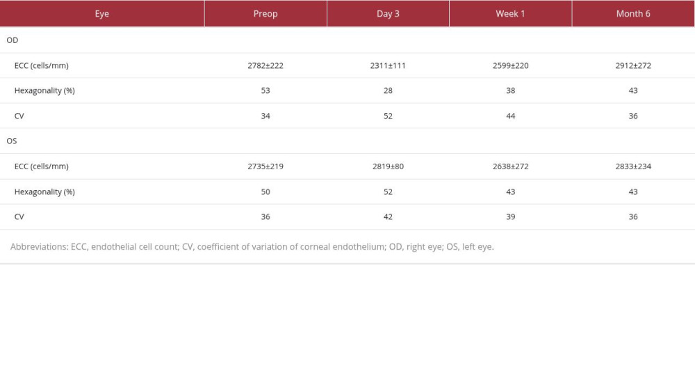
References
1. Chen YI, Chien KL, Wang IJ, An interval-censored model for predicting myopic regression after laser in situ keratomileusis: Invest Ophthalmol Vis Sci, 2007; 48(8); 3516-23
2. Wollensak G, Spoerl E, Seiler T, Riboflavin/ultraviolet-a-induced collagen crosslinking for the treatment of keratoconus: Am J Ophthalmol, 2003; 135(5); 620-27
3. Kymionis GD, Corneal collagen cross linking – PLUS: Open Ophthalmol J, 2011; 5; 10
4. Randleman JB, Woodward M, Lynn MJ, Stulting RD, Risk assessment for ectasia after corneal refractive surgery: Ophthalmology, 2008; 115(1); 37-50
5. Mangioris GF, Papadopoulou DN, Balidis MO, Corneal infiltrates after corneal collagen cross-linking: J Refract Surg, 2010; 26(8); 609-11
6. Güell JL, Verdaguer P, Elies D, Late onset of a persistent, deep stromal scarring after PRK and corneal cross-linking in a patient with forme fruste keratoconus: J Refract Surg, 2014; 30(4); 286-88
7. Mazzotta C, Moramarco A, Traversi C, Accelerated corneal collagen cross-linking using topography-guided UV-A energy emission: Preliminary clinical and morphological outcomes: J Ophthalmol, 2016; 2016; 2031031
8. Gumus K, Acute idiopathic endotheliitis early after corneal cross-linking with riboflavin and ultraviolet-A: Cornea, 2014; 33(6); 630-33
9. Fuchs B, Wendelstein J, Mariacher S, Late onset of a persistent, deep stromal and endothelial scarring after corneal collagen crosslinking for keratoconus: A case report: Eur J Ophthalmol, 2022; 32(6); 3195-200
10. Yurttaser Ocak S, Mangan MS, Endothelial cell loss after accelerated corneal crosslinking using pachymetry-guided hypo-osmolar riboflavin dosing in thin keratoconic corneas: J Cataract Refract Surg, 2021; 47(12); 1530-34
11. Wollensak G, Spoerl E, Reber F, Seiler T, Keratocyte cytotoxicity of riboflavin/UVA-treatment in vitro: Eye (Lond), 2004; 18(7); 718-22
12. Mahmoud MSED, Hassan EE, Abdelhalim AS, Evaluation of corneal endothelium after myopic Laser-Assisted In SituKeratomileusis (LASIK) with accelerated cross-linking (cross-linking) in diabetic patients: Open Ophthalmol J, 2021; 15; 329-37
13. Cingü AK, Sogutlu-Sari E, Cınar Y, Transient corneal endothelial changes following accelerated collagen cross-linking for the treatment of progressive keratoconus: Cutan Ocul Toxicol, 2014; 33(2); 127-31
14. Dong C, Zou D, Duan H, Ex vivo cultivated retinal pigment epithelial cell transplantation for the treatment of rabbit corneal endothelial dysfunction: Eye Vis (Lond), 2023; 10(1); 34
15. Amoozadeh J, Aliakbari S, Behesht-Nejad AH, Confocal microscopy of corneal stroma and endothelium after LASIK and PRK: J Refract Surg, 2009; 25(10 Suppl); S963-67
16. Mohan RR, Hutcheon AE, Choi R, Apoptosis, necrosis, proliferation, and myofibroblast generation in the stroma following LASIK and PRK: Exp Eye Res, 2003; 76(1); 71-87
17. Juda M, Bedliński M, Roszkowska AM, Wierzbowska J, Clinical evaluation of corneal endothelial parameters following laser refractive surgery in myopic eyes: A review: J Clin Med, 2024; 13(6); 1665
18. Al-Karawi AS, Kadhim AS, Correlation of autoimmune response and immune system components in the progression of IgA nephropathy: A comparative study: Hum Immunol, 2024; 85(6); 111181
19. Özbilen KT, Altinkurt E, Ceylan NA, Effect of myopic femtosecond laser-assisted LASIK on anterior chamber inflammation (flare values) and corneal endothelium: A prospective before and after study: J Ophthalmol, 2021; 2021; 2395028
20. Abbondanza M, Wong ZSY, De Felice V, Abbondanza G, Customised peripheral corneal cross-linking (P-CXL) for ultra-thin corneas with stage III and IV keratoconus: Semin Ophthalmol, 2023; 38(7); 630-37
21. Gokhale NS, Corneal endothelial damage after collagen cross-linking treatment: Cornea, 2011; 30(12); 1495-98
22. Al-Essa RS, Turjoman AA, Acute attack of primary angle closure in a highly axially myopic eye: A case report: Am J Case Rep, 2021; 22; e931002
23. Kadhim AS, Al-Karawi AS, Correlation between vitamin D3 Levels, autoantibodies, and antibody-related diseases in patients with Hashimoto’s thyroiditis: Turk J Immunol, 2024; 12(3); 209-19
24. Kadhim HI, Kadhim AS, Exploring the association between obesity, inflammation, and type II diabetes: Insights from body mass index correlation and immune response analysis: Al-Nahrain J Sci, 2024; 27(3); 50-55
Figures
Figure 1. Images showing corneal topography. (A) Preoperative; (B) 2 days after surgery. Abbreviations: OD, right eye; OS, left eye.Figure 2. Images showing corneal endothelial microscopy. (A) Preoperative endothelial microscopy. (B) Corneal endothelial microscopy on postoperative day 3 showing hyporeflective zones on the endothelial surface in both eyes; the right eye (OD), affected by corneal edema, is not clearly visualized. Red arrows indicate the hyporeflective zones. (C) At 1 week postoperatively, the number and area of hyporeflective zones are significantly reduced; however, a few small hyporeflective zones remain visible (blue box in the right eye and white arrow in the left eye [OS]). (D) At 6 months postoperatively, endothelial cell density and percentage of hexagonal cells show further recovery, with no significant hyporeflective zones observed.Figure 3. Images showing slit lamp examination and anterior segment optical coherence tomography (AS-OCT; Anterion, Heidelberg, Germany). (A) right (OD) and (B) left (OS) hyporeflective endothelial changes (indicated by the black arrows) visible on the endothelial surface of both eyes, with corneal edema in the right eye on the third day after surgery; (C) right and (D) left 6 months postoperatively. The corneal endothelial surface is smooth, with a small amount of stromal haze. Corneal OCT showed no significant abnormalities. In Press
Case report
Am J Case Rep In Press; DOI: 10.12659/AJCR.953317
Case report 
Am J Case Rep In Press; DOI: 10.12659/AJCR.954643
Case report 
Am J Case Rep In Press; DOI: 10.12659/AJCR.954074
Case report 
Am J Case Rep In Press; DOI: 10.12659/AJCR.953592
Most Viewed Current Articles
07 Dec 2021 : Case report
28,712,429
DOI :10.12659/AJCR.934347
Am J Case Rep 2021; 22:e934347
06 Dec 2021 : Case report  180,530
DOI :10.12659/AJCR.934406
Am J Case Rep 2021; 22:e934406
21 Jun 2024 : Case report
124,276
DOI :10.12659/AJCR.944371
Am J Case Rep 2024; 25:e944371
08 May 2026 : Case report
95,045
DOI :10.12659/AJCR.952158
Am J Case Rep 2026; 27:e952158