


28 August 2026
: Case report 
Aggressive Sporadic Renal Angiomyolipoma With Tumor Thrombus Extending Into the Inferior Vena Cava, Right Atrium, and Right Ventricle: A Multidisciplinary Case Report
Unusual clinical course, Challenging differential diagnosis, Unusual setting of medical care
Caroline J. Cushman ABCDEF 1*, Kolos K. Nagy EF 1, Rodolfo Martinez D 2, Kaiser R. Tarafdar ABCDEF 3
DOI: 10.12659/AJCR.952664
Am J Case Rep 2026; 27:e952664


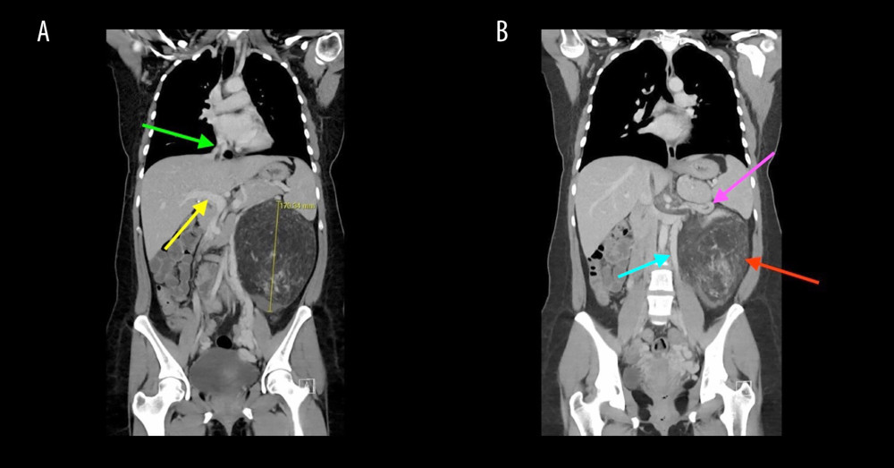
Figure 2 Coronal views of contrast-enhanced computed tomography demonstrating a giant left renal angiomyolipoma measuring 19.5 × 18.0 × 10.5 cm (maximal dimension 17.0 cm) with a contiguous tumor thrombus measuring approximately 18 cm in length extending through the left renal vein into the inferior vena cava and reaching the right atrium. Panel (A) demonstrates the inferior vena cava (green arrow) and portal vein (yellow arrow), shown for anatomic orientation and illustrating the path of tumor thrombus extension toward the heart. Panel (B) demonstrates the primary left renal angiomyolipoma (red arrow) with tumor thrombus extending through the left renal vein (pink arrow). A dilated left ovarian vein varix consistent with secondary pelvic congestion syndrome due to high-flow tumor physiology is also visualized (blue arrow). No infiltrative parenchymal or metastatic disease is identified within the abdomen, pelvis, or chest.