


28 August 2026: Articles 
Aggressive Sporadic Renal Angiomyolipoma With Tumor Thrombus Extending Into the Inferior Vena Cava, Right Atrium, and Right Ventricle: A Multidisciplinary Case Report
Unusual clinical course, Challenging differential diagnosis, Unusual setting of medical care
Caroline J. Cushman ABCDEF 1*, Kolos K. Nagy EF 1, Rodolfo Martinez D 2, Kaiser R. Tarafdar ABCDEF 3
DOI: 10.12659/AJCR.952664
Am J Case Rep 2026; 27:e952664


Abstract
BACKGROUND: Renal angiomyolipoma (AML) is a typically benign mesenchymal tumor with an indolent clinical course. Rarely, AML demonstrate aggressive vascular invasion, most commonly in association with tuberous sclerosis complex (TSC). Intracardiac extension, particularly into the right ventricle, is exceptionally uncommon in sporadic cases and presents significant diagnostic and surgical challenges.
CASE REPORT: A 36-year-old woman with a history of rheumatoid arthritis presented with progressive left upper-quadrant abdominal pain, nausea, and back discomfort. Cross-sectional imaging revealed a massive left renal angiomyolipoma measuring 19.5×18.0×10.5 cm, with a contiguous tumor thrombus extending from the left renal vein through the inferior vena cava into the right atrium, intermittently prolapsing across the tricuspid valve into the right ventricle. Echocardiography confirmed a mobile intracardiac mass without evidence of valvular obstruction. Genetic testing was negative for TSC1 and TSC2 mutations, consistent with a sporadic angiomyolipoma. The patient underwent successful en bloc left radical nephrectomy with complete venous thrombectomy and inferior vena cava reconstruction through a coordinated multidisciplinary approach. Final pathology confirmed complete excision with negative margins and absence of epithelioid features.
CONCLUSIONS: This case was an exceptionally rare presentation of sporadic renal angiomyolipoma with extensive intracardiac extension into the right ventricle. It demonstrates that significant vascular and cardiac involvement can occur even in the absence of TSC-associated genetic mutations or epithelioid histology, thereby challenging traditional assumptions regarding AML behavior. These findings underscore the importance of early recognition, comprehensive imaging, and individualized operative planning in the management of complex AML presentations. Given the potential for aggressive progression in otherwise benign tumors, close long-term radiographic surveillance remains essential.
Keywords: Angiomyolipoma, Multidisciplinary Care, Vascular Neoplasms
Introduction
Angiomyolipoma (AML) is a benign mesenchymal renal tumor composed of variable proportions of blood vessels, smooth muscle, and adipose tissue. Although typically asymptomatic and discovered incidentally, AMLs occasionally demonstrate aggressive behavior, including extrarenal extension, perinephric invasion, and, in rare instances, vascular invasion into the renal vein and inferior vena cava (IVC). Extension into the right atrium is exceedingly uncommon and can lead to significant morbidity, necessitating a coordinated multidisciplinary approach. Right ventricular extension is rarer still and has been reported almost exclusively in patients with tuberous sclerosis complex (TSC). AMLs with IVC and intracardiac extension are reported to occur almost exclusively in females, a pattern consistent with the current case [1]. Here, we present an exceptionally rare case of sporadic renal AML with tumor thrombus extending through the left renal vein, IVC, right atrium, and right ventricle in the absence of TSC1 or TSC2 mutations. This case underscores the importance of recognizing atypical and aggressive presentations of AML and demonstrates the complex interdisciplinary management required for extensive vascular involvement.
Case Report
SURGICAL TECHNIQUE AND INTRAOPERATIVE MANAGEMENT:
The procedure was performed via a midline laparotomy without median sternotomy. Cardiopulmonary bypass was not used; however, extracorporeal circulation was available on standby. Vascular control was obtained at multiple levels prior to cavotomy. The infrarenal inferior vena cava and contralateral renal vein were first isolated and controlled. Proximal control of the central inferior vena cava was achieved distal to the hepatic vein junction, allowing safe isolation of the thrombus-bearing segment while preserving hepatic venous outflow. This stepwise vascular control minimized the risk of tumor embolization and permitted controlled cavotomy.
Sequential clamping of the renal vein and inferior vena cava allowed en bloc extraction of the tumor thrombus through the venous system without entry into the cardiac chambers. To reduce the risk of pulmonary embolization, thrombus manipulation was minimized and extraction was performed under direct visualization. Continuous intraoperative transesophageal echocardiography was used to monitor thrombus position and confirm complete removal during the procedure.
Following thrombectomy, the inferior vena cava was reconstructed primarily without patch grafting. The cavotomy was closed with running nonabsorbable suture, restoring unobstructed caval flow. Hemodynamic changes associated with temporary inferior vena cava occlusion were managed with coordinated anesthetic support including volume resuscitation and vasopressor administration as needed. Intraoperative blood conservation strategies were implemented, and an autologous cell salvage system was available for blood recovery during the procedure.
The patient subsequently underwent en bloc left radical nephrectomy with complete venous thrombectomy. The resected specimen weighed 2111 g (2.11 kg). Final pathology confirmed complete excision with negative surgical margins (Figure 3), and no epithelioid component was identified.
Total operative time was approximately 6.5 hours with an estimated blood loss of 900 mL. No intraoperative arrhythmias or hemodynamic instability occurred. Postoperatively, the patient required mechanical ventilation for 12 hours and was discharged on postoperative day 7 without complications. At 6-month follow-up, she remained asymptomatic with no evidence of recurrence on surveillance CT and echocardiography.
Discussion
Renal angiomyolipoma (AML) is a benign tumor of the kidney, composed of mature adipose tissue, smooth muscle, and blood vessels, generally considered low risk for extension, invasion, or metastasis [1]. AML is the most common benign renal tumor, although they account for 0.3% to 3% of all renal neoplasms [2]. They most often occur as sporadic cases (80%), but they are also associated with inherited conditions, such as tuberous sclerosis and lymphangioleiomyomatosis. In the setting of hereditary conditions, AML tends to be more aggressive, manifesting at an earlier age, having a higher likelihood of bilaterality, and is usually larger at time of diagnosis [2].
AML typically presents as an incidental finding, but flank pain, hematuria, and palpable mass can occur, with retroperitoneal hemorrhage being the primary clinical concern [1]. The average age of presentation is 52 years and it is more common in middle-aged women [2,3]. The majority of AMLs are asymptomatic and remain stable in size, but approximately 9% of AMLs increase in size over time [2]. After reaching a certain size, AMLs are more likely to grow, become symptomatic, and undergo malignant transformation [2].
The adipose-rich component of angiomyolipoma (AML) is readily identifiable on CT and MRI; however, differentiation among histologic subtypes requires careful radiologic–pathologic correlation [4]. Renal AMLs are histologically classified as typical (triphasic) or atypical (monophasic or epithelioid) tumors. Triphasic AMLs consist of varying proportions of abnormal blood vessels, smooth muscle, and mature adipose tissue, whereas monophasic AMLs demonstrate predominance of a single component, with minimal representation of the others [2]. The epithelioid variant, in particular, has been associated with malignant potential and is the most common subtype implicated in extrarenal extension and vascular invasion (Figure 4) [4]. Epithelioid angiomyolipoma is therefore considered a biologically distinct entity with increased risk of aggressive behavior, recurrence, and metastatic potential, prompting recommendations for long-term serial radiographic surveillance, even following complete surgical resection with negative margins.
According to Kheir et al, venous invasion of AML is rare, with only 90 cases reported in the literature, 76 of which involved the inferior vena cava (IVC) [3]. Intracardiac extension in particular has been predominantly reported in female patients [5]. Although up to 75% of individuals with tuberous sclerosis complex (TSC) develop renal angiomyolipomas, very few demonstrate venous invasion, and an even fewer extend into the cardiac chambers [2]. Right-atrial involvement is rare, and right ventricular extension has been described almost exclusively in the setting of TSC-associated AML. The presence of such extensive intracardiac propagation in our patient without TSC1 or TSC2 mutations therefore is an exceptionally unusual biological variant and this may be the first reported case of sporadic AML extending into the right ventricle. The delayed onset of symptoms in this case, despite massive tumor burden extending from the renal vein through the IVC and into the cardiac chambers, is atypical and underscores the necessity for a high index of clinical suspicion and refined diagnostic sensitivity. AMLs demonstrating such extensive vascular and intracardiac propagation are classically associated with tuberous sclerosis complex; therefore, the emergence of this aggressive phenotype in a patient without these mutations challenges conventional paradigms and highlights a uniquely rare biological variant in which significant tumor progression occurs independently of TSC-related genetic dysregulation [5].
Our patient’s history of rheumatoid arthritis raises an additional point of academic interest. While no established association exists between rheumatoid arthritis and AML, this case illustrates that exceptionally sporadic overlap can occur even in the absence of TSC mutations [6]. However, any potential relationship remains speculative, and the current literature does not support a causal or mechanistic link [6].
Previously published case reports of renal angiomyolipoma with venous extension have primarily described tumor thrombus propagation into the renal vein and inferior vena cava, with occasional extension into the right atrium, most often in patients with tuberous sclerosis complex [3–5]. Reports emphasizing intracardiac involvement highlight the importance of careful image selection and radiologic characterization to define tumor extent and guide operative strategy. Compared with these cases, the present case is distinguished by right ventricular extension, absence of TSC1 and TSC2 mutations, and successful heart-sparing en bloc tumor extraction without cardiopulmonary bypass. These features suggest a distinct biological and surgical phenotype and contribute novel insight to the limited existing literature on aggressive sporadic AML behavior.
The extent of vascular involvement, culminating in a tumor thrombus occupying the IVC, right atrium, and intermittently prolapsing across the tricuspid valve, posed the potential for profound hemodynamic and surgical challenges. Only a handful of such cases have been documented, and the degree of intracardiac involvement necessitated the coordinated expertise of cardiovascular, urologic, and vascular surgical teams to mitigate life-threatening risks, including cardiac obstruction, embolization, and sudden circulatory collapse.
Increase in the size of renal angiomyolipomas is associated with a higher likelihood of symptomatic presentation, hemorrhage, and vascular invasion [1]. Surgical intervention or embolization is generally recommended for symptomatic lesions, tumors exceeding 4 cm, lesions with concerning radiographic features, or in women of childbearing age [2]. Renal artery embolization is often employed as a first-line therapy in select cases, with reported success rates exceeding 90% and average tumor size reduction of approximately 38% [2]. However, in the present case, complete nephrectomy was the most appropriate therapeutic option due to the massive tumor size, extensive contiguous vascular and intracardiac extension, and the inability to safely pursue a nephron-sparing approach. Importantly, no epithelioid component was identified on final pathology, demonstrating that aggressive vascular behavior can occur even in the absence of histologic features traditionally associated with malignant potential.
Management of hereditary AML can include mTOR inhibitor therapy in conjunction with surgical or endovascular interventions to counteract dysregulated mTOR signaling arising from TSC1 or TSC2 mutations [2]. However, the absence of TSC mutations in our patient underscores the existence of a rare sporadic AML phenotype capable of aggressive intravascular progression independent of known genetic drivers.
Conclusions
This case is an exceptionally rare presentation of sporadic renal angiomyolipoma characterized by a massive tumor burden measuring 19.5 × 18.0 × 10.5 cm and weighing 2.111 kg, with contiguous tumor thrombus extension through the inferior vena cava into the cardiac chambers. The extent and complexity of vascular invasion necessitated a highly coordinated multidisciplinary approach to diagnosis, operative planning, and management. The absence of TSC1 and TSC2 mutations emphasizes the rarity of this case, as aggressive AML behavior is typically, but not exclusively, associated with tuberous sclerosis complex. Successful excision of the tumor demonstrates the critical role of coordinated, interdisciplinary expertise in managing cases with extensive vascular and intracardiac involvement. Ongoing follow-up will focus on long-term serial radiographic surveillance for recurrence and management of the patient’s underlying conditions. Early recognition, careful planning, and timely intervention remain essential for optimizing outcomes in angiomyolipomas exhibiting atypical and aggressive behavior, especially in patients with an underlying hereditary disposition.
Figures
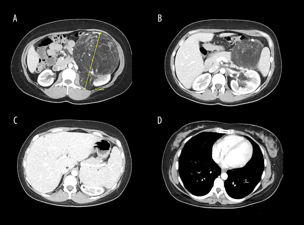 Figure 1. Axial views of contrast-enhanced computed tomography demonstrating a large left renal angiomyolipoma (AML). The tumor measures 16.7 cm in maximal axial dimension and arises from the left kidney (A). Associated tumor thrombus is visualized within the left renal vein causing partial luminal occlusion (B), extending into the inferior vena cava (IVC) (C), and reaching the right atrium adjacent to the tricuspid valve, with intermittent prolapse into the right ventricle (D).
 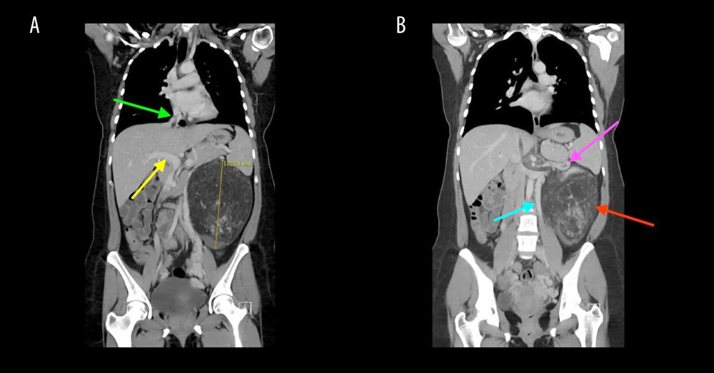 Figure 2. Coronal views of contrast-enhanced computed tomography demonstrating a giant left renal angiomyolipoma measuring 19.5 × 18.0 × 10.5 cm (maximal dimension 17.0 cm) with a contiguous tumor thrombus measuring approximately 18 cm in length extending through the left renal vein into the inferior vena cava and reaching the right atrium. Panel (A) demonstrates the inferior vena cava (green arrow) and portal vein (yellow arrow), shown for anatomic orientation and illustrating the path of tumor thrombus extension toward the heart. Panel (B) demonstrates the primary left renal angiomyolipoma (red arrow) with tumor thrombus extending through the left renal vein (pink arrow). A dilated left ovarian vein varix consistent with secondary pelvic congestion syndrome due to high-flow tumor physiology is also visualized (blue arrow). No infiltrative parenchymal or metastatic disease is identified within the abdomen, pelvis, or chest.
 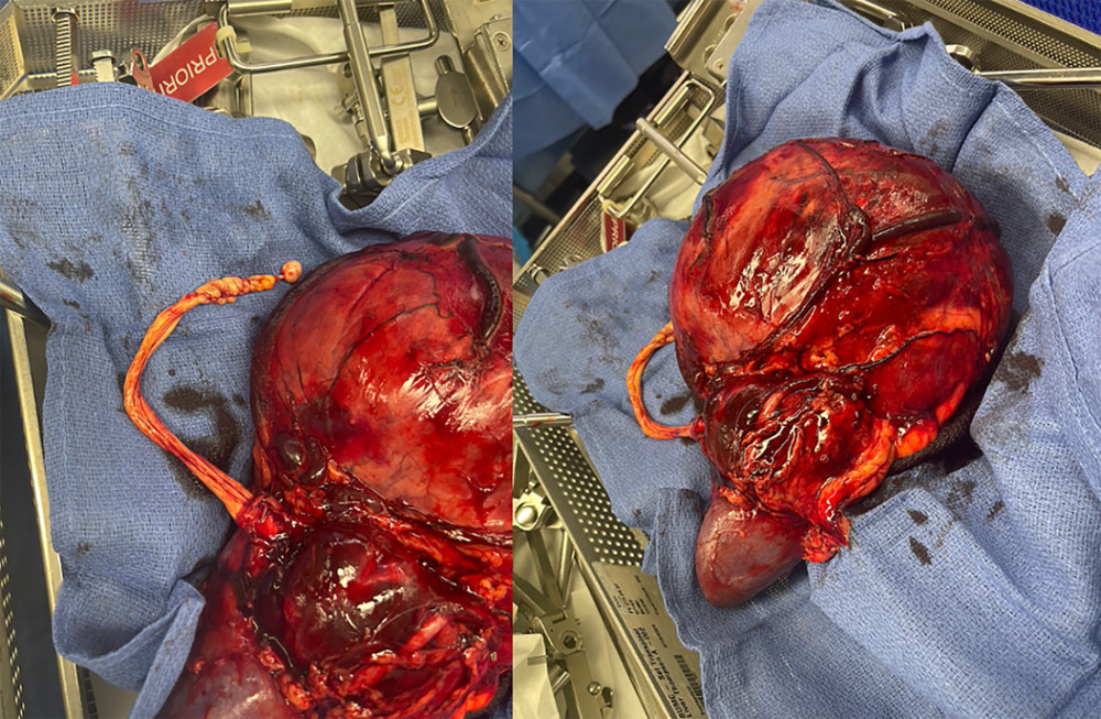 Figure 3. Intraoperative images demonstrating en bloc removal of the left renal angiomyolipoma with complete excision of the extensive tumor thrombus extending from the renal vein through the inferior vena cava.
 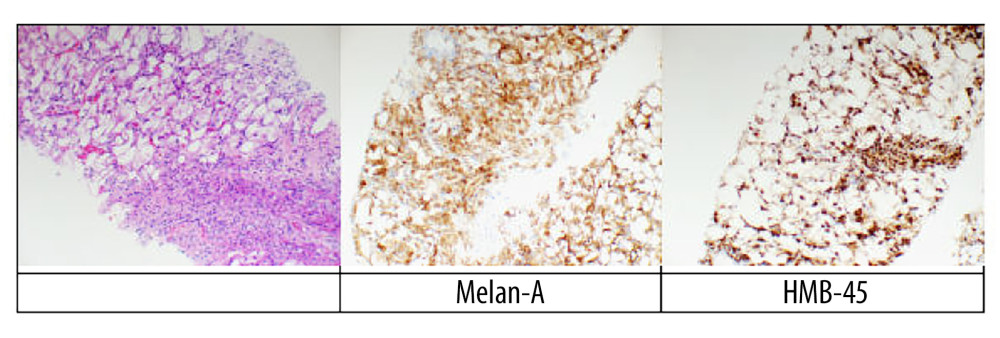 Figure 4. Histopathologic findings of renal angiomyolipoma. Low-power hematoxylin and eosin (H&E) section demonstrates classic triphasic morphology with an admixture of mature adipose tissue, thick-walled blood vessels, and smooth muscle bundles (× 40). Higher-magnification view highlights perivascular epithelioid cells with clear cytoplasm and mild nuclear pleomorphism (× 200). Immunohistochemical staining demonstrates positivity for HMB-45 and smooth muscle actin, confirming the diagnosis of angiomyolipoma.
 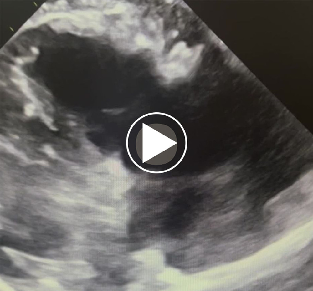 Video 1. Transthoracic echocardiography demonstrating a mobile angiomyolipoma-associated tumor thrombus within the right atrium, intermittently prolapsing across the tricuspid valve into the right ventricle during diastole.
 References
1. Perdue J, Wells A, Patel K, Multidisciplinary approach to the management of renal angiomyolipoma with inferior vena cava thrombus and pulmonary embolism: A case report: J Surg Case Rep, 2024; 2024(3); rjae174
2. Shamam YM, Leslie SW, Renal angiomyolipoma: StatPearls, 2024, Treasure Island, FL, StatPearls Publishing
3. Kheir P, Abdessater M, El Khoury J, Renal angiomyolipoma with IVC thrombus: A case report: Int J Surg Case Rep, 2020; 70; 149-53
4. Jinzaki M, Silverman SG, Akita H, Renal angiomyolipoma: A radiological classification and update on recent developments in diagnosis and management: Abdom Imaging, 2014; 39(3); 588-604
5. Priyadarshini P, Bisoi AK, Chauhan S, Benevolent renal angiomyolipoma with intracardiac extension: A challenge in diagnosis and management: Indian J Surg, 2017; 79(3); 259-61
6. Kronbichler A, Mayer G, Renal involvement in autoimmune connective tissue diseases: BMC Med, 2013; 11; 95
Figures
Figure 1. Axial views of contrast-enhanced computed tomography demonstrating a large left renal angiomyolipoma (AML). The tumor measures 16.7 cm in maximal axial dimension and arises from the left kidney (A). Associated tumor thrombus is visualized within the left renal vein causing partial luminal occlusion (B), extending into the inferior vena cava (IVC) (C), and reaching the right atrium adjacent to the tricuspid valve, with intermittent prolapse into the right ventricle (D).Figure 2. Coronal views of contrast-enhanced computed tomography demonstrating a giant left renal angiomyolipoma measuring 19.5 × 18.0 × 10.5 cm (maximal dimension 17.0 cm) with a contiguous tumor thrombus measuring approximately 18 cm in length extending through the left renal vein into the inferior vena cava and reaching the right atrium. Panel (A) demonstrates the inferior vena cava (green arrow) and portal vein (yellow arrow), shown for anatomic orientation and illustrating the path of tumor thrombus extension toward the heart. Panel (B) demonstrates the primary left renal angiomyolipoma (red arrow) with tumor thrombus extending through the left renal vein (pink arrow). A dilated left ovarian vein varix consistent with secondary pelvic congestion syndrome due to high-flow tumor physiology is also visualized (blue arrow). No infiltrative parenchymal or metastatic disease is identified within the abdomen, pelvis, or chest.Figure 3. Intraoperative images demonstrating en bloc removal of the left renal angiomyolipoma with complete excision of the extensive tumor thrombus extending from the renal vein through the inferior vena cava.Figure 4. Histopathologic findings of renal angiomyolipoma. Low-power hematoxylin and eosin (H&E) section demonstrates classic triphasic morphology with an admixture of mature adipose tissue, thick-walled blood vessels, and smooth muscle bundles (× 40). Higher-magnification view highlights perivascular epithelioid cells with clear cytoplasm and mild nuclear pleomorphism (× 200). Immunohistochemical staining demonstrates positivity for HMB-45 and smooth muscle actin, confirming the diagnosis of angiomyolipoma.Video 1. Transthoracic echocardiography demonstrating a mobile angiomyolipoma-associated tumor thrombus within the right atrium, intermittently prolapsing across the tricuspid valve into the right ventricle during diastole. In Press
Case report 
Am J Case Rep In Press; DOI: 10.12659/AJCR.953317
Case report 
Am J Case Rep In Press; DOI: 10.12659/AJCR.954643
Case report 
Am J Case Rep In Press; DOI: 10.12659/AJCR.954074
Case report
Am J Case Rep In Press; DOI: 10.12659/AJCR.953592
Most Viewed Current Articles
07 Dec 2021 : Case report
28,712,429
DOI :10.12659/AJCR.934347
Am J Case Rep 2021; 22:e934347
06 Dec 2021 : Case report  180,530
DOI :10.12659/AJCR.934406
Am J Case Rep 2021; 22:e934406
21 Jun 2024 : Case report
124,276
DOI :10.12659/AJCR.944371
Am J Case Rep 2024; 25:e944371
08 May 2026 : Case report
95,045
DOI :10.12659/AJCR.952158
Am J Case Rep 2026; 27:e952158