


20 August 2026
: Case report 
Autosomal Dominant Polycystic Kidney Disease Presenting as a Painless Epigastric Mass: A Case Report
Unusual clinical course, Challenging differential diagnosis
Tajul Atiqah Zaulkafali ABCDEF 1, Aiza Nur Izdihar Zainal-Abidin ACDE 1, Alan Basil Peter BCDE 2, Mohamed-Syarif Mohamed-Yassin ACDE 1*
DOI: 10.12659/AJCR.952751
Am J Case Rep 2026; 27:e952751


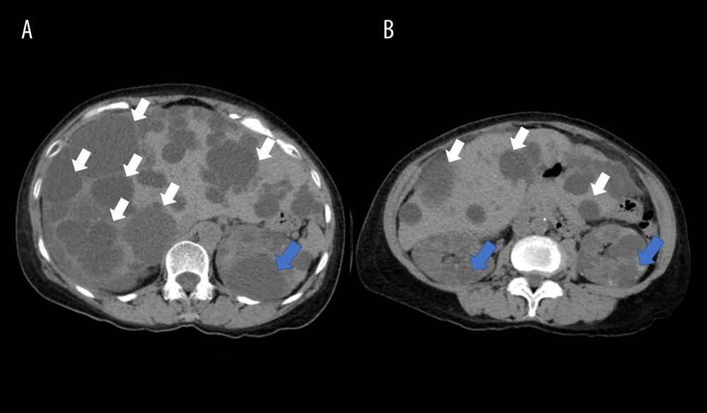
Figure 2 Contrast-enhanced computed tomography of the abdomen. Axial contrast-enhanced computed tomography images (A, B) demonstrate numerous hypodense ovoid lesions of varying sizes throughout the liver (white arrows) and both kidneys (blue arrows), consistent with hepatic and renal cysts.