


20 August 2026: Articles 
Autosomal Dominant Polycystic Kidney Disease Presenting as a Painless Epigastric Mass: A Case Report
Unusual clinical course, Challenging differential diagnosis
Tajul Atiqah Zaulkafali ABCDEF 1, Aiza Nur Izdihar Zainal-Abidin ACDE 1, Alan Basil Peter BCDE 2, Mohamed-Syarif Mohamed-Yassin ACDE 1*
DOI: 10.12659/AJCR.952751
Am J Case Rep 2026; 27:e952751


Abstract
BACKGROUND: Autosomal dominant polycystic kidney disease (ADPKD) is the most common inherited renal disorder, typically presenting with hypertension, hematuria, or progressive renal dysfunction. However, it can have a broad range of initial presentations and, in uncommon cases, may mimic other abdominal disorders. This report presents a case of ADPKD manifesting as an epigastric mass in a patient without a family history, highlighting its importance in the differential diagnosis of upper abdominal masses.
CASE REPORT: A 62-year-old woman presented to our primary care clinic with a 5-day history of a painless epigastric mass and right hypochondriac pain. She had hypertension and dyslipidemia, without a known family history of ADPKD. On examination, a firm mass was palpable in the epigastrium, accompanied by hepatomegaly. Laboratory investigations revealed mild renal insufficiency, microscopic hematuria, and proteinuria. The patient underwent abdominal ultrasonography and contrast-enhanced computed tomography, which demonstrated multiple cysts in both kidneys and the liver, consistent with ADPKD. She was evaluated by the hepatobiliary surgery team, who recommended symptomatic management. She was also assessed by the nephrology team; further evaluation for associated complications, including mitral valve prolapse and intracranial aneurysms, yielded normal findings. She continues regular follow-up with the primary care and nephrology teams for blood pressure optimization and renal function monitoring.
CONCLUSIONS: This case highlights the need to consider ADPKD in patients who present with upper abdominal masses. Early imaging and a multidisciplinary evaluation are essential for establishing an accurate diagnosis and ensuring appropriate management, particularly in patients without a clear family history.
Keywords: autosomal dominant polycystic kidney disease, Case Reports, Hepatomegaly, Tomography, X-Ray Computed, Ultrasonography
Introduction
Autosomal dominant polycystic kidney disease (ADPKD) is the most common inherited renal disease, occurring in approximately 1 in 400 to 1000 live births worldwide [1]. It is characterized by the gradual development of multiple bilateral renal cysts; most patients progress to end-stage renal disease in the fifth or sixth decade of life [2]. ADPKD is caused by mutations in the
ADPKD is mainly a renal disorder but is also associated with several extrarenal manifestations (eg, hepatic cysts, intracranial aneurysms, mitral valve prolapse, and colonic diverticulosis) [4]. Clinical presentations range from incidental findings of asymptomatic renal cysts on imaging to complications such as hypertension, hematuria, urinary tract infections, and kidney failure [1]. In this report, we present an uncommon initial manifestation of ADPKD—a palpable epigastric mass in a woman in her sixties. The primary objective of this report is to highlight the need to consider ADPKD in the differential diagnosis of upper abdominal masses.
Case Report
A 62-year-old Malay woman presented to an academic primary care clinic with a 5-day history of an epigastric mass that she had noticed for the first time. She described the mass as being approximately the size of a ping-pong ball, without a change in size; it became more noticeable when she pressed her abdomen against an object. The mass was painless. She denied vomiting, changes in bowel habits, early satiety, or jaundice. She also reported no urinary symptoms (eg, dysuria, flank pain, or hematuria) and no vaginal bleeding or abnormal discharge. There were no systemic symptoms, including fever, unintentional weight loss, or loss of appetite.
The patient also reported persistent dull, aching pain in the right hypochondriac region. The pain was usually mild (severity rating: 2/10) but occasionally increased to 6/10 with movement or sneezing. It did not radiate and was not related to meals. She could not recall any recent trauma to the area, and there was no history of abdominal surgery.
Her past medical history included well-controlled hypertension and dyslipidemia, both diagnosed in 2017 at the age of 55 years. She was taking amlodipine 5 mg once daily and atorvastatin 10 mg nightly. She was the mother of 7 children. Her family history included a brother who had hematuria and died of an unspecified malignancy, and a sister with an ovarian cyst. To her knowledge, none of her immediate family members had been diagnosed with polycystic kidney disease.
On physical examination, she was alert and comfortable. She was neither pale nor icteric and had no stigmata of chronic liver disease. Her vital signs were stable: blood pressure of 134/88 mm Hg, heart rate of 68 beats per minute, respiratory rate of 16 breaths per minute, temperature of 36.6 °C, and oxygen saturation of 98% on room air. Her abdomen was not distended and was soft on superficial palpation, without guarding. Deep palpation revealed a non-tender, fixed, firm, rounded, well-circumscribed epigastric mass measuring approximately 2 × 2 cm. The mass had a smooth surface. Hepatomegaly was also noted, with a liver span of 20 cm. The liver was smooth and tender. There were no signs of ascites, shifting dullness, or pedal edema. Murphy’s sign was negative, and the spleen was not enlarged. Bowel sounds were present. Respiratory, cardiovascular, and neurological examination findings were unremarkable.
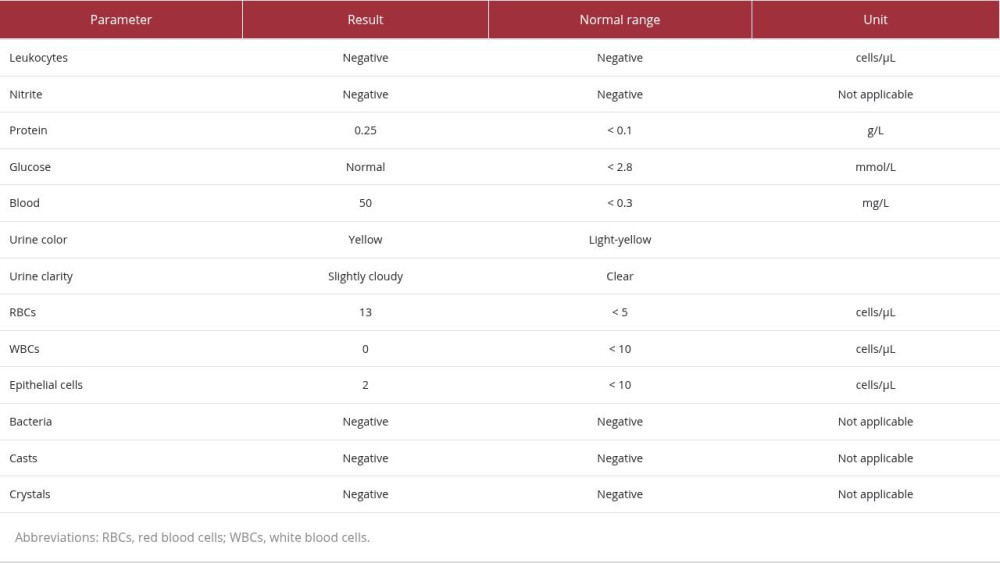
Laboratory tests performed on the same day showed a white blood cell count of 6.38 × 10⁹/L (normal: 4.0–10.0 × 10⁹/L), hemoglobin of 11.9 g/dL (normal: 12–15 g/dL), and platelet count of 292 × 109/L (normal: 150–410 × 109/L). The renal profile revealed a mildly elevated creatinine level of 95 μmol/L (normal: 44–80 μmol/L) and an estimated glomerular filtration rate (eGFR) of 55 mL/min/1.73 m2 (normal: > 60 mL/min/1.73 m2) (Table 1). Other renal parameters were within normal limits (sodium, 141 mmol/L; potassium, 3.7 mmol/L; chloride, 105 mmol/L), indicating the absence of hyperkalemia or metabolic acidosis, which are complications often observed in later stages of ADPKD-related renal failure. Urinalysis showed proteinuria of 0.25 g/L (normal: < 0.1 g/L), hematuria of 50 mg/L (normal: < 0.3 mg/L), and a red blood cell count of 13 cells/μL (normal: < 5 cells/μL), without evidence of pyuria, nitrites, or casts (Table 2). Liver function tests showed mild elevations in gamma-glutamyl transferase at 56 U/L (normal: < 40 U/L) and direct bilirubin at 7.1 μmol/L (normal: 0.0–5.0 μmol/L); other liver parameters remained within normal ranges (Table 1).
Considering the need for early imaging, an abdominal ultrasound was promptly obtained the following week. This assessment revealed multiple cysts of varying sizes in the kidneys and liver; the latter corresponded to the previously palpated epigastric mass (Figure 1). During the examination, the patient experienced discomfort when the radiologist applied pressure to her abdomen with the ultrasound probe. Thus, she was referred back to the primary care clinic for review. Upon consultation, she rated her right hypochondriac pain as 7/10 during movement or sneezing and 6/10 at rest; no new symptoms were present. Given the symptomatic hepatic cysts, she was referred to the hepatobiliary surgery team. They promptly reviewed her case, made a provisional diagnosis of ADPKD, and prescribed tramadol 50 mg (3 times daily) to manage pain related to liver capsule distension. A contrast-enhanced computed tomography scan of the abdomen was arranged for further evaluation, and a follow-up appointment was scheduled within 2 weeks.
A contrast-enhanced computed tomography scan of the abdomen (Figure 2) showed bilaterally enlarged kidneys measuring 14.5 cm on the right and 16.9 cm on the left. Although transverse and anteroposterior diameters were not reported, the kidneys contained numerous cystic lesions of varying sizes, reflecting a high cyst burden. Multiple hepatic cysts were also identified. These findings were consistent with a diagnosis of ADPKD. No surgical intervention was required at this stage; the hepatobiliary surgery team thus referred the patient to the nephrology team for further evaluation and management.
During her visit to the nephrology clinic, the patient no longer reported right hypochondriac pain. Further investigations, including echocardiography to identify mitral valve prolapse and brain computed tomography angiography to screen for intracranial berry aneurysms, were subsequently performed. Echocardiography showed normal mitral valve leaflets, and brain computed tomography angiography demonstrated no evidence of berry aneurysms. At a subsequent follow-up visit, telmisartan 40 mg once daily was added to optimize blood pressure control. Her siblings and children were advised to undergo screening for polycystic kidney disease. At the time of reporting, 1 of her 7 children had been diagnosed with polycystic kidney disease (4 had been screened). She will continue follow-up with the primary care, nephrology, and hepatobiliary surgery teams.
Discussion
The main lesson from this case is that an uncommon presentation of ADPKD can create a potential diagnostic pitfall. When it presents as a localized epigastric mass, it can closely mimic a focal gastric, hepatobiliary, or pancreatic lesion. This diagnostic challenge is further compounded when the patient has no known family history of the disease. Although early imaging led to a prompt and accurate diagnosis in our case, this report highlights the importance of considering ADPKD in the differential diagnosis of upper abdominal masses to avoid diagnostic errors and unnecessary delays.
ADPKD is a genetic disorder characterized by the progressive development of fluid-filled cysts in the kidneys. This multisystem disease can affect organs such as the heart, liver, pancreas, spleen, and arachnoid membranes; it causes progressive renal enlargement and loss of kidney function, ultimately leading to end-stage kidney disease. ADPKD is responsible for approximately 5% to 10% of all cases of end-stage renal failure [5,6].
The estimated prevalence of ADPKD is between 1 in 400 and 1 in 1000 individuals worldwide [6]. The exact prevalence of ADPKD in Malaysia is unknown due to limited epidemiological data. However, the 30th Report of the Malaysian Dialysis and Transplant Registry (2022) indicated that 0.5% of patients requiring dialysis in Malaysia had polycystic kidney disease [7].
ADPKD results from mutations in 2 genes:
This disease is often clinically silent, such that some individuals remain asymptomatic throughout their lives. It typically becomes apparent in the third or fourth decade of life, when patients may present with clinical features such as hypertension (50%–70%) [10,11], hematuria (30%–40%) [12], proteinuria (25%) [13], or impaired kidney function (70%) [11]. Approximately 30% to 70% of patients experience flank pain [14], which can result from renal hemorrhage, obstructive nephrolithiasis, or urinary tract infections [8]. Abdominal masses may also occur in patients with ADPKD due to enlarged polycystic kidneys or cyst-related complications [15]. One such example was reported by Chaudhary and Qian [16], in which the patient displayed acute abdominal pain, abdominal fullness, and ascites; they were subsequently diagnosed with ruptured hepatic cysts associated with ADPKD. Another report by Kumar et al described an older man presenting with a “colossal abdomen” characterized by chronic, progressive, and massive diffuse abdominal distension that arose from giant cysts occupying much of the abdominal cavity [17]. Whereas that case involved diffuse enlargement of the entire abdomen, our patient presented with a focal, firm, well-circumscribed 2 × 2 cm epigastric mass. This focal presentation can mimic localized upper gastrointestinal, hepatobiliary, or pancreatic pathology.
Liver cysts are a common extrarenal manifestation of ADPKD; magnetic resonance imaging analysis has demonstrated their presence in up to 80% of affected individuals by age 30 [15]. Patients with polycystic liver disease may show hepatic dysfunction or abdominal pain resulting from cyst infection or rupture, although many remain asymptomatic [5]. The condition is more prevalent in women, suggesting a hormonal influence; cyst size and number have been shown to increase in response to elevated estrogen levels, such as those associated with oral contraceptive use or estrogen replacement therapy. Additionally, severe cystic liver enlargement is more frequently observed in multiparous women [18]. Estrogen promotes the development and progression of liver cysts by stimulating cholangiocyte proliferation through receptor activation, increasing cyclic-adenosine-monophosphate-mediated fluid secretion, and upregulating vascular endothelial growth factor expression to support cyst vascularization [18,19]. A 2025 longitudinal pilot study by Bazojoo et al demonstrated that pregnancy was significantly associated with an accelerated rate of liver cyst growth [20]. Therefore, in women with multiple pregnancies, cumulative estrogen exposure may accelerate cyst growth and contribute to a greater hepatic cyst burden.
In the present case, the patient’s history of multiparity may have contributed to the development of symptomatic polycystic liver disease. This observation adds to the existing clinical evidence and highlights the importance of counseling women with ADPKD regarding estrogen-related risks, including family planning, contraceptive choices, and postmenopausal hormone replacement therapy. Our patient had no history of contraceptive use and reached menopause at 52 years of age without receiving hormone replacement therapy. Other extrarenal manifestations associated with ADPKD include pancreatic cysts, intracranial aneurysms, cardiac valvular abnormalities, colonic diverticulosis, and hernias [21,22]. Therefore, the patient underwent evaluation with echocardiography and cerebral computed tomography angiography to screen for these complications; the findings were normal.
The diagnosis of ADPKD is usually based on a combination of family history, ultrasonographic and/or computed tomography findings, along with clinical manifestations such as hypertension and renal impairment [5]. However, approximately 25% of patients with ADPKD have no known family history, potentially due to subclinical disease in relatives or de novo mutations [23]. In these individuals, ADPKD can be diagnosed based on clinical evaluation and radiological findings demonstrating bilaterally enlarged kidneys with innumerable cysts (typically ≥10 cysts measuring ≥5 mm), in the absence of features suggesting an alternative cystic disorder [24,25]. In the present case, the patient had no clear family history of ADPKD. Therefore, the diagnosis was established based on clinical findings of hypertension and reduced eGFR, along with imaging findings of bilaterally enlarged kidneys (14.5 cm on the right and 16.9 cm on the left) that contained numerous cystic lesions of varying sizes. Multiple hepatic cysts were also identified; no cysts were detected in other organs, which would suggest an alternative cystic disease.
In the primary care setting, a presentation such as this can create a serious diagnostic pitfall. Because the mass was located in the upper midline abdomen, clinicians may be inclined to prioritize a gastrointestinal workup, including esophagogastroduodenoscopy, to exclude gastric malignancies or subepithelial tumors. This potential diagnostic misdirection is illustrated in the literature. For example, in the case reported by Chaudhary and Qian, the patient presented to a local emergency department with acute abdominal symptoms [16]. Although multiple hepatic and renal cysts were visible during initial imaging studies, she underwent an unremarkable esophagogastroduodenoscopy and an elective laparoscopic cholecystectomy for suspected gallbladder disease. The diagnosis of ADPKD with hepatic cyst rupture was not formally established until she sought a second opinion 5 months later [16]. Our case emphasizes that, without a high index of suspicion, clinicians may focus on endoscopic evaluation while overlooking the systemic nature of ADPKD, particularly when a family history is absent.
The main objectives of ADPKD management are to slow disease progression, reduce symptoms, and prevent complications. Achievement of these goals requires a combination of lifestyle modification, pharmacotherapy, and regular monitoring for disease-related complications. Recommended lifestyle measures include restricting dietary sodium intake; maintaining adequate hydration; and avoiding tobacco, alcohol, and other nephrotoxic substances [13,26]. Optimization of blood pressure control is another key component of ADPKD management because uncontrolled hypertension accelerates kidney damage and increases cerebrovascular risk [27]. Medications that target the renin-angiotensin-aldosterone system, such as angiotensin-converting enzyme inhibitors and angiotensin receptor blockers, are recommended because they can delay disease progression and reduce cardiovascular risk in patients with ADPKD [27]. In the present case, telmisartan, an angiotensin receptor blocker, was added to the patient’s antihypertensive regimen. The vasopressin V2 receptor antagonist tolvaptan has also been shown to slow cyst growth and preserve renal function [25]. However, it is generally reserved for patients at high risk of chronic kidney disease progression, as determined by the Mayo Imaging Classification [25]. Formal Mayo classification requires calculation of height-adjusted total kidney volume. Specific volumetric data, including kidney width and depth, were unavailable in the present case, precluding precise calculation of height-adjusted total kidney volume. Nevertheless, the patient’s clinical profile provided useful prognostic information. The KDIGO 2025 Clinical Practice Guideline acknowledges that kidney length can serve as a practical indicator of disease burden when volumetric measurements are unavailable [9]. In this 62-year-old patient, a left kidney length of 16.9 cm reflected a substantial cyst burden. However, her relatively preserved eGFR of 55 mL/min/1.73 m2 suggested a disease course that may not meet the criteria for rapid progression typically associated with such renal enlargement in younger patients. This clinical assessment supported prioritizing renin-angiotensin-aldosterone system blockade with telmisartan, rather than initiating tolvaptan therapy.
Family screening plays an important role in identifying individuals at risk for ADPKD. In adults with a positive family history, abdominal ultrasonography is recommended as the initial screening modality, using age-specific cyst number criteria to confirm or exclude the diagnosis [9]. Ultrasonography is preferred because it is widely available, safe, and reliable. If ultrasound findings are inconclusive or inconsistent with the clinical presentation, further evaluation via magnetic resonance imaging, computed tomography, or genetic testing may be warranted [9]. In the present case, the patient was advised to inform her children of their potential risk and to encourage screening. At the time of reporting, 4 of her 7 children had undergone abdominal ultrasound screening; her fifth daughter was diagnosed with polycystic kidney disease.
Conclusions
This case report describes an uncommon initial presentation of ADPKD as a palpable epigastric mass, which was subsequently linked to hepatic cysts associated with multiple renal cysts (according to imaging results). The present findings serve as a reminder for clinicians to consider ADPKD in the differential diagnosis of upper abdominal masses. A high index of suspicion and early imaging are essential for timely diagnosis, particularly in patients without a known family history. A multidisciplinary approach involving primary care, radiology, nephrology, and surgery is crucial for the comprehensive evaluation and management of this condition.
Figures
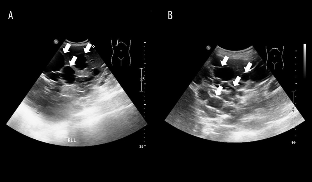 Figure 1. Abdominal ultrasonography. Ultrasound images demonstrate numerous anechoic ovoid lesions (arrows) occupying the majority of the right (A) and left (B) hepatic lobes, consistent with multiple hepatic cysts.
 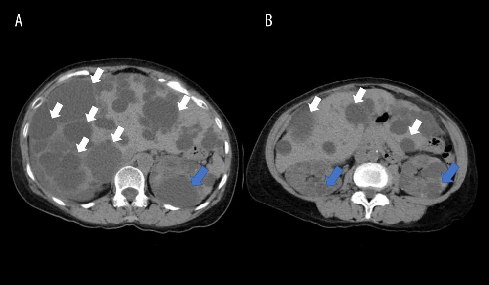 Figure 2. Contrast-enhanced computed tomography of the abdomen. Axial contrast-enhanced computed tomography images (A, B) demonstrate numerous hypodense ovoid lesions of varying sizes throughout the liver (white arrows) and both kidneys (blue arrows), consistent with hepatic and renal cysts.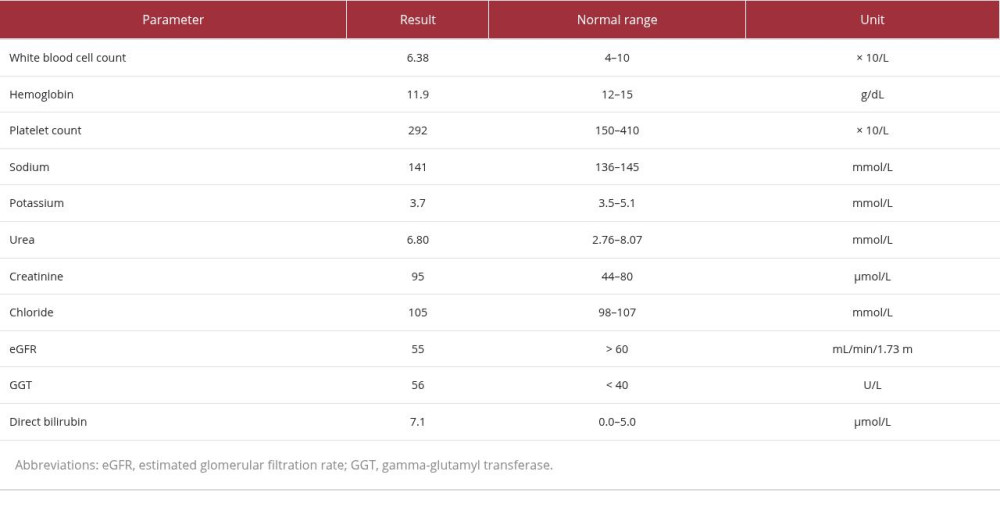
References
1. Chow CL, Ong ACM, Autosomal dominant polycystic kidney disease: Clin Med (Lond), 2009; 9(3); 278-83
2. Theivendran T, Ramachandran A, Rangan G, Drug management of autosomal dominant polycystic kidney disease: Aust Prescr, 2022; 45(5); 167-70
3. Sommerer C, Zeier M, Clinical manifestation and management of ADPKD in Western countries: Kidney Dis (Basel), 2016; 2(3); 120-27
4. Pirson Y, Extrarenal manifestations of autosomal dominant polycystic kidney disease: Adv Chronic Kidney Dis, 2010; 17(2); 173-80
5. Mahboob M, Rout P, Bokhari SRA, Autosomal dominant polycystic kidney disease: StatPearls [Internet], 2024, Treasure Island (FL), StatPearls Publishing Available from: https://www.ncbi.nlm.nih.gov/books/NBK470437/
6. Torres VE, Harris PC, Autosomal dominant polycystic kidney disease: The last 3 years: Kidney Int, 2009; 76(2); 149-68
7. Malaysia Dialysis and Transplant Registry: 30th report of the Malaysian Dialysis & Transplant Registry 2022, 2024, Malaysia Dialysis and Transplant Registry Available from: https://www.msn.org.my/nrr/
8. Torres VE, Harris PC, Pirson Y, Autosomal dominant polycystic kidney disease: Lancet, 2007; 369(9569); 1287-301
9. Kidney Disease: Improving Global Outcomes (KDIGO) ADPKD Work Group, KDIGO 2025 clinical practice guideline for the evaluation, management, and treatment of autosomal dominant polycystic kidney disease (ADPKD): Kidney Int, 2025; 107(2 Suppl); S1-S239
10. Kocyigit I, Taheri S, Eroglu E, Association of OSR-1 with vascular dysfunction and hypertension in polycystic kidney disease: Ther Apher Dial, 2020; 24(1); 64-71
11. Rastogi A, Ameen KM, Al-Baghdadi M, Autosomal dominant polycystic kidney disease: Updated perspectives: Ther Clin Risk Manag, 2019; 15; 1041-52
12. Bhasin B, Alzubaidi M, Velez JCQ, Evaluation and management of gross hematuria in autosomal dominant polycystic kidney disease: A point of care guide for practicing internists: Am J Med Sci, 2018; 356(2); 177-80
13. Chapman AB, Devuyst O, Eckardt KU, Autosomal-dominant polycystic kidney disease (ADPKD): Executive summary from a Kidney Disease: Improving Global Outcomes (KDIGO) Controversies Conference: Kidney Int, 2015; 88(1); 17-27
14. Borghol AH, Munairdjy Debeh FG, Ghanem A, Pain management in autosomal dominant polycystic kidney disease: Clinical challenges and a stepwise algorithmic approach: Kidney360, 2025; 6(9); 1618-31
15. Chebib FT, Torres VE, Autosomal dominant polycystic kidney disease: Core curriculum 2016: Am J Kidney Dis, 2016; 67(5); 792-810
16. Chaudhary S, Qian Q, Acute abdomen and ascites as presenting features of autosomal dominant polycystic kidney disease: World J Hepatol, 2012; 4(12); 394-98
17. Kumar S, Parmar K, Sharma AP, Autosomal dominant polycystic kidney disease presenting as colossal abdomen: Urology, 2020; 142; e39-40
18. Sherstha R, McKinley C, Russ P, Postmenopausal estrogen therapy selectively stimulates hepatic enlargement in women with autosomal dominant polycystic kidney disease: Hepatology, 1997; 26(5); 1282-86
19. Aapkes SE, Bernts LHP, Barten TRM, Estrogens in polycystic liver disease: A target for future therapies?: Liver Int, 2021; 41(9); 2009-19
20. Bazojoo V, Davoudi V, Blumenfeld JD, Effects of pregnancy on liver and kidney cyst growth rates in autosomal dominant polycystic kidney disease: A pilot study: J Clin Med, 2025; 14(11); 3688
21. Shah NK, Huge polycystic kidney weighing 6.24 kg: Med J Dr DY Patil Vidyapeeth, 2018; 11; 165-66
22. Wüthrich RP, Kistler AD, Rodriguez D, Blood pressure control for polycystic kidney disease: Polycystic Kidney Disease, 2015; 95-108, Brisbane (AU), Codon Publications
23. Finnigan NA, Leslie SW, Polycystic kidney disease in adults (archived): StatPearls [Internet], 2023, Treasure Island (FL), StatPearls Publishing Available from: https://www.ncbi.nlm.nih.gov/books/NBK532934/
24. Alves M, Fonseca T, de Almeida EAF, Differential diagnosis of autosomal dominant polycystic kidney disease: Polycystic Kidney Disease, 2015; 3-19, Brisbane (AU), Codon Publications
25. Torres VE, Bennett WM, Autosomal dominant polycystic kidney disease (ADPKD) in adults: epidemiology, clinical presentation, and diagnosis: UpToDate [Internet], 2026, Wolters Kluwer [cited 2026 May 31]. Available from: https://www.uptodate.com/contents/autosomal-dominant-polycystic-kidney-disease-adpkd-in-adults-epidemiology-clinical-presentation-and-diagnosis
26. Sans-Atxer L, Torra R, Fernandez-Llama P, Hypertension in autosomal-dominant polycystic kidney disease (ADPKD): Clin Kidney J, 2013; 6(5); 457-63
27. Khalil I, Faiyaz AF, Hossain I, Akter M, Multisystemic impact of autosomal dominant polycystic kidney disease: A case report highlighting renal, hepatic, and neurological involvement: Radiol Case Rep, 2025; 20(4); 2093-100
Figures
Figure 1. Abdominal ultrasonography. Ultrasound images demonstrate numerous anechoic ovoid lesions (arrows) occupying the majority of the right (A) and left (B) hepatic lobes, consistent with multiple hepatic cysts.Figure 2. Contrast-enhanced computed tomography of the abdomen. Axial contrast-enhanced computed tomography images (A, B) demonstrate numerous hypodense ovoid lesions of varying sizes throughout the liver (white arrows) and both kidneys (blue arrows), consistent with hepatic and renal cysts. In Press
Case report 
Am J Case Rep In Press; DOI: 10.12659/AJCR.953543
Case report 
Am J Case Rep In Press; DOI: 10.12659/AJCR.954216
Case report 
Am J Case Rep In Press; DOI: 10.12659/AJCR.954932
Case report 
Am J Case Rep In Press; DOI: 10.12659/AJCR.954146
Most Viewed Current Articles
07 Dec 2021 : Case report
28,712,695
DOI :10.12659/AJCR.934347
Am J Case Rep 2021; 22:e934347
06 Dec 2021 : Case report  180,721
DOI :10.12659/AJCR.934406
Am J Case Rep 2021; 22:e934406
21 Jun 2024 : Case report
124,419
DOI :10.12659/AJCR.944371
Am J Case Rep 2024; 25:e944371
08 May 2026 : Case report
95,462
DOI :10.12659/AJCR.952158
Am J Case Rep 2026; 27:e952158