


10 September 2026
: Case report 
Delayed Diagnosis of Type 1 Gaucher Disease at Age 15 After Years of Mild Cytopenias and Splenomegaly: A Case Report and Long-Term Follow-Up
Challenging differential diagnosis, Diagnostic / therapeutic accidents, Rare disease
Katarzyna Muras-Szwedziak ABCDEF 1, Jacek Kasznicki ABCDEF 2, Olga Wegner ABCDEF 3, Maciej Wójcik ABCDEF 4, Olga Wojtyczka ABCDEF 4*
DOI: 10.12659/AJCR.952793
Am J Case Rep 2026; 27:e952793


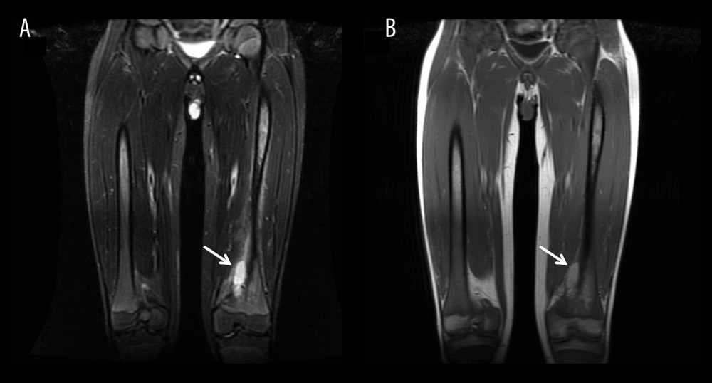
Figure 6 Magnetic resonance imaging findings during acute skeletal presentation.Coronal magnetic resonance imaging of the lower extremities. (A) Short tau inversion recovery sequence demonstrates a heterogeneous area of increased signal intensity within the distal metaphysis and diaphysis of the left femur, suggestive of bone marrow edema or infiltration. (B) Corresponding T1-weighted image shows reduced signal intensity in the same region, indicating replacement of normal bone marrow. White arrows indicate the abnormal marrow lesion within the distal femur. No cortical destruction or periosteal reaction is observed. Overall, the imaging findings are consistent with an acute bone crisis in Gaucher disease.