


10 September 2026: Articles 
Delayed Diagnosis of Type 1 Gaucher Disease at Age 15 After Years of Mild Cytopenias and Splenomegaly: A Case Report and Long-Term Follow-Up
Challenging differential diagnosis, Diagnostic / therapeutic accidents, Rare disease
Katarzyna Muras-Szwedziak ABCDEF 1, Jacek Kasznicki ABCDEF 2, Olga Wegner ABCDEF 3, Maciej Wójcik ABCDEF 4, Olga Wojtyczka ABCDEF 4*
DOI: 10.12659/AJCR.952793
Am J Case Rep 2026; 27:e952793


Abstract
BACKGROUND: Gaucher disease (GD) is the most common lysosomal storage disorder caused by glucocerebrosidase deficiency. Type 1 GD (GD1) often presents with nonspecific manifestations, including splenomegaly, cytopenias, growth impairment, and skeletal involvement, leading to delayed diagnosis and irreversible complications. This report highlights the delayed diagnosis of GD1 in a patient with longstanding mild manifestations and emphasizes the importance of considering GD in patients with unexplained cytopenias and splenomegaly.
CASE REPORT: We describe a male patient with GD1 whose first manifestations appeared in infancy and included splenomegaly. During childhood, persistent cytopenias, hepatosplenomegaly, and growth deceleration were observed; however, the diagnosis remained unrecognized. At age 14, the patient developed severe skeletal pain accompanied by fever, prompting further diagnostic evaluation. Imaging studies revealed bone marrow abnormalities, and bone biopsy demonstrated foamy macrophages but did not establish a definitive diagnosis. At age 15, GD1 was suspected and subsequently confirmed by enzymatic testing, biomarker assessment, and genetic analysis. Enzyme replacement therapy with imiglucerase resulted in clinical improvement, reduced organomegaly, and stabilization of laboratory parameters. The patient subsequently underwent a structured transition from pediatric to adult care without treatment interruption.
CONCLUSIONS: GD should be considered in patients with unexplained splenomegaly and persistent cytopenias, even when early manifestations are mild and nonspecific. Early recognition and disease-specific diagnostic testing may reduce diagnostic delay, facilitate timely treatment initiation, and help prevent irreversible complications. This case also highlights the importance of a structured transition from pediatric to adult care in maintaining long-term treatment continuity.
Keywords: delayed diagnosis, enzyme replacement therapy, Gaucher disease, Lysosomal Storage Diseases, Transitional Care, Case Reports
Introduction
Gaucher disease (GD) is the most common lysosomal storage disorder, with an estimated incidence of approximately 1 in 40 000 to 60 000 live births worldwide [1,2]. It is caused by pathogenic variants in the
The clinical presentation of GD is highly heterogeneous [2,12]. Although the disease is typically classified into 3 main types based on neurological involvement, these categories are not absolute [2,12]. Neuronopathic GD is increasingly regarded as a phenotypic continuum; the lack of a clear diagnostic boundary between GD2 and GD3 can lead to diagnostic and therapeutic uncertainty [2,14,15]. GD1, the most common form, is characterized by the absence of central nervous system involvement and typically presents with hepatosplenomegaly, cytopenias, growth impairment, and skeletal involvement [2,12,16]; all of these features were observed in the present case. The key clinical features of the 3 GD types are summarized in Table 1 [2,5,12,14]. The genotype-phenotype correlation in GD remains unclear, given that the same genetic variant can result in diverse clinical manifestations [12,17,18].
The diagnosis of GD is based on demonstration of low β-glucocerebrosidase activity, most commonly assessed by dried blood spot (DBS)-based enzymatic testing, and confirmation of a pathogenic variant in the
Symptoms of GD can manifest early in childhood and may be detected through basic physical, radiologic, and laboratory examinations; however, their nonspecific nature often leads to delayed diagnosis [23,24]. Because persistent mild cytopenias and splenomegaly are frequently attributed to more common conditions, they may remain insufficiently investigated for prolonged periods, representing a substantial diagnostic gap [23–25]. This delay may result in missed opportunities for early treatment, leading to progressive skeletal involvement, impaired growth, and increased long-term disease burden, as well as unnecessary diagnostic procedures, highlighting the clinical consequences of delayed recognition [23–25]. Similar diagnostic challenges and variable diagnostic pathways have been described in published reports of GD presenting with splenomegaly and cytopenias [16,26]. Early recognition is crucial given the availability of effective disease-specific treatments [2,3].
Current disease-specific treatment options for GD include enzyme replacement therapy (ERT) and substrate reduction therapy (SRT) [2,27]. Available intravenous ERT agents include imiglucerase, velaglucerase alfa, and taliglucerase alfa; oral SRT options include miglustat and eliglustat [2,27]. Treatment choice is individualized according to disease severity, patient characteristics, and treatment eligibility [2,27]. ERT remains the standard therapeutic approach for most pediatric patients and many individuals with clinically significant disease [27].
The aim of this case report is to highlight the delayed diagnosis of GD1 in a patient with longstanding mild and nonspecific manifestations, then emphasize the importance of considering GD in patients presenting with unexplained cytopenias and splenomegaly. The report also illustrates the benefits of ERT and underscores the role of a structured transition from pediatric to adult health care services in maintaining continuity of care.
Case Report
A male infant was delivered at 39 weeks of gestation by elective cesarean section due to advanced maternal age. Family history was unremarkable. The perinatal course was uncomplicated. Immediate postnatal adaptation was appropriate, with an Apgar score of 10 at 1 minute. Birth weight was 3700 g (corresponding to the 78th percentile according to World Health Organization growth charts), and length was 56 cm (97th percentile). On the second day of life, the infant developed mild physiologic jaundice, which did not require medical intervention. He was breastfed on demand and demonstrated adequate weight gain. The clinical course and key diagnostic milestones are summarized in Figure 1.
During the first year of life, the child’s development was appropriate for age, with linear growth tracking between the 93rd and 99th percentiles according to national percentile charts prepared by the Institute of Mother and Child. At 8 months of age, due to recurrent colic and abdominal pain, the patient’s mother consulted a pediatrician. Abdominal ultrasonography demonstrated mild splenomegaly, with a spleen length of approximately 8.5 cm. At that stage, the findings were managed with continued observation, and no further diagnostic workup was undertaken. Subsequent anthropometric measurements showed gradual slowing of linear growth; no early skeletal manifestations were reported at that stage. By 16 months of age, height had declined to the 76th percentile, and by 6 years and 7 months, to the 50th percentile (Figure 2). Additionally, the mother reported delayed motor development, weakness, and night sweats.
Between 8 months and 4 years of age, follow-up abdominal ultrasounds consistently demonstrated hepatosplenomegaly, such that both organs extended 2 to 3 cm below the costal margins. Laboratory tests revealed stable mild anemia with low serum iron, reduced or low-normal mean corpuscular volume (MCV) and mean corpuscular hemoglobin, and a persistently low-normal platelet count (Figures 3–5). No clinically significant bleeding tendency was observed. Iron supplementation was ineffective. These findings were considered consistent with anemia of chronic disease.
At 4 years of age, the patient was admitted to the pediatric gastroenterology, hepatology, and immunology unit for further diagnostic evaluation. Viral infections (eg, Epstein-Barr virus, hepatitis B, hepatitis C, and cytomegalovirus) were ruled out, as were celiac disease, alpha-1 antitrypsin deficiency, and Wilson disease. Doppler assessment revealed no hepatic vascular abnormalities. After the evaluation at 4 years of age, the patient remained under outpatient hematology follow-up with regular clinical monitoring. At age 7, he was admitted to the pediatric oncology and hematology department. Follow-up investigations confirmed the previously identified abnormalities. Normal soluble transferrin receptor levels supported the diagnosis of anemia of chronic disease.
At age 14, the patient presented with severe pain in the left lower limb accompanied by fever. Initial suspicion was directed toward growing pains; however, radiographs of the lower limbs revealed heterogeneous osteosclerotic changes in the distal left femur, without evidence of periosteal reaction. Magnetic resonance imaging demonstrated a heterogeneous bone marrow signal with features suggestive of edema or infiltration, as well as a subperiosteal lesion suggestive of a hematoma (Figure 6). To differentiate between inflammatory and neoplastic etiologies, a biopsy was performed.
The initial histopathologic assessment indicated osteomalacia in the setting of a nonspecific proliferative process; however, no malignant features were identified. Because no definitive diagnosis was established, a second tissue sample was obtained. Subsequent evaluation revealed nonspecific changes, most likely posttraumatic in origin and related to the previous biopsy. The specimen also contained macrophages with morphology characteristic of so-called foam cells; however, no further diagnostic workup for conditions associated with foamy macrophages was conducted at that time. After the first biopsy, prophylactic clindamycin therapy was administered, during which the patient developed a low-grade fever. After the second biopsy, due to persistent fever, broad-spectrum antibiotic therapy with ciprofloxacin and ceftriaxone was initiated.
At age 15, follow-up evaluations confirmed persistence of the previously observed abnormalities. Additionally, a greatly elevated serum ferritin level was detected. Considering the histopathologic findings (foam cells) and evolving clinical presentation, GD1 was suspected.
Enzymatic testing using DBSs revealed undetectable β-glucocerebrosidase activity and a substantially elevated lyso-Gb1 level of 543.4 ng/mL (reference value < 14 ng/mL). Chitotriosidase activity was also increased, measuring 178.5 mmol/mL/hour (reference value < 100 mmol/mL/hour). Molecular analysis, performed by next-generation sequencing on DNA extracted from DBSs, confirmed the presence of a homozygous pathogenic
Taken together, the longstanding history of hepatosplenomegaly; cytopenias; skeletal involvement demonstrated by imaging studies; exclusion of alternative infectious, inflammatory, and neoplastic conditions; greatly reduced β-glucocerebrosidase activity; elevated lyso-Gb1 and chitotriosidase levels; and molecular confirmation of a homozygous pathogenic
As part of the qualification process for ERT, the necessary evaluations were performed, including abdominal ultrasonography (organ measurements are detailed in Table 2; June 2019). Bone mineral density was assessed via dual-energy X-ray absorptiometry. Z scores were within the expected range for age (reference threshold > −2.0) for both the lumbar spine (L1–L4; Z score, +0.5) and whole-body measurement (Z score, −0.1). Given that no neurologic or oculomotor abnormalities were identified during the evaluation, no formal neurologic consultation was required. Following approval for the treatment program, the first infusion of imiglucerase was administered in September 2019, at age 15. The dose was adjusted according to the patient’s body weight and administered intravenously every 2 weeks in a repeating 3-infusion cycle of 2000 U, 2000 U, and 1600 U. The infusions were well tolerated. Concurrently, vitamin D deficiency was diagnosed; supplementation was initiated at 4000 IU/day during autumn and winter and 2000 IU/day during spring and summer.
ERT resulted in pronounced acceleration of growth velocity and subsequent stabilization of growth parameters within higher percentile ranges (Figure 2), indicating improvement in the patient’s overall metabolic status. Serial abdominal ultrasounds demonstrated progressive reductions in liver and spleen size (Table 2), confirming a favorable organ response to treatment, although minor variability in measurement techniques across time points should be considered when interpreting longitudinal changes. Concurrently, bone lesions observed on femoral magnetic resonance imaging remained stable compared with previous assessments.
Ferritin levels decreased relative to pretreatment values but remained at the upper limit of normal (Figure 3). Despite supplementation, iron levels remained stable at the lower limit of normal (Figure 3). In contrast, hemoglobin and MCV values showed a sustained gradual increase after ERT initiation, reaching the upper normal range by the end of follow-up (Figure 4). Platelet counts remained within the lower normal range throughout (a transient decline in 2019–2020 was followed by recovery) (Figure 5).
The patient remained under continuous care at a pediatric oncology and hematology center until age 18, with ERT doses adjusted according to body weight and consistently well tolerated. The transition to adult care was planned over several months through collaboration between the pediatric and adult care teams, as well as consultations with the patient and his family. In 2023, at age 18, the patient was transferred to an adult rare disease center selected by the family, where imiglucerase therapy has continued as day-case infusions during 1-day hospitalizations in an internal medicine unit, with consistently good tolerance. At present, the patient remains asymptomatic, with stable laboratory and imaging findings.
Discussion
This case highlights the diagnostic challenges of GD when early manifestations are mild and nonspecific. The patient presented with a paucisymptomatic, non-neuronopathic form of GD, in which subtle hematologic abnormalities persisted for years prior to the onset of severe skeletal symptoms. Only after the development of severe skeletal manifestations was further diagnostic evaluation conducted, given that inflammatory and neoplastic etiologies were initially considered in the differential diagnosis.
Although GD is the most common lysosomal storage disorder, with type 1 representing approximately 90% of cases [2], its diagnosis is frequently delayed (average delay ~ 4 years) [25]. This pattern is consistent with recent studies highlighting diagnostic delays in GD, particularly among patients with mild and nonspecific early manifestations [23,24]. This delay is largely attributable to the nonspecific nature of early manifestations and their overlap with more common conditions, partly related to chronic immune activation [7,8], which may lead to misdiagnosis as conditions such as juvenile idiopathic arthritis, idiopathic thrombocytopenic purpura, anemia of chronic disease, or benign causes of bone pain [20,23,28]. Importantly, in GD, platelet counts can remain persistently low-normal or only mildly reduced (“gray zone” thrombocytopenia), which should not be considered reassuring and should prompt further diagnostic evaluation when accompanied by splenomegaly [23,25,28]. Thus, among patients with unexplained chronic cytopenias and splenomegaly, GD should be considered in the differential diagnosis, even in the absence of neurologic manifestations [21,29]. This consideration is particularly important before irreversible interventions such as splenectomy are contemplated; splenectomy is associated with significantly worse outcomes in GD [21,30] and can further complicate or delay recognition of the underlying disease, as illustrated by a recently reported case in which GD remained unrecognized for approximately 40 years after childhood splenectomy [31].
Several potential diagnostic opportunities arose during the patient’s clinical course. Mild splenomegaly—identified during infancy—was eventually associated with persistent cytopenias and hepatosplenomegaly. At age 14, the patient developed severe bone pain and fever, which, following the diagnosis of GD, were considered consistent with an acute bone crisis. Histopathologic examination of the bone lesion also revealed macrophages with foamy morphology. Although none of these findings is specific for GD in isolation, their combination could have prompted earlier targeted diagnostic testing. Similar diagnostic challenges have been noted in previous case reports [16,32]. Chen et al described 2 patients with longstanding thrombocytopenia and splenomegaly in whom the diagnosis of GD was delayed by approximately 10 and 20 years, respectively, despite persistent hematologic abnormalities [16]. In contrast, Cullufi et al reported an infant with hepatosplenomegaly, anemia, and thrombocytopenia, in whom early diagnostic investigations (eg, bone marrow examination, biochemical testing, and genetic analysis) led to diagnosis during infancy and initiation of treatment at age 7 months [32]. Although this patient later developed manifestations of a severe neuronopathic form of GD, the initial presentation at diagnosis was similar to observations in the present case. Taken together, these reports illustrate that comparable early manifestations, particularly splenomegaly and cytopenias, may lead to diagnosis within months or remain unrecognized for many years, depending on the diagnostic approach.
To achieve early diagnosis of GD, awareness must be raised among health care professionals across multiple specialties, particularly hematologists, pediatricians, internists, and family physicians [20,28]. In clinical practice, lysosomal storage disorders often remain outside standard diagnostic pathways due to their rarity and nonspecific presentation [20,23,28]. Thus, incorporation of GD into routine diagnostic algorithms, especially for patients with unexplained splenomegaly, persistent cytopenias (particularly thrombocytopenia), or bone abnormalities, may be beneficial [21,29,33]. Tools such as the Gaucher Earlier Diagnosis Consensus scoring system can facilitate risk stratification and prioritization of diagnostic evaluation [33]. Ultimately, improved clinical awareness and structured diagnostic approaches may help shorten the time to diagnosis and reduce the risk of irreversible complications [25,28].
The measurement of β-glucocerebrosidase activity from DBSs is the most commonly implemented first-line screening test for suspected GD [19,22,34]. This approach enables differential diagnosis using a single sample [22,34–36]. When interpreted in the context of the clinical presentation and confirmed by genetic testing, which can also be performed via DBSs, the results are sufficient to establish the diagnosis and qualify the patient for disease-specific treatment [19,22]. Moreover, DBSs can be used to assess chitotriosidase activity, a biomarker of macrophage activation that provides supportive diagnostic information and enables monitoring of disease burden and treatment response in GD [20–22]. For patients with a strong clinical suspicion of GD despite inconclusive or negative DBS results, further diagnostic evaluation should be pursued, including additional enzymatic testing, lyso-Gb1 measurement, and comprehensive molecular analysis of the
Notably, the use of DBS-based diagnostics may reduce the need for invasive procedures such as bone marrow cytology or histopathology; diagnostic confirmation can often be achieved via minimally invasive testing [19,22]. Macrophages with a foamy or Gaucher-like appearance (pseudo-Gaucher cells) may also be observed in various other conditions, including hematologic malignancies and inflammatory, autoimmune, or infectious diseases, limiting the diagnostic value of bone marrow biopsy in this context [19,37]. Moreover, immune-mediated and inflammatory disorders, such as IgG4-related disease, can exhibit overlapping clinical features and macrophage-rich infiltrates [38]. Therefore, histopathologic findings should be interpreted with caution and cannot be considered diagnostic of GD in isolation [19,37].
Although both ERT and SRT are available treatment options for GD, ERT remains the standard of care for most patients [2,27]. For adolescents and young adults, oral SRT is often perceived as an attractive alternative to intravenous therapy; however, current evidence suggests that its use should be deferred until completion of skeletal maturation due to concerns regarding bone development and long-term safety [39,40]. Thus, ERT remains the preferred therapeutic option in this age group [27]. Although ERT involves repeated administration of an exogenous enzyme, clinically significant immune reactions are uncommon in patients with GD1, supporting its favorable safety profile [2,27].
ERT is generally most effective when initiated early because it prevents further organ enlargement, improves hematologic parameters, reduces skeletal complications, and alleviates chronic pain, thus improving quality of life [2,3,12,27,41]. Additionally, ERT may attenuate chronic inflammatory and immune activation, which has been implicated in the increased risk of hematologic malignancies reported in GD [5,12,30,39]. However, the optimal timing of treatment initiation remains a clinical challenge—immediate therapy is not always required in individuals with mild or slowly progressive disease [20,39,40]. In the present case, ERT was linked to improved growth velocity and hemoglobin levels, as well as increased MCV and a slight increase in platelet counts within the normal range, consistent with recent findings of improved erythropoiesis during ERT in the GD context [42]. Ferritin levels remained elevated throughout follow-up, whereas serum iron levels decreased, and liver and spleen size progressively declined, as illustrated in Figures 3–5 and Table 2. Overall, these findings match previously reported clinical responses to ERT in cases of GD [27,30].
Another important issue is the transition from pediatric to adult care, which represents a critical phase in the management of chronic rare diseases such as GD [43,44]. Transition may follow different models, including individual transition, group-based programs, or joint pediatric-adult visits; no universally accepted standard exists [43,45,46]. During this period, young adults face multiple challenges, including assuming responsibility for lifelong therapy; balancing education, employment, and family planning; and coping with the loss of structured pediatric support [43,46]. Poorly planned transition is associated with an increased risk of loss to follow-up and treatment discontinuation [22,26]. Accordingly, transition should be approached as a structured, gradual process based on early preparation, close collaboration between pediatric and adult care teams, patient and family education, and, when needed, psychological support to facilitate autonomy and sustained engagement in care [43,45,46]. In the present case, a structured transition process enabled uninterrupted continuation of therapy and follow-up.
Conclusions
This report highlights the diagnostic challenges of GD in patients with longstanding mild and nonspecific manifestations. In the present case, the nonspecific nature of the initial findings contributed to a prolonged diagnostic delay, emphasizing the importance of considering GD in the differential diagnosis of unexplained cytopenias and splenomegaly. Early diagnosis facilitates timely initiation of disease-specific therapy and may help prevent irreversible complications.
Although delayed diagnosis of GD has been described in the literature, the value of this report lies in the detailed longitudinal documentation of subtle early manifestations, key diagnostic decision points, treatment response, and the transition from pediatric to adult care within a single patient. The report also underscores the importance of a structured transition process in maintaining continuity of treatment and long-term follow-up.
Figures
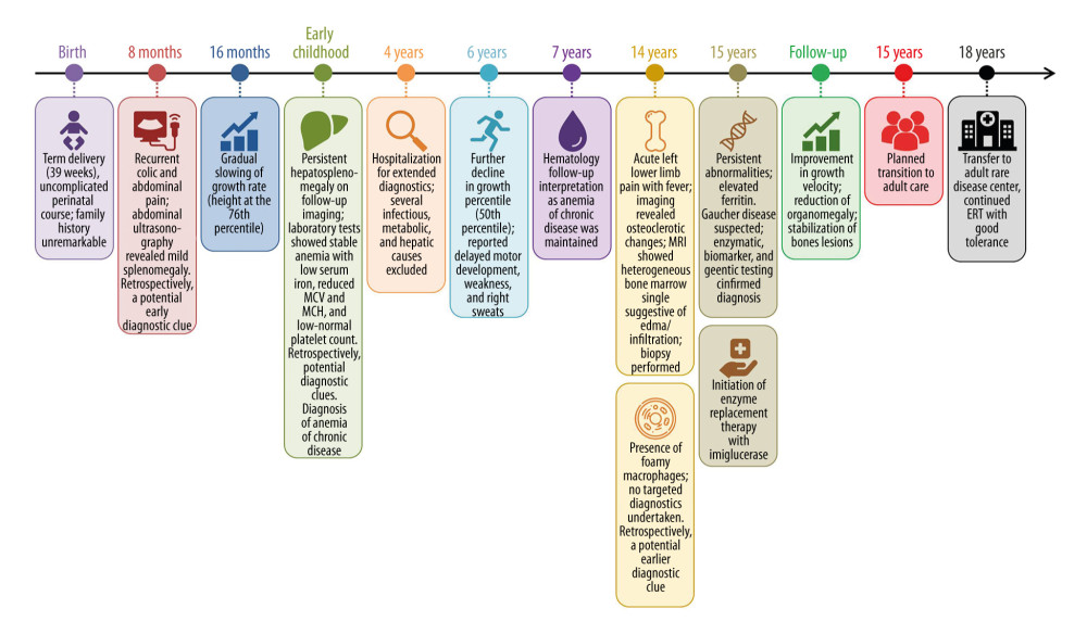 Figure 1. Clinical timeline from first manifestations to diagnosis, treatment initiation, and transition to adult care.Timeline of the patient’s clinical course, diagnostic evaluation, and treatment, highlighting key diagnostic milestones and potential earlier diagnostic clues. Abbreviations: ERT, enzyme replacement therapy; MCH, mean corpuscular hemoglobin; MCV, mean corpuscular volume; MRI, magnetic resonance imaging.
 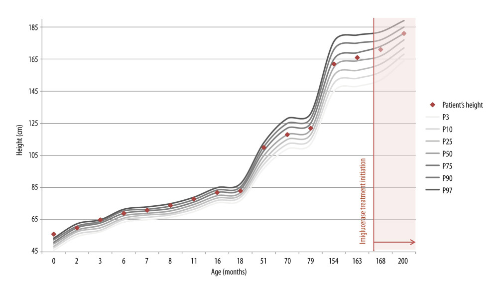 Figure 2. Growth trajectory before and after diagnosis of Gaucher disease.Patient’s height measurements plotted on Polish reference growth charts: Institute of Mother and Child charts for boys aged 0–5 years and Polish OLA/OLAF growth charts for older age groups.
 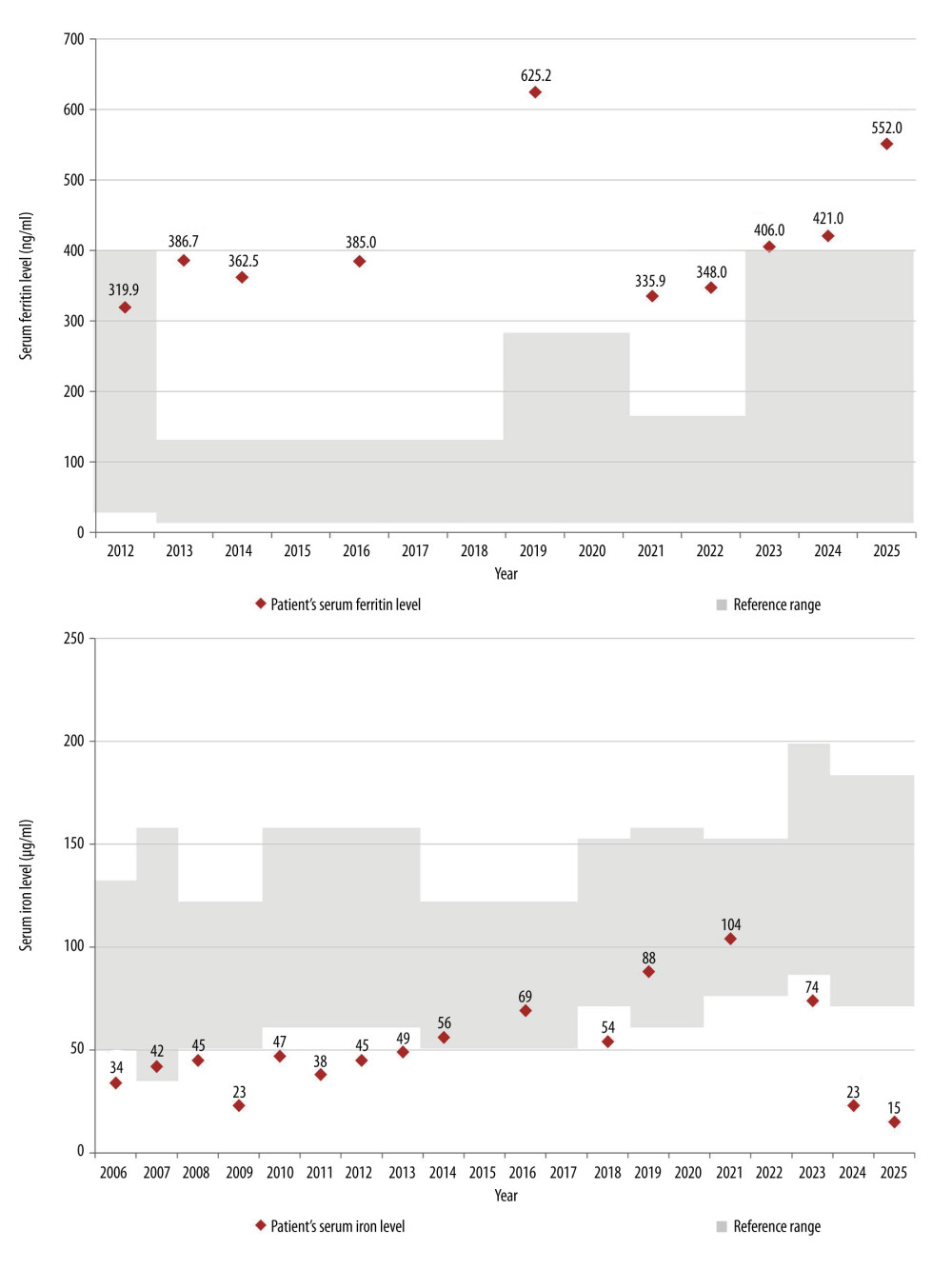 Figure 3. Longitudinal changes in iron metabolism parameters during follow-up.Longitudinal changes in the patient’s serum ferritin and serum iron levels over time. Reference ranges are indicated by shaded areas and were derived from the laboratory reports available at each time point. Given that laboratory assessments were performed in different laboratories over the years, age-specific reference ranges varied between measurements and thus may appear nonuniform across the figure. Data labels represent the patient’s individual measurements at each time point.
 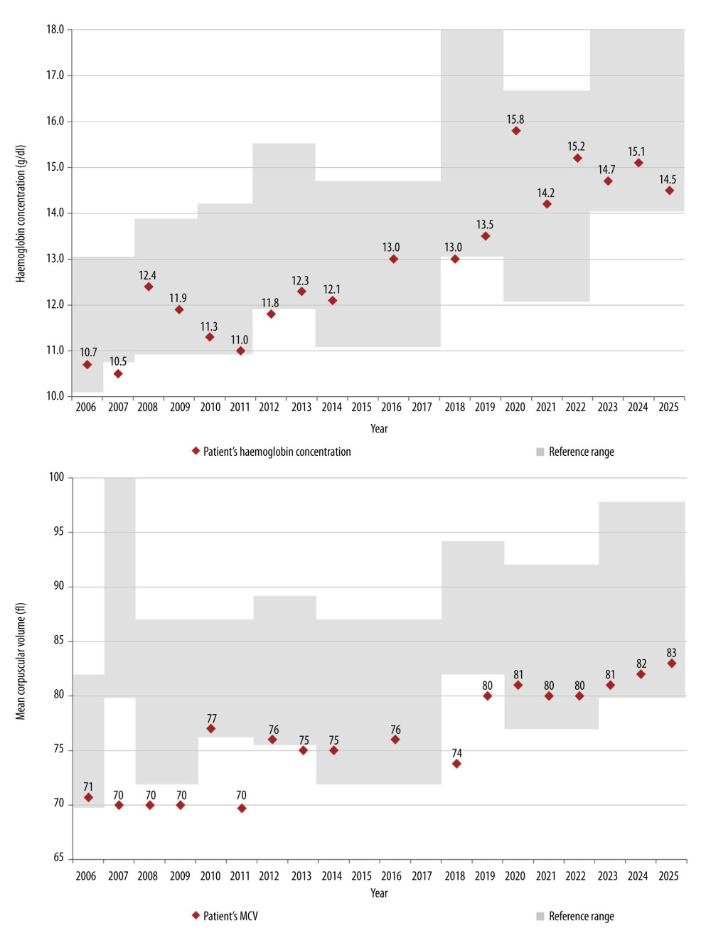 Figure 4. Longitudinal changes in hematologic parameters during follow-up.Longitudinal changes in the patient’s hemoglobin concentration and mean corpuscular volume (MCV) over time. Reference ranges are indicated by shaded areas and were derived from the laboratory reports available at each time point. Given that laboratory assessments were performed in different laboratories over the years, age-specific reference ranges varied between measurements and thus may appear nonuniform across the figure. Data labels represent the patient’s individual measurements at each time point.
 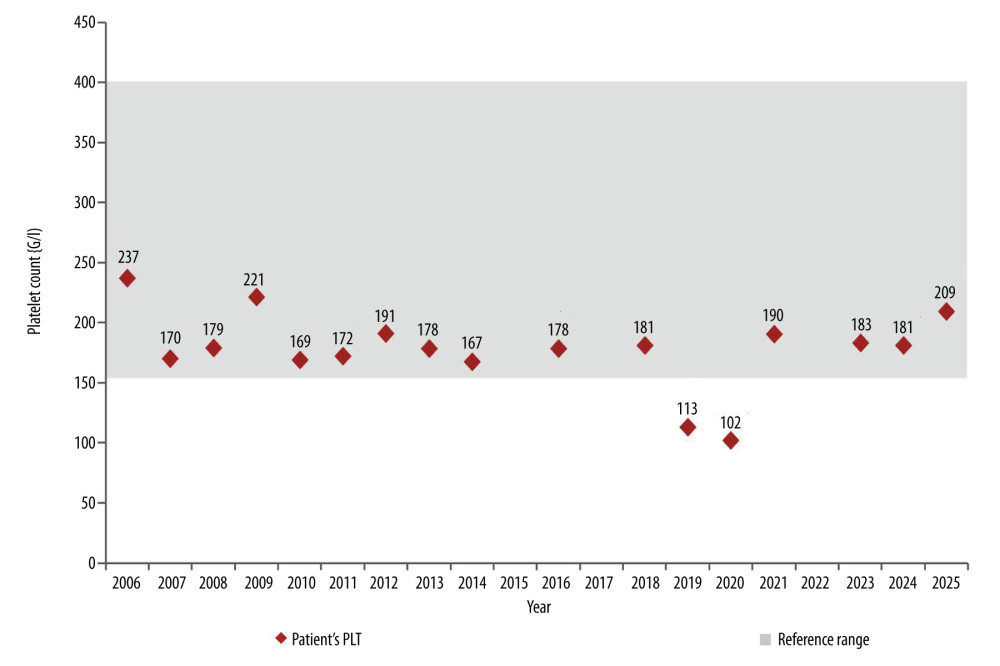 Figure 5. Longitudinal changes in platelet count during follow-up.Longitudinal changes in the patient’s platelet count over time. Reference ranges are indicated by shaded areas and were derived from the laboratory reports available at each time point. Data labels represent the patient’s individual values at each time point.
 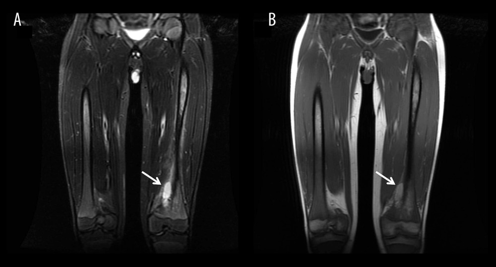 Figure 6. Magnetic resonance imaging findings during acute skeletal presentation.Coronal magnetic resonance imaging of the lower extremities. (A) Short tau inversion recovery sequence demonstrates a heterogeneous area of increased signal intensity within the distal metaphysis and diaphysis of the left femur, suggestive of bone marrow edema or infiltration. (B) Corresponding T1-weighted image shows reduced signal intensity in the same region, indicating replacement of normal bone marrow. White arrows indicate the abnormal marrow lesion within the distal femur. No cortical destruction or periosteal reaction is observed. Overall, the imaging findings are consistent with an acute bone crisis in Gaucher disease.
 References
1. Wang M, Li F, Zhang J, Global epidemiology of Gaucher disease: An updated systematic review and meta-analysis: J Pediatr Hematol Oncol, 2022; 44(8); e1154-62
2. Stirnemann J, Belmatoug N, Camou F, A review of Gaucher disease pathophysiology, clinical presentation and treatments: Int J Mol Sci, 2017; 18(2); 441
3. Gary SE, Ryan E, Steward AM, Sidransky E, Recent advances in the diagnosis and management of Gaucher disease: Expert Rev Endocrinol Metab, 2018; 13(2); 107-18
4. Elleder M, Foamy transformed Gaucher cells: Zentralbl Pathol, 1992; 138(1); 47-50
5. Sidransky E, Gaucher disease: Insights from a rare Mendelian disorder: Discov Med, 2012; 14(77); 273-81
6. Cox TM, Cachón-González MB, The cellular pathology of lysosomal diseases: J Pathol, 2012; 226(2); 241-54
7. Panicker LM, Miller D, Awad O, Gaucher iPSC-derived macrophages produce elevated levels of inflammatory mediators and serve as a new platform for therapeutic development: Stem Cells, 2014; 32(9); 2338-49
8. Xu YH, Jia L, Quinn B, Global gene expression profile progression in Gaucher disease mouse models: BMC Genomics, 2011; 12; 20
9. Simpson WL, Imaging of Gaucher disease: World J Radiol, 2014; 6(9); 657-68
10. Burrow TA, Sun Y, Prada CE, CNS, lung, and lymph node involvement in Gaucher disease type 3 after 11 years of therapy: Clinical, histopathologic, and biochemical findings: Mol Genet Metab, 2015; 114(2); 233-41
11. Willemsen R, Tybulewicz VLJ, Sidransky E, A biochemical and ultrastructural evaluation of the type 2 Gaucher mouse: Mol Chem Neuropathol, 1995; 24(2–3); 179-92
12. Baris HN, Cohen IJ, Mistry PK, Gaucher disease: The metabolic defect, pathophysiology, phenotypes and natural history: Pediatr Endocrinol Rev, 2014; 12(Suppl 1); 72-81
13. Rosenbaum H, Sidransky E, Cholelithiasis in patients with Gaucher disease: Blood Cells Mol Dis, 2002; 28(1); 21-27
14. Schiffmann R, Sevigny J, Rolfs A, The definition of neuronopathic Gaucher disease: J Inherit Metab Dis, 2020; 43(5); 1056-59
15. Goker-Alpan O, Schiffmann R, Park JK, Phenotypic continuum in neuronopathic Gaucher disease: An intermediate phenotype between type 2 and type 3: J Pediatr, 2003; 143(2); 273-76
16. Chen B, Dai W, Xu Y, Two delayed-diagnosis case reports of long-lasting thrombocytopenia with splenomegaly: Medicine (Baltimore), 2024; 103(34); e39354
17. Koprivica V, Stone DL, Park JK, Analysis and classification of 304 mutant alleles in patients with type 1 and type 3 Gaucher disease: Am J Hum Genet, 2000; 66(6); 1777-86
18. Sidransky E, Gaucher disease: Complexity in a “simple” disorder: Mol Genet Metab, 2004; 83(1–2); 6-15
19. Dardis A, Michelakakis H, Rozenfeld P, Patient-centered guidelines for the laboratory diagnosis of Gaucher disease type 1: Orphanet J Rare Dis, 2022; 17(1); 1-15
20. Mistry PK, Cappellini MD, Lukina E, A reappraisal of Gaucher disease—diagnosis and disease management algorithms: Am J Hematol, 2011; 86(1); 110-15
21. Hughes DA, Pastores GM, Gaucher disease, 2000, Seattle (WA), University of Washington, Seattle [updated 2023 Dec 7]
22. Dinur T, Bauer P, Beetz C, Gaucher disease diagnosis using lyso-Gb1 on dry blood spot samples: Time to change the paradigm?: Int J Mol Sci, 2022; 23(3); 1627
23. Nishimura S, Ma C, Sidransky E, Ryan E, Obstacles to early diagnosis of Gaucher disease: Ther Clin Risk Manag, 2025; 21; 93-101
24. Perez Y, Belmatoug N, Bengherbia M, Yousfi K, Lioger B, Misdiagnosis of Gaucher disease in real life: Retrospective study of the French Gaucher disease registry: Rev Med Interne, 2023; 44(2); 55-61
25. Mistry PK, Sadan S, Yang R, Consequences of diagnostic delays in type 1 Gaucher disease: The need for greater awareness among hematologists-oncologists and an opportunity for early diagnosis and intervention: Am J Hematol, 2007; 82(8); 697-701
26. Suris JC, Akre C, Key elements for, and indicators of, a successful transition: An international Delphi study: J Adolesc Health, 2015; 56(6); 612-18
27. Van Rossum A, Holsopple M, Enzyme replacement or substrate reduction? A review of Gaucher disease treatment options: Hosp Pharm, 2016; 51(7); 553-63
28. Mehta A, Belmatoug N, Bembi B, Exploring the patient journey to diagnosis of Gaucher disease from the perspective of 212 patients with Gaucher disease and 16 Gaucher expert physicians: Mol Genet Metab, 2017; 122(3); 122-29
29. Camou F, Berger MG, Gaucher disease, state of the art and perspectives: J Intern Med, 2025; 298(3); 155-72
30. Weinreb NJ, Goldblatt J, Villalobos J, Long-term clinical outcomes in type 1 Gaucher disease following 10 years of imiglucerase treatment: J Inherit Metab Dis, 2013; 36(3); 543-53
31. Wu M, Yang Z, Gaucher disease masked by childhood splenectomy: A forty-year diagnostic delay: Pol Arch Intern Med, 2026; 136(3); 17233
32. Cullufi P, Velmishi V, Dervishi E, Phenotypic spectrum of type 2–3 Gaucher disease: A case study in the Balkan genotype: Am J Case Rep, 2026; 27; e948999
33. Revel-Vilk S, Shalev V, Gill A, Assessing the diagnostic utility of the Gaucher Earlier Diagnosis Consensus (GED-C) scoring system using real-world data: Orphanet J Rare Dis, 2024; 19; 71
34. Miyamoto T, Iino M, Komorizono Y, Screening for Gaucher disease using dried blood spot tests: A Japanese multicenter, cross-sectional survey: Intern Med, 2021; 60(5); 699-707
35. Gelb MH, Lukacs Z, Ranieri E, Schielen PCJI, Newborn screening for lysosomal storage disorders: methodologies for measurement of enzymatic activities in dried blood spots: Int J Neonatal Screen, 2019; 5(1); 1
36. Orsini JJ, Martin MM, Showers AL, Lysosomal storage disorder 4+1 multiplex assay for newborn screening using tandem mass spectrometry: Application to a small-scale population study for five lysosomal storage disorders: Clin Chim Acta, 2012; 413(15–16); 1270-73
37. Gören Şahin D, Üsküdar Teke H, Karagülle M, Gaucher cells or pseudo-Gaucher cells: That’s the question: Turk J Hematol, 2014; 31(4); 428-29
38. Mahajan VS, Mattoo H, Deshpande V, IgG4-related disease: Annu Rev Pathol, 2014; 9; 315-47
39. Piran S, Nimmo GAM, Chaboureau A, The natural history of Gaucher disease type 1 in 31 patients over a median of 15 years: A retrospective study: Intern Med J, 2024; 54(10); 1661-68
40. Dinur T, Zimran A, Becker-Cohen M, Long-term follow-up of 103 untreated adult patients with type 1 Gaucher disease: J Clin Med, 2019; 8(10); 1662
41. Di Rocco M, Dionisi Vici C, Burlina A, Screening for lysosomal diseases in a selected pediatric population: The case of Gaucher disease and acid sphingomyelinase deficiency: Orphanet J Rare Dis, 2023; 18(1); 1-13
42. Motta I, Delbini P, Scaramellini N, Enzyme replacement therapy improves erythropoiesis and iron dysregulation in Gaucher disease: Ann Hematol, 2024; 103(12); 5113-21
43. Weinreb NJ, Goker-Alpan O, Kishnani PS, The diagnosis and management of Gaucher disease in pediatric patients: Where do we go from here?: Mol Genet Metab, 2022; 136(1); 4-21
44. Stepien KM, Žnidar I, Kieć-Wilk B, Transition of patients with Gaucher disease type 1 from pediatric to adult care: Results from two international surveys of patients and health care professionals: Front Pediatr, 2024; 12; 1439236
45. Blum RWM, Garell D, Hodgman CH, Transition from child-centered to adult health care systems for adolescents with chronic conditions: J Adolesc Health, 1993; 14(7); 570-76
46. Crowley R, Wolfe I, Lock K, Improving the transition between paediatric and adult health care: A systematic review: Arch Dis Child, 2011; 96(6); 548-53
Figures
Figure 1. Clinical timeline from first manifestations to diagnosis, treatment initiation, and transition to adult care.Timeline of the patient’s clinical course, diagnostic evaluation, and treatment, highlighting key diagnostic milestones and potential earlier diagnostic clues. Abbreviations: ERT, enzyme replacement therapy; MCH, mean corpuscular hemoglobin; MCV, mean corpuscular volume; MRI, magnetic resonance imaging.Figure 2. Growth trajectory before and after diagnosis of Gaucher disease.Patient’s height measurements plotted on Polish reference growth charts: Institute of Mother and Child charts for boys aged 0–5 years and Polish OLA/OLAF growth charts for older age groups.Figure 3. Longitudinal changes in iron metabolism parameters during follow-up.Longitudinal changes in the patient’s serum ferritin and serum iron levels over time. Reference ranges are indicated by shaded areas and were derived from the laboratory reports available at each time point. Given that laboratory assessments were performed in different laboratories over the years, age-specific reference ranges varied between measurements and thus may appear nonuniform across the figure. Data labels represent the patient’s individual measurements at each time point.Figure 4. Longitudinal changes in hematologic parameters during follow-up.Longitudinal changes in the patient’s hemoglobin concentration and mean corpuscular volume (MCV) over time. Reference ranges are indicated by shaded areas and were derived from the laboratory reports available at each time point. Given that laboratory assessments were performed in different laboratories over the years, age-specific reference ranges varied between measurements and thus may appear nonuniform across the figure. Data labels represent the patient’s individual measurements at each time point.Figure 5. Longitudinal changes in platelet count during follow-up.Longitudinal changes in the patient’s platelet count over time. Reference ranges are indicated by shaded areas and were derived from the laboratory reports available at each time point. Data labels represent the patient’s individual values at each time point.Figure 6. Magnetic resonance imaging findings during acute skeletal presentation.Coronal magnetic resonance imaging of the lower extremities. (A) Short tau inversion recovery sequence demonstrates a heterogeneous area of increased signal intensity within the distal metaphysis and diaphysis of the left femur, suggestive of bone marrow edema or infiltration. (B) Corresponding T1-weighted image shows reduced signal intensity in the same region, indicating replacement of normal bone marrow. White arrows indicate the abnormal marrow lesion within the distal femur. No cortical destruction or periosteal reaction is observed. Overall, the imaging findings are consistent with an acute bone crisis in Gaucher disease. Tables
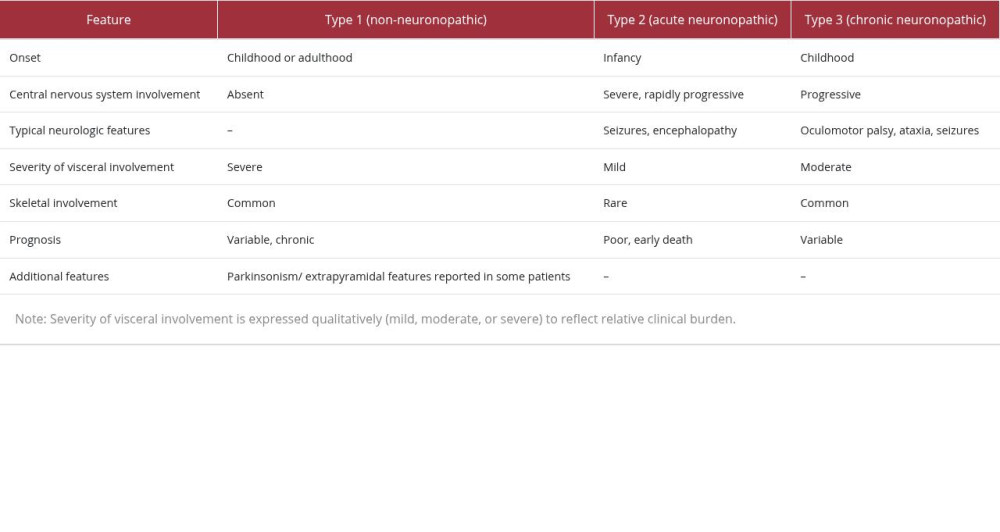 Table 1. Clinical classification and key features of Gaucher disease [2,5,12,14].
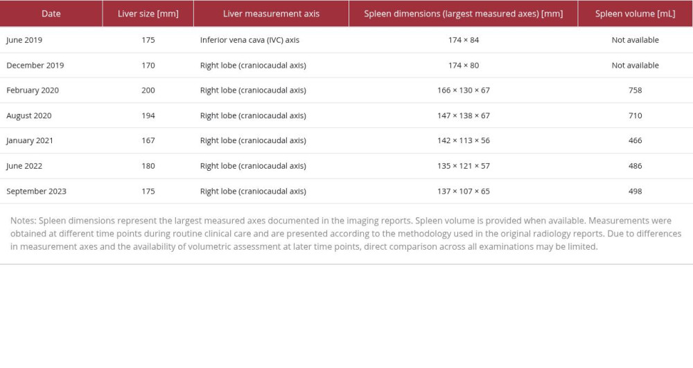 Table 2. Longitudinal measurements of liver and spleen size obtained via routine imaging follow-up.Table 1. Clinical classification and key features of Gaucher disease [2,5,12,14].Table 2. Longitudinal measurements of liver and spleen size obtained via routine imaging follow-up.
 In Press
Case report 
Am J Case Rep In Press; DOI: 10.12659/AJCR.953539
Case report 
Am J Case Rep In Press; DOI: 10.12659/AJCR.954530
Case report 
Am J Case Rep In Press; DOI: 10.12659/AJCR.954296
Case report 
Am J Case Rep In Press; DOI: 10.12659/AJCR.954629
Most Viewed Current Articles
07 Dec 2021 : Case report  28,708,227
DOI :10.12659/AJCR.934347
Am J Case Rep 2021; 22:e934347
06 Dec 2021 : Case report  180,327
DOI :10.12659/AJCR.934406
Am J Case Rep 2021; 22:e934406
21 Jun 2024 : Case report  124,051
DOI :10.12659/AJCR.944371
Am J Case Rep 2024; 25:e944371
08 May 2026 : Case report
94,430
DOI :10.12659/AJCR.952158
Am J Case Rep 2026; 27:e952158