


13 September 2026
: Case report 
Intravitreal Onchocerca sp. With Concomitant Cerebral MRI Lesion: A Case Report
Challenging differential diagnosis, Rare disease
Supanji ABCDEF 1,2,3,4*, Hifdza F. Felisha ABDEF 1,5, Dewi F. Romdhoniyyah DEF 1,2, Syania Nursawitri DEF 1, Sekar Syahriza DEF 1, Izzati A.K. Farzana DEF 1, Yasmin H. Basalamah DEF 1,2, Sarah Rizqia Indrayanti BDEF 3, Nia Milastuti BDEF 3, Karimah Amalia BCDEF 6,7, Nurhuda H. Setyawan BCDEF 6,7, Ajib Diptyanusa BCDEF 8, Tridjoko Hadianto CDEF 8, Sekar Satiti DEF 9,10, Mohammad Eko Prayogo BDEF 11,3, Firman S. Wardhana BDEF 11,2,3, Muhammad B. Sasongko BDEF 11,2,3, Raden R.U.N.W. Astuti CDEF 12, Yusuf Ridwan CDEF 13
DOI: 10.12659/AJCR.952921
Am J Case Rep 2026; 27:e952921


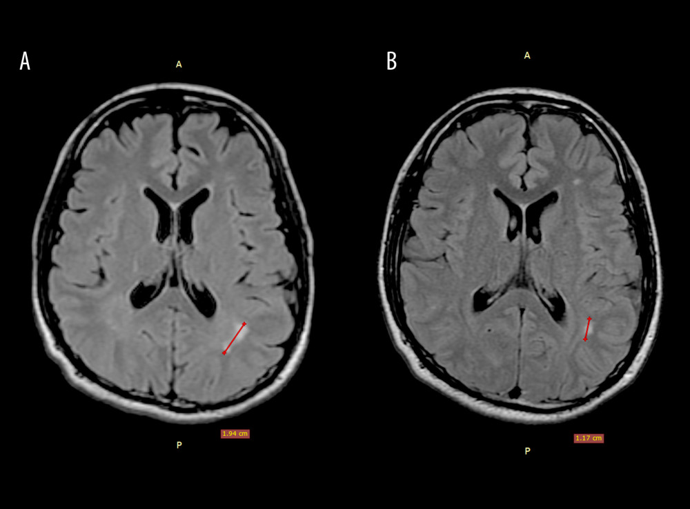
Figure 2 Brain magnetic resonance images of a 24-year-old man with vitreous Onchocerca lupi infection. (A) Hyperintense lesion in the white matter of the left parietal lobe on T2-weighted fluid-attenuated inversion recovery (FLAIR) sequences; (B) follow-up magnetic resonance image at 9 months demonstrating persistence of the lesion, with a decrease in size.