


13 September 2026: Articles 
Intravitreal Onchocerca sp. With Concomitant Cerebral MRI Lesion: A Case Report
Challenging differential diagnosis, Rare disease
Supanji ABCDEF 1,2,3,4*, Hifdza F. Felisha ABDEF 1,5, Dewi F. Romdhoniyyah DEF 1,2, Syania Nursawitri DEF 1, Sekar Syahriza DEF 1, Izzati A.K. Farzana DEF 1, Yasmin H. Basalamah DEF 1,2, Sarah Rizqia Indrayanti BDEF 3, Nia Milastuti BDEF 3, Karimah Amalia BCDEF 6,7, Nurhuda H. Setyawan BCDEF 6,7, Ajib Diptyanusa BCDEF 8, Tridjoko Hadianto CDEF 8, Sekar Satiti DEF 9,10, Mohammad Eko Prayogo BDEF 11,3, Firman S. Wardhana BDEF 11,2,3, Muhammad B. Sasongko BDEF 11,2,3, Raden R.U.N.W. Astuti CDEF 12, Yusuf Ridwan CDEF 13
DOI: 10.12659/AJCR.952921
Am J Case Rep 2026; 27:e952921


Abstract
BACKGROUND: Onchocerca lupi is a zoonotic filarial nematode that primarily infects canids. Human infection is rare and typically involves the eye or subcutaneous tissue. O. lupi infection should be considered in cases of unresolved uveitis or neurological symptoms. We report a diagnostically challenging case of human ocular O. lupi infection with possible neurological involvement.
CASE REPORT: A 24-year-old man from Jakarta, Indonesia, presented with a 1-month history of blurred vision, floaters, left ocular discomfort, and intermittent headaches. He did not respond to initial treatment for uveitis. Further examination revealed a mobile, thread-like, whitish worm in the left vitreous. Pars plana vitrectomy enabled removal of an intravitreal worm morphologically most consistent with adult male O. lupi, although definitive species-level confirmation was not possible in the absence of molecular testing. The patient’s ocular inflammation resolved postoperatively, but his visual acuity remained poor. Due to persistent headaches, brain magnetic resonance imaging (MRI) was performed; a T1-hypointense and T2-weighted fluid-attenuated inversion recovery (FLAIR)-hyperintense white matter lesion was evident in the left parietal lobe. The lesion persisted on 9-month follow-up MRI, and its etiology was uncertain; a causal relationship with the parasitic infection could not be established.
CONCLUSIONS: We describe the first reported case of intravitreal O. lupi infection with a concurrent cerebral MRI lesion. This infection should be considered in the differential diagnosis of unresolved uveitis in endemic areas to facilitate timely diagnosis and help prevent irreversible visual damage.
Keywords: Nematode Infections, Onchocerca, Onchocerciasis, Ocular
Introduction
Clinical manifestations of human zoonotic onchocerciasis range from asymptomatic infection to severe disease. Ocular involvement may present as conjunctivitis, ocular pain, lacrimation, or subcutaneous nodules containing adult worms [8]. If left untreated, severe cases can result in vision impairment or chronic inflammation [9]. Diagnosis is challenging due to the rarity of the condition and its similarity to other filarial infections. Thus, diagnostic confirmation often requires histological examination or molecular testing [10].
Treatment of human zoonotic onchocerciasis typically involves surgical removal of nodules or worms, supplemented by antiparasitic therapy such as ivermectin. However, complete resolution can be difficult to achieve in ocular cases [11]. Given its zoonotic potential,
Case Report
We present the case of a 24-year-old man from Jakarta, Indonesia, who resided in an apartment and reported no regular contact with animals. While living in Solo, Central Java, he developed ocular symptoms in his left eye over the course of 1 month, including blurred vision, hair-like floaters, and ocular discomfort. Concurrently, he began experiencing intermittent headaches. He reported no ocular pain, redness, or discharge and had no history of ocular trauma or inflammation. He was otherwise medically fit and well.
Despite a prior diagnosis of uveitis and treatment at multiple clinics, the patient’s ocular symptoms progressively worsened, prompting him to seek a second opinion at our institution. Comprehensive ophthalmic examination revealed severely reduced visual acuity in the left eye (6/60, without improvement on pinhole testing), a positive relative afferent pupillary defect, and retinal and subretinal lesions. A mobile, whitish, thread-like parasite was identified in the vitreous of the left eye, prompting urgent surgical intervention.
Pars plana vitrectomy was performed to remove the parasite, followed by postoperative antibiotic prophylaxis. Microscopic examination of the extracted parasite using an Olympus light microscope (CX21FS1, Olympus Corp, Tokyo, Japan) revealed a mature white worm measuring 12 mm × 320 μm. Examination with an Olympus binocular microscope (CX23, Olympus Corp) at 40 × objective magnification demonstrated cuticular ridges and striae, the absence of a buccal cavity, and a short posterior spicule. These morphological features were most consistent with adult male
Given the patient’s history of chronic headaches, brain magnetic resonance imaging (MRI) was performed. This imaging investigation revealed a hyperintense white matter lesion in the left parietal lobe on T2-weighted fluid-attenuated inversion recovery (FLAIR) sequences, with corresponding hypointensity on T1-weighted images (Figure 2A). Consultations with Neurology and Infectious Disease specialists were conducted to evaluate the need for further intervention and the possible clinical significance of the
Based on the MRI findings, cerebral vasculitis and cytomegalovirus (CMV) infection were considered in the differential diagnosis. CMV encephalitis was considered unlikely given the absence of systemic or ophthalmic manifestations of CMV infection, the patient’s immunocompetent status, and spontaneous clinical improvement without antiviral therapy. However, formal serological testing was not performed, and CMV infection could not be definitively excluded. Immune-mediated vasculitis was also considered less likely because there were no focal neurological deficits, systemic features suggestive of vasculitis, or history of immunosuppressive therapy; the lesion improved without specific immunosuppressive treatment.
At the 9-month follow-up, the patient reported gradual improvement in his headaches. Repeat brain MRI demonstrated that the hyperintense white matter lesion in the left parietal lobe on T2-FLAIR sequences persisted but had substantially decreased in size (Figure 2B). Because the patient’s clinical presentation was inconsistent with ischemic stroke, we considered the possibility that the brain lesion reflected migration of the worm or its metabolites across the blood-brain barrier (BBB); however, such migration remains speculative and was not supported by direct evidence. Although the lesion may have contributed to the patient’s headaches, a definitive causal relationship could not be established. The patient’s headaches improved with nonsteroidal anti-inflammatory drugs.
Discussion
This case report describes a severe and novel presentation of zoonotic
Human infection with
Our patient provides a pertinent illustration of these diagnostic challenges. His blurred vision and floaters were consistent with recognized ocular manifestations of
A key finding in the present case was the lack of visual improvement after successful pars plana vitrectomy and worm extraction. This finding suggests that the pathological process, including mechanical injury and presumed toxic or inflammatory metabolite-induced chorioretinitis [10,11], had caused irreversible retinal damage before surgery. Such an outcome underscores the potentially devastating consequences of intraocular nematode infection and the importance of early intervention.
Our parasitological examination indicated that the worm was most consistent with adult male
The most intriguing aspect of the present case was the possible neurological involvement. After parasite removal, the patient’s headaches gradually improved over 9 months, accompanied by a reduction in lesion size on follow-up MRI. The hyperintense MRI lesion suggested possible central nervous system involvement. We cautiously hypothesize that either the parasite or its inflammatory metabolites may have crossed the BBB and contributed to lesion formation; however, this interpretation remains speculative in the absence of histopathological or cerebrospinal fluid confirmation.
Neurotropic behavior has been documented in other nematodes, including
Definitive diagnosis of neuroparasitic infection typically requires histopathological examination or cerebrospinal fluid analysis [20]. However, these invasive investigations were not feasible in the present case. Although the clinical and radiological findings support possible central nervous system involvement, they are insufficient to establish a causal relationship. Accordingly, long-term neurological follow-up is warranted.
Other potential causes of a solitary white matter lesion, including demyelinating disease and alternative infectious etiologies, were also explored. Demyelinating disease was considered less likely because there was no history of transient neurological episodes, no multifocal lesions on MRI, and no clinical course typical of multiple sclerosis or related disorders. Furthermore, there were no clinical or laboratory findings suggestive of other neurotropic infections, such as tuberculosis, toxoplasmosis, or fungal disease. Nevertheless, in the absence of cerebrospinal fluid analysis or brain biopsy, these alternative diagnoses cannot be definitively excluded, and the lesion etiology remains uncertain.
Some limitations of this case should be acknowledged. Given the lack of molecular confirmation of species identification and the absence of histopathological or cerebrospinal fluid evidence of cerebral infection, any suggestion of neurotropic dissemination or BBB crossing remains speculative. Additionally, serological testing for CMV infection was not performed, and neither cerebrospinal fluid analysis nor brain biopsy was undertaken, both of which would have been necessary to definitively exclude infectious and inflammatory causes of the intracranial lesion.
Despite these limitations, this case broadens the recognized spectrum of ocular involvement in human
Future research should focus on developing accessible diagnostic assays for rare pathogens. Additional clinical and experimental studies are also needed to clarify the neurotropic potential of
Conclusions
We report the first case of intravitreal infection most consistent with
Figures
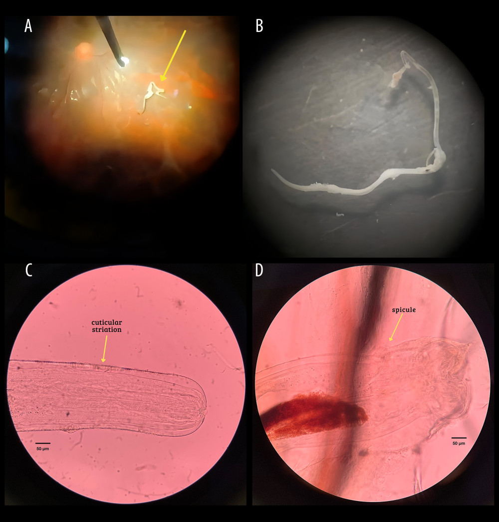 Figure 1. Parasite identification. (A) Intravitreal worm observed during the vitrectomy procedure; (B) cuticular striations at the anterior end of Onchocerca lupi; (C) short posterior spicule of O. lupi; (D) thin, thread-like, white adult O. lupi worm (12 mm × 320 μm).
 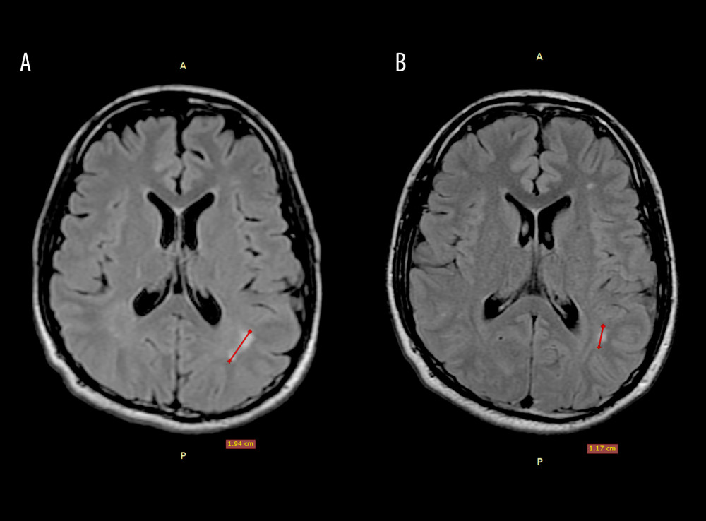 Figure 2. Brain magnetic resonance images of a 24-year-old man with vitreous Onchocerca lupi infection. (A) Hyperintense lesion in the white matter of the left parietal lobe on T2-weighted fluid-attenuated inversion recovery (FLAIR) sequences; (B) follow-up magnetic resonance image at 9 months demonstrating persistence of the lesion, with a decrease in size.
 References
1. Eberhard ML, Ostovar GA, Chundu K: Am J Trop Med Hyg, 2013; 88(3); 601-5
2. Eberhard ML, Ortega Y, Dial S: Vet Parasitol, 2000; 90(4); 333-38
3. Otranto D, Eberhard ML, Zoonotic helminths affecting the human eye: Parasit Vectors, 2011; 4; 41
4. Rojas A, Morales-Calvo F, Salant H: Yale J Biol Med, 2021; 94(2); 331-41
5. Grácio AJ, Richter J, Komnenou AT, Grácio MA: Parasitol Res, 2015; 114(7); 2401-13
6. Cambra-Pellejà M, Gandasegui J, Balaña-Fouce R: Pathogens, 2020; 9(9); 761
7. Otranto D, Giannelli A, Scotty Trumble N: Parasit Vectors, 2015; 8; 89
8. Das D, Ramachandra V, Islam S, Update on pathology of ocular parasitic disease: Indian J Ophthalmol, 2016; 64(11); 794-802
9. Hotterbeekx A, Namale Ssonko V, Oyet W: Brain Res Bull, 2019; 145; 39-44
10. Otranto D, Sakru N, Testini G: Am J Trop Med Hyg, 2011; 84(1); 55-58
11. Cantey PT, Weeks J, Edwards M: Clin Infect Dis, 2016; 62(6); 778-83
12. de Almeida M, Nascimento FS, Mathison BA: Am J Trop Med Hyg, 2020; 103(4); 1556-62
13. Otranto D, Dantas-Torres F, Giannelli A: Emerg Infect Dis, 2013; 19(12); 2000-3
14. Wesołowska M, Zając-Pytrus H, Masny A: Parasit Vectors, 2020; 13(1); 61
15. Rustam SNRN, Hadi UK, Soviana S: Jurnal Veteriner, 2019; 20(4); 511-18 [in Indonesian]
16. Hassan HK, Bolcen S, Kubofcik J: Emerg Infect Dis, 2015; 21(5); 789-96
17. Harlimton LMK, Wahyuni S, Reza A, Human ocular thelaziasis: A case report from Indonesia: Case Rep Ophthalmol, 2022; 13(2); 446-52
18. Mowlavi G, Farzbod F, Kheirkhah A: J Helminthol, 2014; 88(2); 250-55
19. Gyasi ME, Okonkwo ON, Tripathy K, Onchocerciasis. [Updated 2023 Aug 25]: StatPearls [Internet], 2025, Treasure Island (FL), StatPearls Publishing Available from: https://www.ncbi.nlm.nih.gov/books/NBK559027/
20. McGarry JW, Carrozza R, Bradley C: BMC Vet Res, 2022; 18(1); 66
Figures
Figure 1. Parasite identification. (A) Intravitreal worm observed during the vitrectomy procedure; (B) cuticular striations at the anterior end of Onchocerca lupi; (C) short posterior spicule of O. lupi; (D) thin, thread-like, white adult O. lupi worm (12 mm × 320 μm).Figure 2. Brain magnetic resonance images of a 24-year-old man with vitreous Onchocerca lupi infection. (A) Hyperintense lesion in the white matter of the left parietal lobe on T2-weighted fluid-attenuated inversion recovery (FLAIR) sequences; (B) follow-up magnetic resonance image at 9 months demonstrating persistence of the lesion, with a decrease in size. In Press
Case report 
Am J Case Rep In Press; DOI: 10.12659/AJCR.953317
Case report 
Am J Case Rep In Press; DOI: 10.12659/AJCR.954643
Case report 
Am J Case Rep In Press; DOI: 10.12659/AJCR.954074
Case report 
Am J Case Rep In Press; DOI: 10.12659/AJCR.953592
Most Viewed Current Articles
07 Dec 2021 : Case report
28,712,429
DOI :10.12659/AJCR.934347
Am J Case Rep 2021; 22:e934347
06 Dec 2021 : Case report  180,530
DOI :10.12659/AJCR.934406
Am J Case Rep 2021; 22:e934406
21 Jun 2024 : Case report
124,276
DOI :10.12659/AJCR.944371
Am J Case Rep 2024; 25:e944371
08 May 2026 : Case report
95,045
DOI :10.12659/AJCR.952158
Am J Case Rep 2026; 27:e952158