


15 September 2026
: Case report 
Pheochromocytoma Masquerading as Acute Coronary Syndrome Complicated by Cardiogenic Shock: A CARE-Compliant Case Report
Unusual clinical course, Challenging differential diagnosis
Songyan Zhang BC 1, Ying Cui EF 1, Shucheng Li ADG 1*DOI: 10.12659/AJCR.952994
Am J Case Rep 2026; 27:e952994


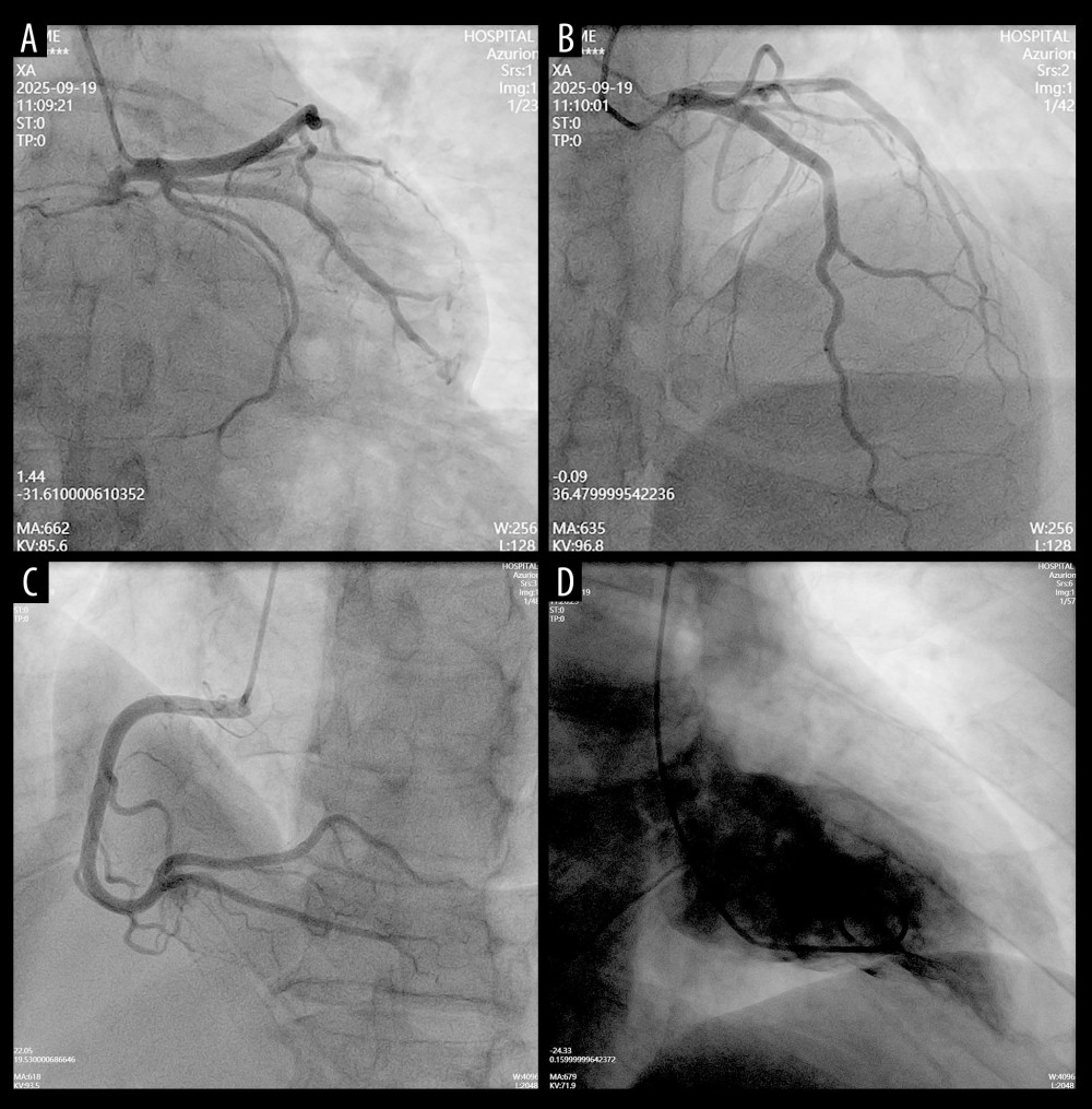
Figure 3 Coronary angiography findings(A–D) Selective coronary angiograms demonstrating no significant stenosis of the left anterior descending (A), left circumflex (B), or right coronary artery (C). An incidental small fistula from the right coronary artery to the right ventricle is noted (C, arrow). Left ventriculography (D) demonstrates normal chamber size and contractility, excluding obstructive coronary artery disease and supporting the diagnosis of myocardial infarction with non-obstructive coronary arteries (MINOCA).