


15 September 2026: Articles 
Pheochromocytoma Masquerading as Acute Coronary Syndrome Complicated by Cardiogenic Shock: A CARE-Compliant Case Report
Unusual clinical course, Challenging differential diagnosis
Songyan Zhang BC 1, Ying Cui EF 1, Shucheng Li ADG 1*DOI: 10.12659/AJCR.952994
Am J Case Rep 2026; 27:e952994


Abstract
BACKGROUND: Pheochromocytoma is a rare neuroendocrine tumor arising from chromaffin cells of the adrenal medulla that can manifest as catecholamine-induced cardiomyopathy mimicking acute coronary syndrome. Misdiagnosis is common, particularly when cardiogenic shock and elevated troponin levels occur in the absence of obstructive coronary artery disease. We describe pheochromocytoma masquerading as acute coronary syndrome complicated by cardiogenic shock, highlighting key diagnostic clues and management strategies.
CASE REPORT: A 59-year-old man with a 10-year history of intermittent chest tightness exhibited acute cardiogenic shock, peak troponin I level of 5.48 ng/mL, and N-terminal pro-B-type natriuretic peptide level of 19 800 pg/mL. Initial electrocardiography showed sinus tachycardia and T-wave abnormalities. Echocardiography revealed a reduced left ventricular ejection fraction of 40% with regional wall motion abnormalities. Coronary angiography demonstrated no significant stenosis, consistent with myocardial infarction with non-obstructive coronary arteries (MINOCA). Plasma free normetanephrine and metanephrine levels were 653.9 pg/mL (>680 times the upper limit of normal) and 378.5 pg/mL (>630 times the upper limit of normal), respectively. Abdominal computed tomography identified a right adrenal mass. After 2 weeks of preoperative alpha-blockade with phenoxybenzamine and volume expansion, the patient underwent laparoscopic adrenalectomy. Histopathology confirmed pheochromocytoma. Postoperatively, cardiac function normalized and symptoms completely resolved.
CONCLUSIONS: Pheochromocytoma should be suspected in patients with MINOCA and unexplained cardiogenic shock, particularly when accompanied by labile blood pressure. Substantially elevated plasma metanephrine and adrenal imaging facilitate early diagnosis. Catecholamine-induced cardiomyopathy is reversible after tumor resection. Multidisciplinary management, from hemodynamic stabilization to definitive surgery, is essential.
Keywords: acute coronary syndrome, cardiogenic shock, Case Reports, Endocrinology, Myocardial Infarction, Pheochromocytoma
Introduction
Pheochromocytoma, a neuroendocrine tumor arising from chromaffin cells of the adrenal medulla, affects approximately 0.2 to 0.6 per 100 000 individuals annually [1]. The classic clinical triad consists of episodic headache, sweating, and palpitations; approximately 10% of patients initially present with atypical cardiovascular manifestations, which are easily misdiagnosed as acute coronary syndrome [2]. Excessive catecholamine release directly induces myocardial injury and coronary vasospasm, leading to catecholamine-induced cardiomyopathy characterized by elevated troponin levels, regional wall motion abnormalities, and left ventricular dysfunction [3]. This cardiac manifestation of pheochromocytoma poses substantial diagnostic challenges, particularly when patients present with elevated troponin levels, ST-segment changes, and left ventricular dysfunction in the absence of coronary artery stenosis, a phenotype consistent with myocardial infarction with non-obstructive coronary arteries (MINOCA) [4]. We report a case of pheochromocytoma that initially manifested as acute coronary syndrome complicated by cardiogenic shock. By reviewing the diagnostic and therapeutic process, we emphasize the importance of broadening the differential diagnosis in patients with unexplained cardiovascular collapse and alert clinicians to the possibility of pheochromocytoma, thus facilitating early recognition and appropriate management of similar cases [5].
Case Report
PATIENT INFORMATION:
The patient was a 59-year-old Han Chinese man. His relevant medical history included intermittent chest tightness and dyspnea for more than 10 years, previously attributed to an unspecified cardiac etiology with poor response to symptomatic treatment. No relevant family history was reported. Before admission to a local hospital, he had received aminophylline, methylprednisolone, deslanoside (a cardiac glycoside), furosemide, and nesiritide (a recombinant B-type natriuretic peptide), all with poor therapeutic response.
TIMELINE:
More than 10 years prior to the first admission to our hospital, the patient developed intermittent chest tightness and dyspnea following emotional stress. These episodes lasted 3 to 5 minutes and were relieved by rest. During this period, he underwent multiple unsuccessful treatments at local hospitals. One day before the first admission, he experienced acute deterioration characterized by generalized fatigue, diffuse pain, dyspnea with cough and expectoration, orthopnea, and substernal chest pain radiating to the back. He was diagnosed with heart failure and pneumonia at a local hospital.
On the day of the first admission, he presented to the emergency department of our hospital and was transferred to the intensive care unit for management of cardiogenic shock. Initial diagnoses included myocardial injury, with a differential diagnosis of acute coronary syndrome versus myocarditis, New York Heart Association class IV heart failure, severe pneumonia, influenza A (H1N1) infection, and right adrenal incidentaloma of unknown etiology. During days 1 to 7 of the first admission, he received noninvasive ventilation, anti-infective therapy, antiplatelet therapy, and diuretic treatment. His troponin levels gradually declined, but persistent hypotension required dopamine support. The patient refused coronary angiography and was discharged against medical advice.
Three months before the second admission, he developed recurrent dyspnea at rest accompanied by palpitations and dizziness. During the second admission, he was rehospitalized; pheochromocytoma was confirmed via biochemical and radiologic evaluation. Coronary angiography showed no significant stenosis, and blood pressure lability was managed with phenoxybenzamine. One month after discharge from the second admission, he was admitted to the urology department for definitive surgical management. After 2 weeks of preoperative alpha-blockade and volume expansion, he underwent laparoscopic right adrenalectomy and had a favorable postoperative recovery.
CLINICAL FINDINGS:
At the first admission, the patient’s vital signs in the emergency department were as follows: temperature, 36.4 °C; heart rate, 146 beats/minute; respiratory rate, 33 breaths/minute; blood pressure, 113/96 mm Hg; and oxygen saturation, 96% on 5 L/minute oxygen via face mask. General examination indicated that the patient was conscious but had an altered mental status (confusion and agitation) with generalized cold, clammy skin. Cardiovascular examination revealed no pathologic murmurs and low peripheral skin temperature. Respiratory examination demonstrated bilateral moist rales with diminished breath sounds in the left lung. Examination of the extremities showed no edema but scattered ecchymoses around the knees.
At the second admission, blood pressure was highly labile, ranging from 76/54 mm Hg to 198/95 mm Hg. Cardiovascular examination revealed tachycardia with palpitations.
DIAGNOSTIC ASSESSMENT:
Biochemical investigations at the first admission showed the following results. Troponin I peaked at 5.48 ng/mL (reference, < 0.3 ng/mL), indicating substantial elevation. N-terminal pro-B-type natriuretic peptide peaked at 16 500 pg/mL (reference, ≤ 300 pg/mL) upon initial admission, indicating severe elevation. The white blood cell count was 14.68 × 10⁹/L (reference, 3.50–9.50 × 10⁹/L), indicating leukocytosis. Hemoglobin was 195 g/L (reference, 130–175 g/L), indicating polycythemia. The neutrophil percentage was 87.1% (reference, 40.0%–75.0%), indicating neutrophilia. C-reactive protein was 94.89 mg/L (reference, 0–10 mg/L), indicating substantial elevation. D-dimer was 3.37 μg/mL (reference, < 0.55 μg/mL), indicating elevation. Arterial lactate was 6.5 mmol/L (reference, 0.5–1.6 mmol/L), indicating severe hyperlactatemia.
At the second admission, troponin I was 1.45 ng/mL, remaining elevated above the reference range of less than 0.3 ng/mL. N-terminal pro-B-type natriuretic peptide was 19 800 pg/mL, indicating severe elevation. At 8: 00 AM, adrenocorticotropic hormone was 9.32 pg/mL (within the normal range; reference, 7.20–63.30 pg/mL), whereas cortisol was above 1750 nmol/L (reference, 166–507 nmol/L), indicating substantial elevation. At midnight, adrenocorticotropic hormone was 37.85 pg/mL (within the normal range; reference, 7.20–63.30 pg/mL), whereas cortisol remained above 1750 nmol/L (reference, 166–507 nmol/L), indicating persistent substantial elevation.
Confirmatory testing showed a plasma free normetanephrine level of 653.9 pg/mL (reference, 0.90–0.96 pg/mL), representing more than 680 times the upper limit of normal. Plasma free metanephrine was 378.5 pg/mL (reference, 0.50–0.60 pg/mL), representing more than 630 times the upper limit of normal. The combined plasma free normetanephrine and metanephrine level was 1032.4 pg/mL, indicating substantial elevation. Urinary normetanephrine was 358.4 μg per 24 hours (reference, < 460 μg per 24 hours), which was within the normal range. Urinary metanephrine was 538.7 μg per 24 hours (reference, < 302 μg per 24 hours), representing approximately 1.8 times the upper limit of normal. The combined urinary normetanephrine and metanephrine level was 897.1 μg per 24 hours, indicating elevation. Urinary dopamine, epinephrine, and norepinephrine levels were 338, 280, and 479.5 μg per 24 hours, respectively.
IMAGING AND SPECIAL EXAMINATIONS:
Electrocardiography (ECG) showed the following findings. At the first admission (Figure 1A), ECG demonstrated sinus tachycardia (heart rate, 131 beats/minute), abnormal Q waves in leads V1 and V2, left axis deviation, poor R-wave progression, and T-wave abnormalities. At the second admission (Figure 1B), ECG demonstrated sinus rhythm (heart rate, 73 beats/minute), P-wave broadening, and QT interval prolongation (474 ms).
Bedside echocardiography at the first admission showed a left ventricular diameter of 47 mm, regional wall motion abnormalities, a reduced left ventricular ejection fraction of 40%, and mild to moderate mitral regurgitation. Preoperative echocardiography demonstrated a left ventricular diameter of 52 mm and a normalized left ventricular ejection fraction of 62%. Abdominal computed tomography (Figure 2) revealed a right adrenal mass exhibiting imaging features consistent with pheochromocytoma.
Coronary angiography (Figure 3A–3D) demonstrated no significant coronary artery stenosis, an incidental right coronary artery-to-right ventricular fistula, and normal left ventriculography [5].
Gross pathological examination (Figure 4) revealed a cystic-solid tumor measuring 5 × 4 × 3 cm with a 2-cm rupture. The cut surface showed a dark red central region and a pale yellow peripheral region, with attached irregular tissue measuring 4 × 3 × 1.5 cm. Histopathologic examination demonstrated a trabecular and small nested growth pattern, abundant eosinophilic cytoplasm, nuclear pleomorphism, a richly vascularized stroma with prominent sinusoidal vessels, and extensive hemorrhage. The final diagnosis was pheochromocytoma of the right adrenal gland [6].
DIFFERENTIAL DIAGNOSIS:
Acute coronary syndrome was initially considered because of the patient’s chest pain, elevated troponin levels, ST-segment changes, and regional wall motion abnormalities. However, this diagnosis was excluded by the absence of significant coronary artery stenosis on angiography and a wall motion pattern that did not correspond to a single coronary vascular territory.
Viral myocarditis was also considered because of the concurrent influenza infection, elevated troponin levels, and reduced left ventricular ejection fraction. However, the absence of typical prodromal symptoms, prolonged recurrent clinical course, and lack of supporting evidence from endomyocardial biopsy argued against this diagnosis.
Takotsubo cardiomyopathy was included in the differential diagnosis because of the emotional trigger and the presence of wall motion abnormalities. However, it was considered unlikely because characteristic apical ballooning was absent and the left ventricular dysfunction was persistent rather than transient.
The diagnosis of pheochromocytoma was ultimately confirmed by the substantially elevated plasma metanephrine levels, presence of a right adrenal mass, strongly labile blood pressure, and complete resolution of cardiac dysfunction after tumor resection.
THERAPEUTIC INTERVENTION:
During the first admission, supportive care included noninvasive ventilatory support, anti-infective therapy with coverage for influenza A, antiplatelet therapy with aspirin, diuresis with furosemide, and inotropic support with intravenous dopamine after metoprolol and nitroglycerin were discontinued due to hypotension. The patient declined coronary angiography and was discharged against medical advice.
During the second admission, definitive management began with acute hemodynamic stabilization. Intravenous sodium nitroprusside was initiated at 10 μg/minute but was ineffective. Intravenous phenoxybenzamine was then administered as a 5-mg bolus followed by a continuous infusion at 150 μg/minute. During hospitalization in the urology department, oral phenoxybenzamine (20 mg, 3 times daily) was administered for preoperative preparation, along with volume expansion using crystalloids and colloids for 2 weeks. The patient subsequently underwent retroperitoneal laparoscopic partial right adrenalectomy with lysis of perirenal adhesions. During tumor manipulation, the systolic blood pressure transiently increased to 190 mm Hg. A tumor measuring 5 × 4 × 3 cm was successfully resected. Postoperatively, hemodynamic monitoring demonstrated stable blood pressure without the need for vasoactive support.
FOLLOW-UP AND OUTCOMES:
After preoperative medical optimization, the patient’s blood pressure was 104/62 mm Hg, hemoglobin was 118 g/L, troponin was negative, and the N-terminal pro-B-type natriuretic peptide level had decreased to 21 pg/mL. Postoperatively, he remained hemodynamically stable without further hypertensive crises. At the 3-month follow-up, echocardiography demonstrated complete normalization of left ventricular ejection fraction and regional wall motion. At the 6-month follow-up, he remained free of chest tightness and palpitations, with normal blood pressure and complete recovery of cardiac function.
Discussion
MECHANISM OF CATECHOLAMINE-INDUCED MYOCARDIAL INJURY:
Cardiac manifestations of pheochromocytoma involve a complex pathophysiology that extends beyond simple coronary vasospasm. Direct myocardial toxicity occurs when excessive catecholamines, particularly norepinephrine, activate β1-adrenergic receptors, triggering intracellular calcium overload, myocyte necrosis, and inflammatory infiltration that histologically resembles myocarditis [4]. This mechanism explains our patient’s reduced left ventricular ejection fraction of 40% and regional wall motion abnormalities despite patent coronary arteries [7,8].
Microvascular dysfunction and coronary vasospasm occur when catecholamines induce substantial constriction of the epicardial coronary arteries and intramyocardial microvasculature, resulting in myocardial ischemia without fixed coronary stenosis, a phenotype consistent with MINOCA [2].
A supply-demand mismatch also contributes to myocardial injury because sudden catecholamine surges greatly increase myocardial oxygen demand by elevating blood pressure and heart rate, precipitating ischemia when oxygen demand exceeds supply.
DIAGNOSTIC CLUES: SIGNIFICANCE OF BLOOD PRESSURE LABILITY:
A characteristic clinical feature of pheochromocytoma is paroxysmal hypertension alternating with hypotension or shock over a short period. Hypertensive crises occur when massive catecholamine release causes intense vasoconstriction and pronounced cardiac stimulation. In contrast, hypotension and shock can arise from several mechanisms, including vascular paralysis after chronic catecholamine exposure, absolute hypovolemia caused by fluid extravasation secondary to prolonged vasoconstriction, and cardiogenic shock resulting from catecholamine-induced cardiomyopathy [9,10].
Our patient exhibited this classic pattern, with blood pressure ranging from 76/54 mm Hg to 198/95 mm Hg, a systolic variation of 122 mm Hg. Such prominent blood pressure lability should prompt immediate evaluation for secondary causes of hypertension.
UNIQUE FEATURES OF THIS CASE:
Unlike previously reported cases of pheochromocytoma-induced cardiomyopathy, which typically involve transient myocardial injury and rapid recovery after tumor resection, our patient had a 10-year history of recurrent symptoms and repeated misdiagnoses before ultimately presenting with cardiogenic shock. This prolonged clinical course highlights the insidious nature of atypical presentations. Furthermore, the extreme elevation of plasma metanephrines (> 680 times the upper limit of normal for normetanephrine and > 630 times the upper limit of normal for metanephrine) and the development of cardiogenic shock in the context of MINOCA without obstructive coronary artery disease represent an extreme phenotype that has rarely been documented. The combination of prolonged diagnostic delay, severe biochemical abnormalities, and complete reversal of cardiac dysfunction after tumor resection, with left ventricular ejection fraction improving from 40% to 62% and N-terminal pro-B-type natriuretic peptide decreasing from 19 800 pg/mL to 21 pg/mL, makes this case particularly instructive. It emphasizes that pheochromocytoma should be considered even in patients with longstanding cardiac symptoms and angiographically normal coronary arteries.
COMPARISON WITH PREVIOUSLY REPORTED CASES:
It has been reported that fewer than 10% of patients with pheochromocytoma initially present with acute coronary syndrome; most experience transient myocardial injury and recovery of cardiac function within 1 to 3 months after tumor resection. In contrast, our patient experienced recurrent symptoms for 10 years and was repeatedly misdiagnosed before the onset of cardiogenic shock. This case highlights the potential for substantial diagnostic delay in atypical pheochromocytoma and underscores the need for heightened clinical vigilance.
CLINICAL IMPLICATIONS:
In patients who presenting with MINOCA, pheochromocytoma, vasculitis, and coronary embolism should be systematically excluded [11]. In patients with refractory heart failure despite angiographically normal coronary arteries, plasma metanephrine measurements and adrenal imaging should be performed. In cases of unexplained shock that arise in patients with a history of hypertension, catecholamine withdrawal after chronic catecholamine excess should be considered. In patients with labile blood pressure and episodic symptoms, 24-hour urinary or plasma metanephrine testing should be obtained, followed by adrenal computed tomography or magnetic resonance imaging as indicated [1,12].
MULTIDISCIPLINARY MANAGEMENT:
Successful management required coordinated care across multiple specialties. Cardiology provided hemodynamic stabilization and coronary angiography to exclude acute coronary syndrome. Endocrinology established the biochemical diagnosis and optimized the patient preoperatively. Urology performed surgical resection after appropriate alpha-blockade, and anesthesiology managed intraoperative catecholamine surges.
The transition from symptomatic cardiovascular support to definitive treatment with alpha-adrenergic blockade followed by surgical resection was critical [2]. Postoperative normalization of cardiac function and normalization of biomarkers, with an N-terminal pro-B-type natriuretic peptide level of 21 pg/mL, confirmed that treatment of the underlying cause resulted in complete recovery [6,10].
IMPLICATIONS FOR FUTURE RESEARCH AND CLINICAL PRACTICE:
This case suggests that plasma metanephrine testing and adrenal imaging should be performed early in patients with recurrent chest tightness, unexplained heart failure, or cardiogenic shock—even in the absence of the classic triad of pheochromocytoma—to avoid misdiagnosis and inappropriate treatment. Future retrospective studies of atypical cardiovascular manifestations of pheochromocytoma may help establish an early recognition scoring system to improve diagnostic accuracy.
Conclusions
Pheochromocytoma can present as life-threatening acute coronary syndrome complicated by cardiogenic shock, requiring a high index of clinical suspicion to avoid misdiagnosis. This case demonstrates that a broad differential diagnosis is essential in patients who present with MINOCA and unexplained cardiogenic shock. Pronounced blood pressure lability is a hallmark of pheochromocytoma; prompt measurement of plasma or urinary metanephrines, along with adrenal imaging, is essential for diagnosis. Catecholamine-induced cardiomyopathy is reversible after appropriate tumor resection and management. Multidisciplinary care, from acute hemodynamic stabilization to definitive surgery, is the cornerstone of treatment. Early recognition of such atypical presentations is crucial for improving patient outcomes [2].
Figures
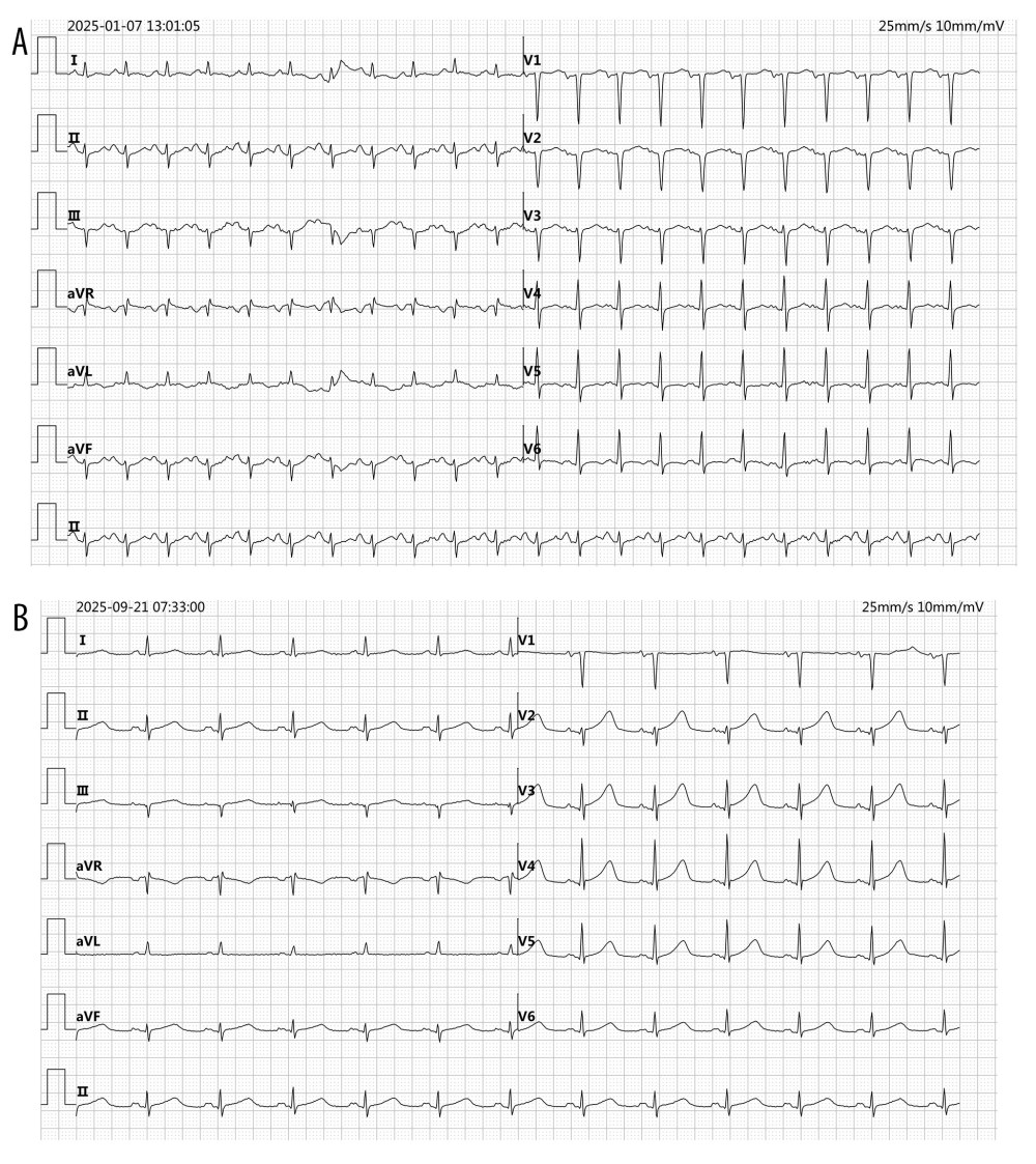 Figure 1. Electrocardiographic findings(A) Initial admission ECG demonstrating sinus tachycardia (131 bpm), abnormal Q waves in leads V1–V2, left axis deviation, poor R-wave progression, and T-wave abnormalities, suggestive of myocardial ischemia or injury. (B) ECG obtained during the second admission showing sinus rhythm (73 bpm), P-wave broadening, and a prolonged QT interval (474 ms), consistent with ongoing electrical instability in the context of catecholamine excess.
 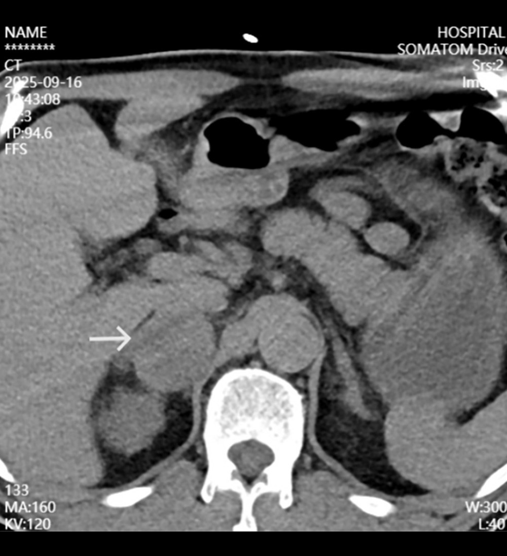 Figure 2. Abdominal computed tomographyAxial contrast-enhanced computed tomography scan revealing a well-defined right adrenal mass (arrow) exhibiting heterogeneous enhancement and imaging features characteristic of pheochromocytoma.
 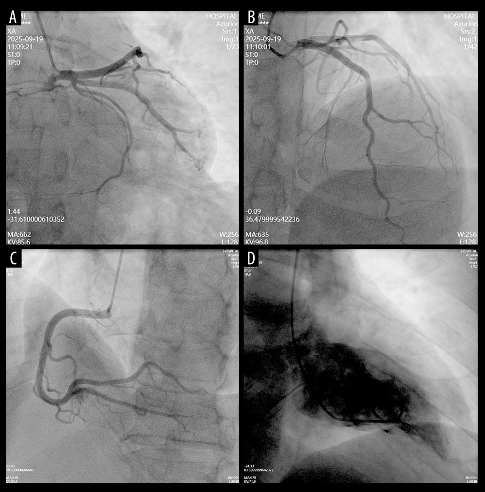 Figure 3. Coronary angiography findings(A–D) Selective coronary angiograms demonstrating no significant stenosis of the left anterior descending (A), left circumflex (B), or right coronary artery (C). An incidental small fistula from the right coronary artery to the right ventricle is noted (C, arrow). Left ventriculography (D) demonstrates normal chamber size and contractility, excluding obstructive coronary artery disease and supporting the diagnosis of myocardial infarction with non-obstructive coronary arteries (MINOCA).
 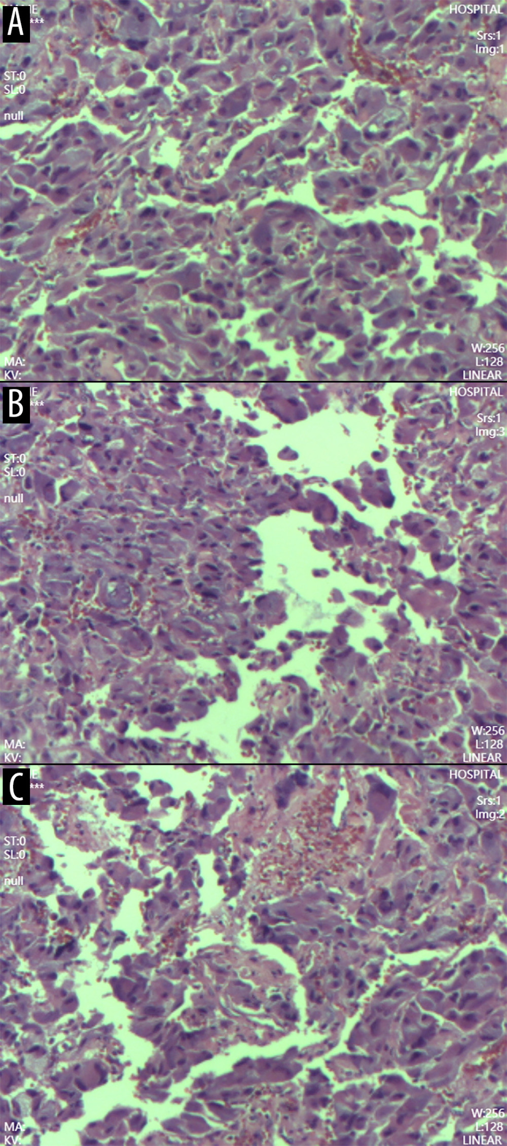 Figure 4. Pathological examination of the resected adrenal tumor(A) Gross specimen showing a cystic-solid adrenal tumor measuring 5 × 4 × 3 cm, with a central dark red hemorrhagic area, a peripheral pale yellow rim, and a 2-cm rupture. (B) Low-power photomicrograph (hematoxylin and eosin stain) demonstrating a trabecular and nested (Zellballen) growth pattern characteristic of pheochromocytoma. (C) High-power photomicrograph showing tumor cells with abundant eosinophilic cytoplasm, nuclear pleomorphism, and prominent sinusoidal vasculature. Extensive intratumoral hemorrhage is present. These histopathologic features confirm the diagnosis of right adrenal pheochromocytoma.
 References
1. Lenders JW, Eisenhofer G, Mannelli M, Phaeochromocytoma: Lancet, 2005; 366(9486); 665-75
2. Zhang R, Gupta D, Albert SG, Pheochromocytoma as a reversible cause of cardiomyopathy: Analysis and review of the literature: Int J Cardiol, 2017; 249; 319-23
3. Manger WM, The protean manifestations of pheochromocytoma: Horm Metab Res, 2009; 41(9); 658-63
4. Sanchez-Recalde A, Costero O, Oliver JM, Pheochromocytoma-related cardiomyopathy: inverted Takotsubo contractile pattern: Circulation, 2006; 113(17); e738-39
5. Pasupathy S, Air T, Dreyer RP, Systematic review of patients presenting with suspected myocardial infarction and nonobstructive coronary arteries: Circulation, 2015; 131(10); 861-70
6. Brouwers FM, Lenders JW, Eisenhofer G, Pheochromocytoma as an endocrine emergency: Rev Endocr Metab Disord, 2003; 4(2); 121-28
7. Kurioka H, Takahashi K, Miyake Y, Pheochromocytoma-induced Takotsubo-like cardiomyopathy: Heart Vessels, 2015; 30(4); 527-32
8. Y-Hassan S, De Bacquer D, Torp-Pedersen C, Pheochromocytoma and stress cardiomyopathy: A different pathogenesis than generally assumed?: Eur Heart J, 2018; 39(22); 1987-89
9. Whitelaw BC, Prague JK, Mustafa OG, Phaeochromocytoma crisis: Clin Endocrinol (Oxf), 2014; 80(1); 13-22
10. Ghadri JR, Wittstein IS, Prasad A, International expert consensus document on Takotsubo syndrome (part I): Clinical characteristics, diagnostic criteria, and pathophysiology: Eur Heart J, 2018; 39(22); 2032-46
11. Lenders JW, Duh QY, Eisenhofer G, Pheochromocytoma and paraganglioma: An Endocrine Society clinical practice guideline: J Clin Endocrinol Metab, 2014; 99(6); 1915-42
12. Pelliccia F, Kaski JC, Crea F, Pathophysiology of Takotsubo syndrome: Circulation, 2017; 135(24); 2426-41
Figures
Figure 1. Electrocardiographic findings(A) Initial admission ECG demonstrating sinus tachycardia (131 bpm), abnormal Q waves in leads V1–V2, left axis deviation, poor R-wave progression, and T-wave abnormalities, suggestive of myocardial ischemia or injury. (B) ECG obtained during the second admission showing sinus rhythm (73 bpm), P-wave broadening, and a prolonged QT interval (474 ms), consistent with ongoing electrical instability in the context of catecholamine excess.Figure 2. Abdominal computed tomographyAxial contrast-enhanced computed tomography scan revealing a well-defined right adrenal mass (arrow) exhibiting heterogeneous enhancement and imaging features characteristic of pheochromocytoma.Figure 3. Coronary angiography findings(A–D) Selective coronary angiograms demonstrating no significant stenosis of the left anterior descending (A), left circumflex (B), or right coronary artery (C). An incidental small fistula from the right coronary artery to the right ventricle is noted (C, arrow). Left ventriculography (D) demonstrates normal chamber size and contractility, excluding obstructive coronary artery disease and supporting the diagnosis of myocardial infarction with non-obstructive coronary arteries (MINOCA).Figure 4. Pathological examination of the resected adrenal tumor(A) Gross specimen showing a cystic-solid adrenal tumor measuring 5 × 4 × 3 cm, with a central dark red hemorrhagic area, a peripheral pale yellow rim, and a 2-cm rupture. (B) Low-power photomicrograph (hematoxylin and eosin stain) demonstrating a trabecular and nested (Zellballen) growth pattern characteristic of pheochromocytoma. (C) High-power photomicrograph showing tumor cells with abundant eosinophilic cytoplasm, nuclear pleomorphism, and prominent sinusoidal vasculature. Extensive intratumoral hemorrhage is present. These histopathologic features confirm the diagnosis of right adrenal pheochromocytoma. In Press
Case report
Am J Case Rep In Press; DOI: 10.12659/AJCR.953317
Case report 
Am J Case Rep In Press; DOI: 10.12659/AJCR.954643
Case report 
Am J Case Rep In Press; DOI: 10.12659/AJCR.954074
Case report 
Am J Case Rep In Press; DOI: 10.12659/AJCR.953592
Most Viewed Current Articles
07 Dec 2021 : Case report
28,712,429
DOI :10.12659/AJCR.934347
Am J Case Rep 2021; 22:e934347
06 Dec 2021 : Case report  180,530
DOI :10.12659/AJCR.934406
Am J Case Rep 2021; 22:e934406
21 Jun 2024 : Case report
124,276
DOI :10.12659/AJCR.944371
Am J Case Rep 2024; 25:e944371
08 May 2026 : Case report
95,045
DOI :10.12659/AJCR.952158
Am J Case Rep 2026; 27:e952158