


15 September 2026
: Case report 
Pheochromocytoma Masquerading as Acute Coronary Syndrome Complicated by Cardiogenic Shock: A CARE-Compliant Case Report
Unusual clinical course, Challenging differential diagnosis
Songyan Zhang BC 1, Ying Cui EF 1, Shucheng Li ADG 1*DOI: 10.12659/AJCR.952994
Am J Case Rep 2026; 27:e952994


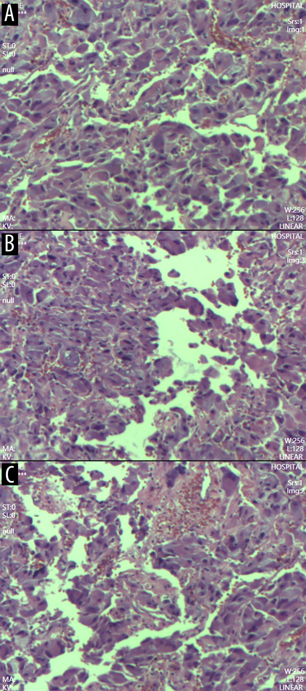
Figure 4 Pathological examination of the resected adrenal tumor(A) Gross specimen showing a cystic-solid adrenal tumor measuring 5 × 4 × 3 cm, with a central dark red hemorrhagic area, a peripheral pale yellow rim, and a 2-cm rupture. (B) Low-power photomicrograph (hematoxylin and eosin stain) demonstrating a trabecular and nested (Zellballen) growth pattern characteristic of pheochromocytoma. (C) High-power photomicrograph showing tumor cells with abundant eosinophilic cytoplasm, nuclear pleomorphism, and prominent sinusoidal vasculature. Extensive intratumoral hemorrhage is present. These histopathologic features confirm the diagnosis of right adrenal pheochromocytoma.