


10 August 2026
: Case report 
A 42-Year-Old Woman Presenting With Pheochromocytoma Multisystem Crisis Associated With Bilateral Pheochromocytomas Due to Multiple Endocrine Neoplasia Type 2A
Management of emergency care, Rare disease
Akira Umemura AEFG 1*, Ai Chida BCD 2, Shigenori Kan BCD 3, Saki Kuroda BCD 2, Eriko Yoshida BCD 2, Toshie Segawa BCD 2, Yutaka Hasegawa AF 2, Yoshihiko Takahashi AF 2, Yasushi Ishigaki AEG 2, Ayaka Sato CD 4, Naoki Yanagawa CD 4, Akira Sasaki AEG 1
DOI: 10.12659/AJCR.953137
Am J Case Rep 2026; 27:e953137


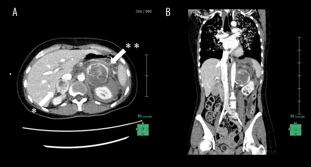
Figure 2 Enhanced CT examination on arrival(A) An enhanced CT examination detected bilateral adrenal gland tumors with 30 mm of the right side (white arrow*) and 60 mm of the left side in size (white arrow**). The tumor margins showed marked enhancement, while the interior of the bilateral tumors was low density, reflecting internal necrosis. These CT findings led to the diagnosis of bilateral pheochromocytomas. (B) Fluid collection was mainly observed around the left adrenal tumor. This fluid collection was caused by the rupture of the left adrenal tumor due to internal necrosis.