


10 August 2026: Articles 
A 42-Year-Old Woman Presenting With Pheochromocytoma Multisystem Crisis Associated With Bilateral Pheochromocytomas Due to Multiple Endocrine Neoplasia Type 2A
Management of emergency care, Rare disease
Akira Umemura AEFG 1*, Ai Chida BCD 2, Shigenori Kan BCD 3, Saki Kuroda BCD 2, Eriko Yoshida BCD 2, Toshie Segawa BCD 2, Yutaka Hasegawa AF 2, Yoshihiko Takahashi AF 2, Yasushi Ishigaki AEG 2, Ayaka Sato CD 4, Naoki Yanagawa CD 4, Akira Sasaki AEG 1
DOI: 10.12659/AJCR.953137
Am J Case Rep 2026; 27:e953137


Abstract
BACKGROUND: Multiple endocrine neoplasia type 2A (MEN 2A) is a rare autosomal-dominant genetic syndrome characterized by mutations in the RET gene and tumors of endocrine glands, including pheochromocytomas. Pheochromocytoma is a benign primary endocrine tumor of the adrenal glands that produces catecholamines and in rare cases is complicated by pheochromocytoma multisystem crisis (PMC), a life-threatening endocrine emergency caused by severe catecholamine overproduction. This report describes a 42-year-old woman presenting with PMC associated with bilateral pheochromocytomas and MEN2A.
CASE REPORT: The patient presented with a sudden onset of back pain and dyspnea with severe shock. An enhanced computed tomography examination revealed bilateral adrenal gland tumors, and she was diagnosed with PMC complicated by multiple organ failure and catecholamine-induced cardiomyopathy. We introduced simultaneous veno-venous extracorporeal membrane oxygenation and continuous hemodiafiltration. A definitive operation for PMC was performed on hospital day 12, with the intent of complete resection of bilateral pheochromocytomas. The patient was weaned from continuous hemodiafiltration on hospital day 21st day and was finally weaned from artificial ventilation on hospital day 30. Genetic examination revealed a pathogenic RET mutation (Cys634Arg, C634R), leading to the diagnosis of MEN2A. She is now undergoing rehabilitation, with intensive surveillance for thyroid medullary carcinoma and hyperparathyroidism.
CONCLUSIONS: PMC can easily lead to multiple organ failure within a few days. A multidisciplinary approach to PMC can rescue these patients by making surgical pheochromocytoma resection possible. If synchronous bilateral pheochromocytomas are detected, MEN2A should be suspected.
Keywords: Adrenal Gland Neoplasms, Case Reports, crisis, Endocrinology, Multiple Endocrine Neoplasia Type 2a, Pheochromocytoma
Introduction
Pheochromocytomas are relatively rare neuroendocrine tumors that arise from chromaffin cells of the adrenal medulla can secrete high concentrations of catecholamines. Pheochromocytomas are stratified as idiopathic and hereditary diseases, with multiple endocrine neoplasia (MEN) syndrome being the most important hereditary disease. MEN presents as at least 2 endocrine gland tumors and is categorized into 3 types: MEN1, MEN2A, and MEN2B [1]. MEN2A, the predominant phenotype of MEN2, is caused by germline mutations in the RET proto-oncogene, which predisposes carriers to an increased risk of developing medullary thyroid cancer, primary hyperparathyroidism, and pheochromocytoma [2].
Pheochromocytomas usually cause paroxysmal episodes of hypertension, palpitations, headaches, hyperthermia, and sweating. However, in rare cases, these tumors can also cause pheochromocytoma multisystem crisis (PMC), which is characterized by excessive, continuous, and uncontrollable catecholamine release [3,4]. PMC is a highly fatal complication of pheochromocytoma; therefore, a multidisciplinary treatment strategy should be established immediately for patients with this condition.
PMC caused by synchronous bilateral pheochromocytomas is extremely rare and potentially fatal. Because PMC can rapidly progress to life-threatening multiple organ failure due to excessive catecholamine release, prompt diagnosis and multidisciplinary management are essential for patient survival [5]. This report describes a 42-year-old woman presenting with PMC associated with bilateral pheochromocytomas and MEN 2A.
Case Report
The patient had been suspected of having unstable angina at the previous center. A blood test for serum catecholamines performed at that time revealed serum epinephrine, norepinephrine, and dopamine levels of 4300 pg/mL, 3900 pg/mL, and 30 pg/mL, respectively, but her high catecholamine levels had not subsequently been followed up. Five years after the previous diagnosis of unstable angina, she experienced a sudden onset of back pain and dyspnea and was taken to our hospital in an ambulance. Vital signs upon arrival in the ambulance were as follows: heart rate of 140 bpm, non-invasive blood pressure of 74/48 mm Hg, and 65% percutaneous oxygen saturation with high-flow oxygen. We started a multidisciplinary approach at the emergency room just after arrival of the ambulance. An initial workup revealed that severe pulmonary edema might have induced severe hypoxemia due to acute cardiac dysfunction, followed by multiple organ dysfunction. Her results for blood gas analysis, the usual laboratory data, and her chest X-ray are shown in Table 1 and Figure 1. An enhanced computed tomography (CT) examination also revealed bilateral adrenal gland tumors, with a 30-mm tumor on the right side and a 60-mm tumor on the left side (Figure 2). Some fluid collection was evident around the left adrenal gland tumor and kidney, indicating a rupture of the left adrenal gland tumor (Figure 2). Severe elevations of serum catecholamines (Table 1) and CT findings suggested that the bilateral adrenal tumors were simultaneous pheochromocytomas. Transthoracic echocardiography also revealed a severe decrease in the left ventricular ejection fraction (LVEF) to 30% and a left ventricular end-diastolic diameter over 55 mm. Therefore, her acute heart failure led to the diagnosis of catecholamine-induced cardiomyopathy (CIC) [6]. From these initial workup studies, she was diagnosed with PMC due to bilateral pheochromocytomas. Then, she was immediately moved to the intensive care unit to perform further specialized treatments.
We simultaneously performed intubation and initiated artificial ventilation; however, her systemic oxygenation did not improve, and her circulatory insufficiency due to CIC became severe. Therefore, we introduced veno-venous extracorporeal membrane oxygenation (VV-ECMO) to stabilize her systemic oxygenation and circulation. We were unable to reverse her metabolic acidosis with our initial treatments, so we simultaneously introduced continuous hemodiafiltration (CHDF) using a polymethyl methacrylate membrane. We then focused on controlling her tachycardia and catecholamine levels using phentolamine mesylate, landiolol hydrochloride, and isosorbide dinitrate. She was removed from VV-ECMO on hospital day 5, and her LVEF dramatically improved to 52%. We tried to use conservative treatments to control the PMC caused by the severe hyper-catecholamine condition; however, her organ dysfunctions, such as acute lung and kidney injury, continued. To make a breakthrough to rescue the patient, definitive surgery for PMC removal was planned hospital day 12 to complete resection of the bilateral pheochromocytomas.
She was placed in a supine position under general anesthesia, with strict intraoperative blood pressure control. We started the operation using a midline incision of the upper abdomen. To resect the left-sided pheochromocytoma, we dissected the gastrocolic and gastrosplenic ligament and taped the pancreas because the left-sided pheochromocytoma was enlarged beyond the inferior border of the pancreas. We successfully resected the left-sided pheochromocytoma (Figure 3). We then simultaneously resected the right-sided pheochromocytoma. The direct blood pressure decreased below 50 mm Hg immediately after complete division of the drainage veins of both pheochromocytomas; therefore, we initiated systemic administration of catecholamines. The operating time was 256 minutes, and the intraoperative blood loss was 198 mL. Intensive therapies were continued after surgery, and the patient was weaned from CHDF on hospital day 21 and was finally weaned from artificial ventilation on hospital day.
The cut surface of the resected specimens revealed necrotization of the inner sides of the bilateral tumors due to their proliferative potential (Figure 4). Histopathological examination revealed nested tumor cells surrounded by fibrovascular septa (also termed Zellballen), with central necrosis of the tumor (Figure 5). Immunohistochemistry staining revealed that the tumor cells were positive for chromogranin A and synaptophysin and that the Ki-67 index was lower than 1% (Figure 5). The Pheochromocytoma of the Adrenal Gland Scale Score (PASS) was 4, and the Grading System for Adrenal Pheochromocytoma and Paraganglioma (GAPP) score was 4 [7,8]. Genetic examination revealed a pathogenic variation of
The patient is now undergoing rehabilitation with intensive surveillance for thyroid medullary carcinoma and hyperparathyroidism. Her clinical course from admission to recovery from PMC is shown in Figure 6.
Discussion
We successfully treated a patient with PMC induced by bilateral pheochromocytomas due to MEN2A. Bilateral adrenal tumors, severe multiple organ dysfunction and the aberrant levels of serum catecholamine levels promptly led to diagnosis of PMC. Immediate multidisciplinary approaches, including surgical resection, contributed to the successful rescue of this patient. Synchronous bilateral pheochromocytomas are extremely rare; therefore, MEN2A should be suspected and simultaneous multimodal evaluations are required.
Firstly, a unilateral pheochromocytoma often poses diagnostic difficulties, especially in left-side pheochromocytoma, due to several adjacent organs. Enhanced computed tomography (CT) usually shows a heterogenous enhancement in the arterial phase and central necrosis as typical findings of pheochromocytoma, but there have been some reports of cystic pheochromocytoma [9]. The overlapping anatomical structures and cystic lesions should be carefully evaluated, including clinical symptoms, when there are adrenal gland tumors. This case was easily diagnosed based on the presence of bilateral adrenal gland tumors, typical CT findings, and clinical symptoms such as CIC.
In this case report, we demonstrated that prompt multidisciplinary approaches, including definitive resection of bilateral pheochromocytomas, can provide feasible and reasonable treatment for patients with severe PMC. The standard and definitive treatment for pheochromocytoma is generally surgical resection; however, PMC leads to a potentially lethal condition that eliminates radical surgery as an option due to uncontrollable endocrinological disorders. The mortality rate for patients with PMC treated with conservative therapy alone is over 40%; therefore, the best practice for rescuing a patient with PMC is the immediate establishment of a multidisciplinary approach to stabilize the general condition, followed by early surgical resection of the pheochromocytoma [4,10,11].
Previous reports have demonstrated that PMC associated with pheochromocytoma remains highly fatal despite advances in intensive care management; however, reports describing PMC caused by bilateral pheochromocytomas in MEN2A remain limited.
At present, PMC has no comprehensive worldwide diagnostic criteria, so delays may occur in its diagnosis and initiation of disease-specific intensive care. In 2023, Ando et al reported the following novel diagnostic criteria: (1) acute onset, (2) no surgical intervention performed for pheochromocytoma, (3) pheochromocytoma confirmed via examination, and (4) any form of serious organ dysfunction [4]. The following symptoms are also considered to be related to PMC: (1) labile hypertension/hypotension, (2) multiple organ failure (organ damage involving at least 2 organs), (3) fever, and (4) encephalopathy [4]. Because our case met these diagnostic criteria, we started initial intensive treatments and had a successful outcome.
Our patient had a
Pheochromocytomas accompanying MEN2A are generally benign; however, a few reports have demonstrated the existence of malignant pheochromocytomas in MEN2A, with an estimated prevalence of 3% of all tumors investigated [14]. The PASS and GAPP algorithms are broadly used as histopathological diagnostic criteria by the current WHO classification for predicting malignant potential. Stenman et al demonstrated that the 2 most noticeable positive PASS factors (≥ 4) in MEN2A pheochromocytomas were “large nests/diffuse growth” and “high cellularity” [15]. Conversely, no relapse or metastasis occurred in their cohort during the follow-up period [15]. This discrepancy may reflect different
The onset age of pheochromocytoma is much younger in patients with MEN2A than in patients with nonsyndromic pheochromocytoma. The life-threatening nature of MEN2A usually involves the progression of medullary thyroid cancer; however, pheochromocytomas can also be a life-threatening factor in patients with MEN2A if PMC occurs because of the drastic multiple organ damage that happens within a few days [16]. Every physician recognizes that the definitive treatment for PMC is surgical resection as soon as possible. However, Kakoi et al also reported that surgery on the day following admission is not always associated with improved outcomes [10]. Based on these previous insights, critically ill patients should receive immediate multidisciplinary treatment for PMC and sequential surgical resection after their general condition is stabilized.
Because this report describes a single case, it is unclear if there is a direct causal relationship between the
Conclusions
We successfully treated a patient with PMC induced by bilateral pheochromocytomas due to MEN2A. The onset and progression of PMC is fulminant, and PMC can easily lead to multiple organ failure within a few days. Using a multidisciplinary approach to treat PMC can make surgical pheochromocytoma resection possible and help rescue these patients. If synchronous bilateral pheochromocytomas are detected, MEN2A should be suspected and comprehensive endocrinological and genetic evaluations should be promptly performed. Moreover, patients and their families can subsequently undergo genetic counseling, along with intensive follow-up for MEN2A.
Figures
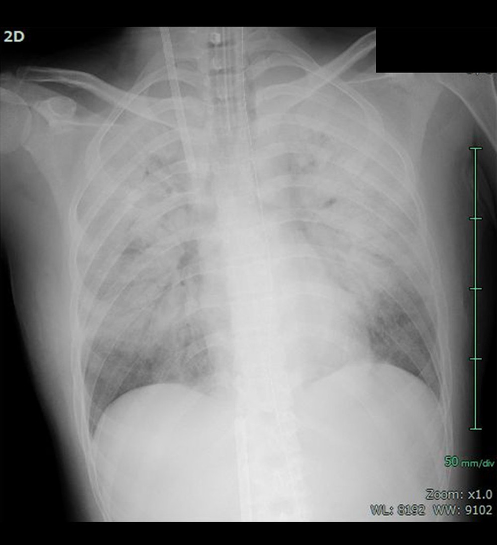 Figure 1. Chest X-ray examination on arrivalThe chest X-ray revealed that severe bilateral infiltrative shadows reflecting severe hypoxemia, acute respiratory distress syndrome, and multiple organ dysfunction.
 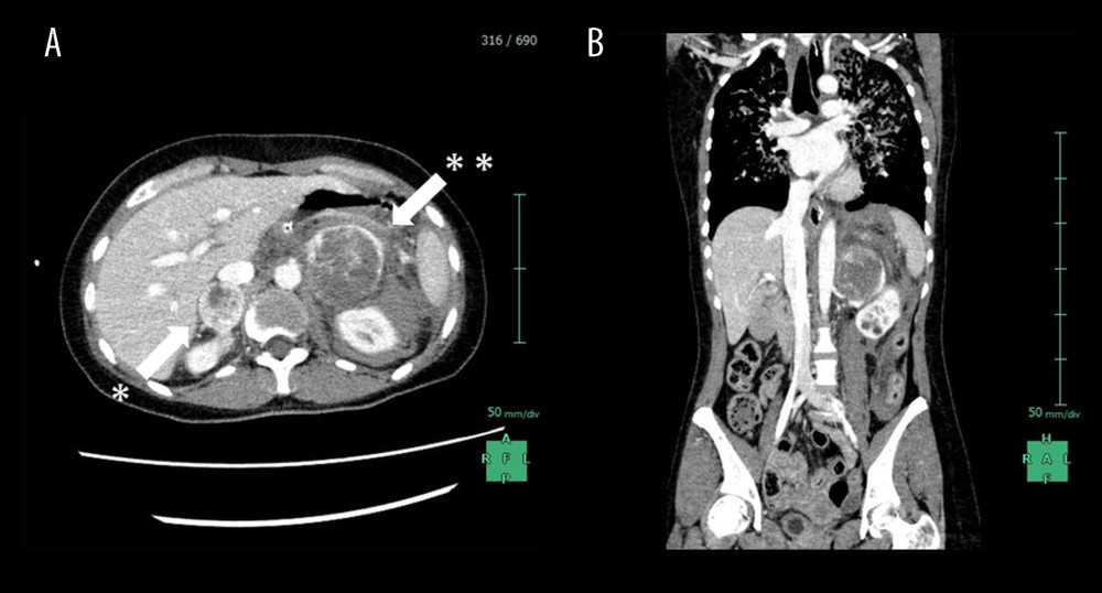 Figure 2. Enhanced CT examination on arrival(A) An enhanced CT examination detected bilateral adrenal gland tumors with 30 mm of the right side (white arrow*) and 60 mm of the left side in size (white arrow**). The tumor margins showed marked enhancement, while the interior of the bilateral tumors was low density, reflecting internal necrosis. These CT findings led to the diagnosis of bilateral pheochromocytomas. (B) Fluid collection was mainly observed around the left adrenal tumor. This fluid collection was caused by the rupture of the left adrenal tumor due to internal necrosis.
 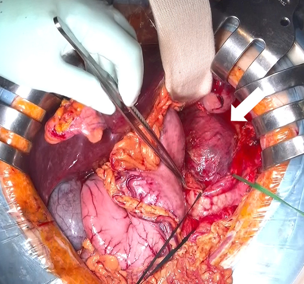 Figure 3. Intraoperative finding of the left-sided pheochromocytomaThe left-sided pheochromocytoma (whit arrow) was enlarged beyond the inferior border of the pancreas. We successfully performed left adrenalectomy without combined resection of the distal pancreas.
 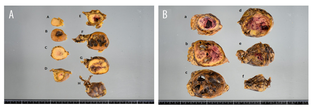 Figure 4. Macroscopic findings of the resected specimens(A) The size of right-side tumor was 70 × 40 mm, and the tumor had a fibrous capsule. Necrosis was observed in the center of the tumor. Internal necrosis was a typical finding of pheochromocytoma. (B) The size of left-side tumor was 80 × 65 mm, and the tumor had a fibrous capsule. Necrosis and intratumoral hemorrhage were observed inside the tumor. Internal necrosis and intratumoral hemorrhage were also typical findings of bulky pheochromocytoma.
 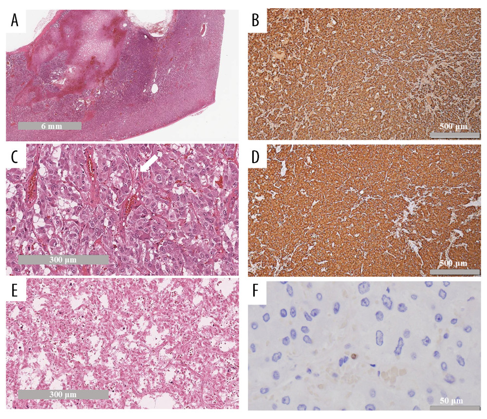 Figure 5. Histopathological findings of the resected specimens(A) The tumor was composed of eosinophilic and basophilic cells, and had a fibrous capsule (HE stain, low-power field). (B) The tumor shows a trabecular or alveolar (Zellballen) architecture, with nests separated by peripheral capillaries (white arrow). The cells showed pleomorphism, and the cytoplasm showed finely granular, amphophilic staining with a range from basophilic to acidophilic (HE stain, high-power field). (C) The center part of the tumor was massively necrotizing (HE stain, high-power field). (D) Tumor cells were strongly positive for chromogranin A, which is a highly useful marker of neuroendocrine granules. (Immunohistochemistry, high-power field). (E) Tumor cells were also strongly positive for synaptophysin (Immunohistochemistry, high-power field). (F) Ki-67 index was < 1% (immunohistochemistry, high-power field).
 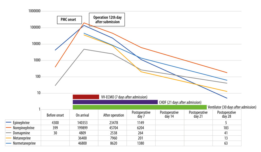 Figure 6. Timeline and summary of this caseThis timeline shows both the acute-phase treatment course and the levels of catecholamines and their metabolites until 4 weeks after admission. Catecholamines and their metabolite dramatically decreased after operation. The patient was weaned from CHDF on hospital day 21 and was finally weaned from artificial ventilation on hospital day 30.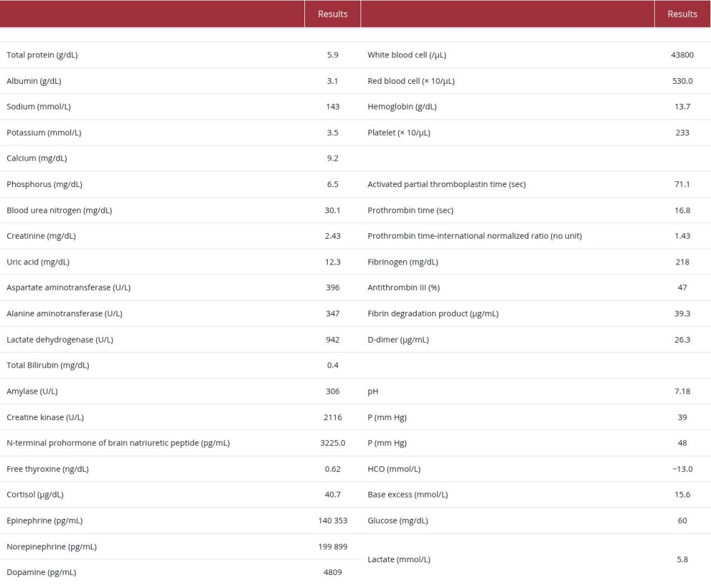
References
1. Machens A, Elwerr M, Lorenz K, 100-Year evolution of precision medicine and surgery for multiple endocrine neoplasia type 2A: Endocrine, 2020; 68(2); 368-76
2. Machens A, Lorenz K, Sekulla C, Molecular epidemiology of multiple endocrine neoplasia 2: Implications for RET screening in the new millenium: Eur J Endocrinol, 2013; 168(3); 307-14
3. Newell K, Prinz RA, Braithwaite S, Brooks M, Pheochromocytoma crisis: Am J Hypertens, 1988; 1(3 Pt 3); 189S-91S
4. Ando Y, Ono Y, Sano A, Clinical characteristics and outcomes of pheochromocytoma crisis: A literature review of 200 cases: J Endocrinol Invest, 2022; 45(12); 2313-28
5. Scholten A, Cisco RM, Vriens MR, Pheochromocytoma crisis is not a surgical emergency: J Clin Endocrinol Metab, 2013; 98(2); 581-91
6. Santos JRU, Brofferio A, Viana B, Pacak K, Catecholamine-induced cardiomyopathy in pheochromocytoma: How to manage a rare complication in a rare disease?: Horm Metab Res, 2019; 51(7); 458-69
7. Thompson LD, Pheochromocytoma of the Adrenal gland Scaled Score (PASS) to separate benign from malignant neoplasms: A clinicopathologic and immunophenotypic study of 100 cases: Am J Surg Pathol, 2002; 26(5); 551-66
8. Kimura N, Takayanagi R, Takizawa N, Pathological grading for predicting metastasis in phaeochromocytoma and paraganglioma: Endocr Relat Cancer, 2014; 21(3); 405-14
9. Huang H, Xu D, Chen Z, Huang J, Diagnostic challenges of abdominal tumors without typical imaging features: From misdiagnosis of pancreatic cystadenoma to adrenal pheochromocytoma and the intraoperative response: Am J Case Rep, 2025; 26; e950574
10. Kakoki K, Miyata Y, Shida Y, Pheochromocytoma multisystem crisis treated with emergency surgery: A case report and literature review: BMC Res Notes, 2015; 8; 758
11. Rosas AL, Kasperlik-Zaluska AA, Papierska L, Pheochromocytoma crisis induced by glucocorticoids: A report of four cases and review of the literature: Eur J Endocrinol, 2008; 158(3); 423-29
12. Castinetti F, Eng C, Genotype/phenotype correlations in multiple endocrine neoplasia type 2: Endocr Relat Cancer, 2024; 31(12); e240139
13. Mucha L, Leidig-Bruckner G, Frank-Raue KGerman study group for rare thyroid cancer, Phaeochromocytoma in multiple endocrine neoplasia type 2: RET codon-specific penetrance and changes in management during the last four decades: Clin Endocrinol (Oxf), 2017; 87(4); 320-26
14. Welander J, Söderkvist P, Gimm O, Genetics and clinical characteristics of hereditary pheochromocytomas and paragangliomas: Endocr Relat Cancer, 2011; 18(6); R253-76
15. Stenman A, Zedenius J, Juhlin CC, Over-diagnosis of potential malignant behavior in MEN 2A-associated pheochromocytomas using the PASS and GAPP algorithms: Langenbecks Arch Surg, 2018; 403(6); 785-90
16. Li Y, Jin D, Shen L, Huang Y, Anesthesia and outcome of 33 surgeries in 24 multiple endocrine neoplasia type 2A (MEN2A) patients: A national rare disease center’s experience: Front Endocrinol (Lausanne), 2022; 13; 905963
Figures
Figure 1. Chest X-ray examination on arrivalThe chest X-ray revealed that severe bilateral infiltrative shadows reflecting severe hypoxemia, acute respiratory distress syndrome, and multiple organ dysfunction.Figure 2. Enhanced CT examination on arrival(A) An enhanced CT examination detected bilateral adrenal gland tumors with 30 mm of the right side (white arrow*) and 60 mm of the left side in size (white arrow**). The tumor margins showed marked enhancement, while the interior of the bilateral tumors was low density, reflecting internal necrosis. These CT findings led to the diagnosis of bilateral pheochromocytomas. (B) Fluid collection was mainly observed around the left adrenal tumor. This fluid collection was caused by the rupture of the left adrenal tumor due to internal necrosis.Figure 3. Intraoperative finding of the left-sided pheochromocytomaThe left-sided pheochromocytoma (whit arrow) was enlarged beyond the inferior border of the pancreas. We successfully performed left adrenalectomy without combined resection of the distal pancreas.Figure 4. Macroscopic findings of the resected specimens(A) The size of right-side tumor was 70 × 40 mm, and the tumor had a fibrous capsule. Necrosis was observed in the center of the tumor. Internal necrosis was a typical finding of pheochromocytoma. (B) The size of left-side tumor was 80 × 65 mm, and the tumor had a fibrous capsule. Necrosis and intratumoral hemorrhage were observed inside the tumor. Internal necrosis and intratumoral hemorrhage were also typical findings of bulky pheochromocytoma.Figure 5. Histopathological findings of the resected specimens(A) The tumor was composed of eosinophilic and basophilic cells, and had a fibrous capsule (HE stain, low-power field). (B) The tumor shows a trabecular or alveolar (Zellballen) architecture, with nests separated by peripheral capillaries (white arrow). The cells showed pleomorphism, and the cytoplasm showed finely granular, amphophilic staining with a range from basophilic to acidophilic (HE stain, high-power field). (C) The center part of the tumor was massively necrotizing (HE stain, high-power field). (D) Tumor cells were strongly positive for chromogranin A, which is a highly useful marker of neuroendocrine granules. (Immunohistochemistry, high-power field). (E) Tumor cells were also strongly positive for synaptophysin (Immunohistochemistry, high-power field). (F) Ki-67 index was < 1% (immunohistochemistry, high-power field).Figure 6. Timeline and summary of this caseThis timeline shows both the acute-phase treatment course and the levels of catecholamines and their metabolites until 4 weeks after admission. Catecholamines and their metabolite dramatically decreased after operation. The patient was weaned from CHDF on hospital day 21 and was finally weaned from artificial ventilation on hospital day 30. In Press
Case report 
Am J Case Rep In Press; DOI: 10.12659/AJCR.953555
Case report
Am J Case Rep In Press; DOI: 10.12659/AJCR.953169
Case report
Am J Case Rep In Press; DOI: 10.12659/AJCR.954027
Case report
Am J Case Rep In Press; DOI: 10.12659/AJCR.954380
Most Viewed Current Articles
07 Dec 2021 : Case report  28,713,109
DOI :10.12659/AJCR.934347
Am J Case Rep 2021; 22:e934347
06 Dec 2021 : Case report  180,916
DOI :10.12659/AJCR.934406
Am J Case Rep 2021; 22:e934406
21 Jun 2024 : Case report
124,581
DOI :10.12659/AJCR.944371
Am J Case Rep 2024; 25:e944371
08 May 2026 : Case report
95,842
DOI :10.12659/AJCR.952158
Am J Case Rep 2026; 27:e952158