


10 August 2026
: Case report 
A 42-Year-Old Woman Presenting With Pheochromocytoma Multisystem Crisis Associated With Bilateral Pheochromocytomas Due to Multiple Endocrine Neoplasia Type 2A
Management of emergency care, Rare disease
Akira Umemura AEFG 1*, Ai Chida BCD 2, Shigenori Kan BCD 3, Saki Kuroda BCD 2, Eriko Yoshida BCD 2, Toshie Segawa BCD 2, Yutaka Hasegawa AF 2, Yoshihiko Takahashi AF 2, Yasushi Ishigaki AEG 2, Ayaka Sato CD 4, Naoki Yanagawa CD 4, Akira Sasaki AEG 1
DOI: 10.12659/AJCR.953137
Am J Case Rep 2026; 27:e953137


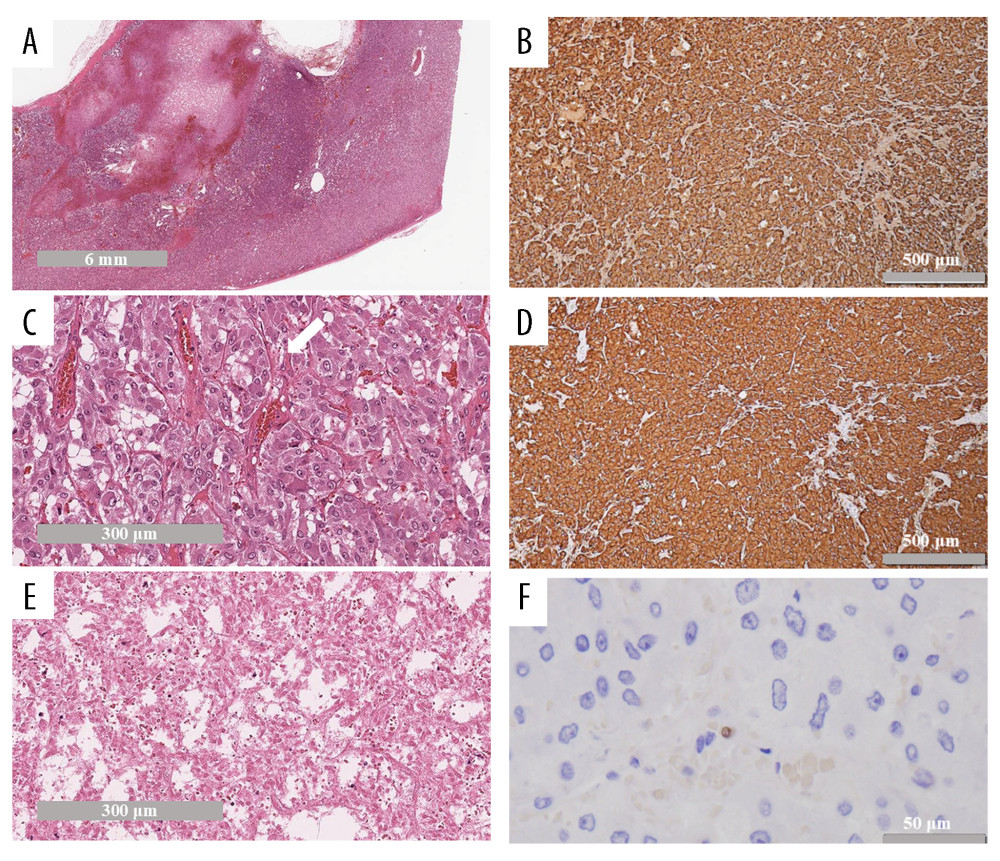
Figure 5 Histopathological findings of the resected specimens(A) The tumor was composed of eosinophilic and basophilic cells, and had a fibrous capsule (HE stain, low-power field). (B) The tumor shows a trabecular or alveolar (Zellballen) architecture, with nests separated by peripheral capillaries (white arrow). The cells showed pleomorphism, and the cytoplasm showed finely granular, amphophilic staining with a range from basophilic to acidophilic (HE stain, high-power field). (C) The center part of the tumor was massively necrotizing (HE stain, high-power field). (D) Tumor cells were strongly positive for chromogranin A, which is a highly useful marker of neuroendocrine granules. (Immunohistochemistry, high-power field). (E) Tumor cells were also strongly positive for synaptophysin (Immunohistochemistry, high-power field). (F) Ki-67 index was < 1% (immunohistochemistry, high-power field).