


21 September 2026
: Case report 
EVT-Inspired Simplified Negative-Pressure Nasoesophageal Drainage for Cervical Abscess Secondary to Esophageal Perforation
Management of emergency care
Lisha Yi AEF 1*, Linquan Zheng BDE 2, Zelong Huang BC 2, Yuling Yang DE 3DOI: 10.12659/AJCR.953266
Am J Case Rep 2026; 27:e953266


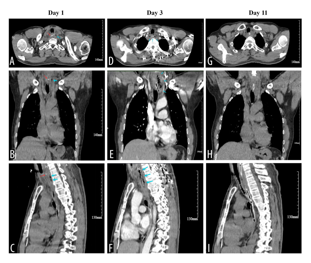
Figure 1 Serial computed tomography (CT) images demonstrating the evolution of cervical esophageal perforation with abscess formation. (A–C) Pre-admission CT images showing the fish bone (red arrows) impacted in the cervical esophagus with periesophageal air (blue arrows). (D–F) CT images on day 3 of hospitalization (48 hours after admission) showing abscess expansion. (D) Axial view demonstrating the abscess measuring 43 × 20 mm at its maximal cross-section. (F) Sagittal view showing a craniocaudal length of approximately 48 mm; the blue arrows indicate the abscess cavity. (G–I) CT images on day 11 of hospitalization (8 days after initiation of simplified negative-pressure nasoesophageal drainage) showing resolution of the abscess with residual esophageal wall edema.