


01 March 2026: Articles 
EVT-Inspired Simplified Negative-Pressure Nasoesophageal Drainage for Cervical Abscess Secondary to Esophageal Perforation
Management of emergency care
Lisha Yi AEF 1*, Linquan Zheng BDE 2, Zelong Huang BC 2, Yuling Yang DE 3DOI: 10.12659/AJCR.953266
Am J Case Rep 2026; 27:e953266


Abstract
BACKGROUND: Esophageal foreign body perforation is a critical emergency needing prompt diagnosis and intervention. The innovative esophageal vacuum therapy (EVT) has been proven effective in the treatment of esophageal perforations and fistulas. However, its high cost and technical complexity limit its widespread use in primary care facilities.
CASE REPORT: A 52-year-old man presented after 4 days of odynophagia and 1 day of fever following accidental fish bone ingestion. Computed tomography (CT) demonstrated cervical esophageal perforation with soft-tissue emphysema. Endoscopy revealed a 5-mm penetrating ulcer on the left esophageal wall, 20 cm from the incisors, with purulent discharge. The patient initially declined nasogastric decompression and was treated conservatively with nil per os, intravenous antibiotics, proton pump inhibitors, and parenteral nutrition. After 48 hours, clinical deterioration occurred with worsening pain. The patient then consented to EVT-inspired simplified negative-pressure nasoesophageal drainage: a 16-Fr nasogastric tube with 3 side holes was blindly inserted to the mid-esophagus and connected to a disposable negative pressure drainage ball. A second CT performed the same day confirmed abscess expansion (43×20×48 mm) and tube positioning. Daily drainage decreased and symptoms improved. Eight days later, CT confirmed abscess resolution. Endoscopy revealed perforation epithelialization, and a residual ulcer was closed with 1 clip. The patient resumed oral intake and was discharged on day 18.
CONCLUSIONS: In this case, EVT-inspired simplified negative-pressure nasoesophageal drainage demonstrated effectiveness in controlling local infection and avoiding surgery for cervical esophageal perforation with a contained abscess. This easily reproducible technique may serve as a temporary bridging therapy when standard EVT or surgery is unavailable.
Keywords: Abscess, Case Reports, Esophageal Perforation, Foreign Bodies, Negative-Pressure Wound Therapy
Introduction
Esophageal foreign body is a common emergency, with most cases occurring in the upper (cervical) esophagus [1]. A timely diagnosis followed by prompt endoscopic removal of the esophageal foreign body typically results in a swift recovery. Conversely, delayed presentation or the continuation of eating after impaction are primary contributors to the development of complications. Due to the absence of a serosal layer in the esophagus, perforation can lead to rapid dissemination of inflammation to surrounding structures, potentially resulting in mediastinitis, cervical abscess, or sepsis. The reported mortality rates range from 10% to 40% for esophageal perforation [2] and up to 30% to 50% for mediastinitis complicating an esophageal leak [1]. Once a cervical abscess develops, the infection can extend along fascial planes. Despite the combined use of broad-spectrum antibiotics, nil per os, and surgical or percutaneous drainage, 10% to 30% of patients still require secondary surgical repair [3].
Traditional nasogastric tubes (NGTs) are primarily utilized for gastric decompression and do not facilitate continuous local drainage or infection control. Over the past decade, esophageal vacuum therapy (EVT) employing a “sponge-suction” system has been developed to effectively clear purulent exudate, reduce wall edema, and promote granulation tissue formation, achieving healing rates of 80% to 90% in cases of esophageal leaks or perforations [4–7]. However, EVT necessitates repeated endoscopic sponge exchanges, incurs significant costs, and presents technical challenges, thereby restricting its accessibility in primary healthcare facilities.
By adopting the local negative pressure concept from EVT, we modified a standard NGT into a bedside “esophageal decompression tube”. This was achieved by placing factory-manufactured side holes at the distal tube segment adjacent to the perforation site and applying continuous low-pressure suction. This minimally invasive approach was intended to facilitate local infection control. We report a case of esophageal perforation with cervical abscess successfully managed using an EVT-inspired simplified nasoesophageal decompression tube. This approach may serve as a temporary bridging strategy toward definitive therapy when standard EVT or surgical expertise is unavailable.
Case Report
A 52-year-old male patient presented with a sore throat that had developed 4 days prior following the accidental ingestion of a fish bone. The severity of the sore throat progressively increased, and he experienced the onset of fever 1 day before presentation, with a peak temperature of 38.9 °C. An emergency computed tomography (CT) scan revealed esophageal perforation caused by the foreign body, accompanied by peri-esophageal air (Figure 1A–1C). Consequently, he was referred to a tertiary care facility. An urgent endoscopic procedure was conducted to successfully extract the fish bone, revealing a rupture measuring nearly 5 mm accompanied by purulent discharge (Figure 2A–2C). Although inpatient treatment was recommended, the patient declined and opted for admission to a secondary hospital closer to his residence. To facilitate clarity, the chronological sequence of clinical events, laboratory findings, imaging studies, and interventions is presented in Table 1.
Upon admission, laboratory test results revealed a white blood cell (WBC) count of 22.78 × 10⁹/L, a C-reactive protein (CRP) level of 113.38 mg/L, and a serum potassium concentration of 3.17 mmol/L; other biochemical parameters were within the reference range. The patient initially declined conventional nasogastric tube decompression and was treated with nil per os, intravenous proton pump inhibitors (PPI, omeprazole 40 mg twice daily), antibiotics (cefotaxime 2 g every 8 hours; ornidazole 1 g every 12 hours), and parenteral nutrition. PPI was administered specifically to suppress gastric acid secretion and thereby reduce the chemical injury to the periesophageal tissues from refluxed gastric contents passing through the perforation defect [1]. After 2 days, the WBC count decreased to 11.19 × 10⁹/L and the CRP level decreased to 88.3 mg/L. Although the fever subsided, the patient continued to experience neck pain and developed new symptoms of chest tightness and throat pain, which were slightly alleviated with oxygen therapy.
After we fully communicated the limitations of conservative therapy, the patient consented to EVT-inspired simplified negative-pressure nasoesophageal drainage (SNPND). A 16-French polyvinyl chloride NGT (Jiangsu Yangzhou Huaguan Technology Development Co, Ltd, Yangzhou, China) with 3 factory-manufactured side holes (each 5.3 mm in diameter, arranged in a 180° spiral configuration, as shown in Figure 3C) was blindly inserted into the mid-esophagus, approximately 30 cm from the nares. The proximal end was connected to a disposable negative pressure drainage ball (capacity: 1000 mL; Yada, Jiangsu Huadong Medical Device Industrial Co, Ltd, China) to enable generalized intraluminal decompression rather than targeted perforation occlusion. Negative pressure was generated by compressing the drainage ball to half its resting volume and sealing the 1-way valve, with re-compression performed every 6 hours. According to the bench testing with a digital manometer (Smart Sensor AS510A, ARCO Electronics Limited), the disposable drainage ball generated approximately −14.3 mm Hg when fully compressed and −2.9 mm Hg at half-compression. The tube was secured to the nasal septum and cheek using adhesive tape with a safety pin, with additional slack allowance to prevent alar necrosis. Immediately upon initiation of suction, yellow purulent fluid was observed in the tube, confirming active drainage (Figure 3A). On the same day, a second contrast-enhanced CT was performed to assess abscess progression and confirm the position of the blindly placed tube. The CT demonstrated expansion of the cervical abscess, with a maximal cross-section of 43 × 20 mm and a craniocaudal length of approximately 48 mm, accompanied by mural edema and air-fluid collection adjacent to the left esophageal wall. The distal end of the tube was located in the mid-esophagus, corresponding to the level of the T8 vertebra (Figure 1D–1F). The daily drainage output subsequently showed a consistent decrease, and the patient’s symptoms gradually improved. Throughout the 8-day SNPND course, the patient underwent daily monitoring for clinical symptoms (hematemesis, melena, worsening pain), tube position, and drainage fluid characteristics; no mucosal adhesion, bleeding, hematoma, or tube displacement was observed. A schematic diagram of the modified tube is shown in Figure 3B, 3C.
On day 8 of SNPND therapy, the patient’s WBC count normalized to 5.63 × 10⁹/L, and CRP level decreased to 7.32 mg/L. Subsequent chest CT demonstrated a regression of the peri-esophageal infection; however, wall edema persisted, and the continuity of the left esophageal wall could not be definitively assessed (Figure 1G–1I). Due to the unavailability of a barium swallow test, a sedated endoscopy was conducted at the patient’s request. The endoscopy revealed a solitary ulcer located 18 to 20 cm from the incisors on the left esophageal wall, characterized by a purplish skin tag at its distal edge (Figure 2D, 2E). Although no active bleeding or frank leakage was observed, the residual mucosal defect with its epithelial gap was deemed incompletely healed. Given the patient’s strong request for early oral feeding, endoscopic closure was performed to mechanically approximate the wound edges and facilitate safe dietary advancement. The defect was successfully closed using 1 endoscopic clip (Jiangsu Vedkang Medical Science and Technology Co, Ltd). The clip was deployed along the longitudinal axis of the ulcer, with its distal jaw positioned at the distal edge adjacent to the purplish skin tag, to achieve focal mucosal apposition (Figure 2F). Subsequently, the patient resumed oral fluid intake 2 hours after the procedure and was discharged after an 18-day hospitalization. The patient transitioned to a normal diet within 1 week. At the 1-month follow-up, he remained asymptomatic, with normal oral intake and no evidence of recurrent infection. As of April 2026 (approximately 8 months after discharge in August 2025), the patient remains asymptomatic, with normal oral intake, and no dysphagia, fever, or neck pain has been reported. Elective endoscopic re-evaluation was not performed, as the patient lacked clinical indications and declined invasive reassessment given his symptom-free status. Long-term clinical follow-up is ongoing.
To facilitate clarity, the chronological sequence of clinical events, laboratory findings, imaging studies, and interventions is presented in Table 1.
Discussion
The prognosis of esophageal foreign body perforation is significantly influenced by the timing of diagnosis [8,9]. Many patients, particularly older adults or patients with pre-existing conditions, often exhibit subtle or atypical early symptoms, which can result in delayed diagnosis and treatment [10,11]. The standard management of esophageal perforation with contained leak typically involves a combination of nil per os, broad-spectrum intravenous antibiotics, PPIs, and either surgical drainage or endoscopic intervention [1]. Nonoperative management is successful in many contained esophageal perforations [12]. Nonoperative management fails in the presence of persistent leak, enlarging mediastinal or cervical abscess, or worsening sepsis. Clinical deterioration after 48 to 72 hours warrants surgical intervention [13]. In the present case, the initial 48-hour conservative course resulted in partial biochemical improvement but failed to achieve anatomical closure, as evidenced by persistent throat pain, newly developed chest tightness, and progressive abscess expansion. Through full discussion with the patient, we opted for EVT-inspired SNPND. The subsequent insertion of the SNPND coincided with definitive clinical and radiological resolution, which supports the causal role of the drainage procedure. This illustrates that EVT-inspired SNPND may serve as an effective bridge therapy for esophageal perforation complicated by cervical abscess.
In the management of perforations in the hypopharynx or cervical esophagus, primary treatment strategies typically encompass NGT decompression, fasting, and conservative anti-infective therapy [3]. Traditional gastrointestinal decompression involves the insertion of the nasogastric tube tip into the stomach, which primarily mitigates gastric reflux but does not provide direct drainage for cervical esophageal perforations. EVT represents one of the most promising advancements in the management of esophageal perforations, particularly in complex cases characterized by large abscess cavities or severe contamination [14]. This technique entails the insertion of a specially designed porous sponge, via endoscopy, into the perforated cavity or adjacent area, which is then connected to an external negative pressure source through the nasogastric tube. The application of continuous negative pressure facilitates the effective drainage of pus and necrotic tissue, controls infection, reduces the size of the cavity by collapsing the esophageal wall, and stimulates the growth of granulation tissue, thereby expediting the healing process [15,16]. Loske et al [17] reported emergency endoscopic negative pressure therapy for an 8-cm spontaneous esophageal perforation using a single-lumen NGT-like open-pore film drain, establishing the feasibility of intraluminal EVT for long-segment perforation. However, our SNPND differed fundamentally from Loske’s approach in terms of tube design (conventional PVC tube with discrete side holes vs purpose-built open-pore film drain), placement method (blind transnasal insertion vs endoscopic guidance), and negative pressure system (manually compressed drainage ball vs electronically regulated vacuum pump). Similarly, Yoon et al [18] described negative-pressure wound therapy for a cervical esophageal perforation with abscess. In a directly analogous clinical scenario, Trocchia et al used percutaneous incision and drainage with a polyurethane foam dressing connected to a portable NPWT device [19]. Our approach differs in its intraluminal rather than extraluminal application, avoiding cervical incision entirely.
In this study, we adapted EVT principles by using a conventional 16-Fr nasogastric tube with 3 factory-manufactured side holes and connecting it to a disposable negative pressure drainage ball for continuous low-pressure drainage. This approach facilitated localized debridement and infection control while preventing the downward spread of infection to mediastinitis or empyema. Although simplified, this method likely operates through mechanisms similar to those of EVT. Unlike traditional EVT, our approach obviates the need for specialized polyurethane sponges and frequent endoscope changes, rendering it particularly suitable for resource-limited settings or as an interim treatment. However, it is important to note that excessive negative pressure can lead to complications such as esophageal mucosal adhesion, pressure-induced bleeding, or perforation [20]. Our method emphasizes compressing the drainage ball to approximately half its resting volume during use, with re-compression performed every 6 hours, or earlier if the ball re-expands to greater than 50% capacity. We performed bench testing of the disposable drainage ball using a digital manometer. The device generated a negative pressure of −1.906 kPa (approximately −14.3 mm Hg) when fully compressed and only −0.384 kPa (approximately −2.9 mm Hg) when compressed to half volume. These measured values are substantially lower than the standard EVT therapeutic range of −50 to −125 mm Hg [6], and even below the threshold typically considered necessary for active wound collapse and sponge-mediated granulation tissue stimulation. This discrepancy represents a major technical limitation of our improvised device. Given these measured values, the clinical efficacy observed in this case likely did not rely on the classic EVT mechanisms of high-pressure luminal collapse and forced wound apposition. Instead, we hypothesize that the therapeutic benefit was derived from 3 primary mechanisms: (1) continuous fluid evacuation that prevents the accumulation of saliva, purulent material, and esophageal secretions within the abscess cavity; (2) passive luminal decompression that maintains a low-pressure conduit favoring drainage along the path of least resistance; and (3) prevention of downstream contamination that reduces the volume of potentially infected material capable of propagating into the mediastinum. In essence, our device functioned more as a low-pressure sump drain than as a true vacuum-assisted closure system. The simplified approach may be most appropriate for contained cervical perforations with an established internal-external drainage tract, where gravity and natural pressure gradients already favor egress, and where the primary therapeutic need is maintenance of a patent drainage pathway rather than active cavity collapse. It is unlikely to be effective for large, thick-walled abscesses or thoracic perforations requiring mechanical collapse of the cavity to achieve healing. While EVT systems offer more precise visual monitoring and control of negative pressure, they are also more costly. The absence of real-time pressure monitoring and precise regulation in our manually operated system remains a significant limitation compared with electronically regulated EVT systems. Future prospective studies should prioritize continuous digital manometry as a mandatory safety endpoint to define the optimal pressure parameters, compression intervals, and alarm thresholds for this improvised device. Future iterations of this concept would require a purpose-built negative pressure source capable of generating and sustaining pressures within the true EVT range (−50 to −125 mm Hg) if the full mechanistic benefits of wound apposition and accelerated granulation are to be realized.
Throughout our patient’s treatment, no complications such as bleeding, recurrent perforation, or lumen stenosis were observed, suggesting that this pressure range was well tolerated in this single case. However, the safety profile of this simplified technique remains incompletely defined, and the absence of adverse events in a single patient cannot establish generalizable safety. Based on our observations in this case, we hypothesize that the following features may indicate readiness to discontinue SNPND: (1) clinical: resolution of pain, dysphagia, and fever for at least 48 hours; (2) laboratory: normalization of WBC count and CRP to near-baseline levels; (3) drainage: reduction in daily output to less than 10 mL of non-purulent fluid; and (4) imaging: CT confirmation of abscess cavity collapse with no residual air-fluid collection. In this patient, all 4 criteria were met by day 11, prompting endoscopic reassessment and clip deployment. We emphasize that these are provisional, empirically derived thresholds from a single case and require validation in prospective studies before they can be recommended as standardized protocols.
In this instance, the perforation was minor and not associated with extensive mediastinal necrosis or sepsis. The abscess cavity was connected to the esophageal lumen, facilitating the creation of an “internal-external communication pathway” via negative pressure drainage. This case was considered suitable for SNPND based on the following characteristics: (1) contained perforation without free mediastinal contamination; (2) cervical esophageal location permitting safe transnasal access; (3) hemodynamic stability; (4) localized abscess without massive mediastinal involvement; and (5) patient refusal of standard nasogastric decompression, creating need for an alternative minimally invasive strategy. Nonetheless, the applicability of this method to other esophageal locations or cases with varying perforation sizes necessitates further clinical observation and validation.
This case report represents a single instance without a control group. Consequently, formal patient selection criteria, contraindications, and a comparative decision algorithm incorporating standard EVT, percutaneous drainage, and surgical intervention cannot be established from this report alone. These elements represent critical directions for future prospective research. Such studies should investigate optimal negative pressure parameters, side hole design, and dwell time, as well as facilitate comparative analyses of efficacy and cost between SNPND and established alternatives. Importantly, vigilant monitoring of infection indicators and imaging changes remains essential during this simplified negative pressure therapy, to prevent delays in surgical treatment.
Regarding resource considerations, the unavailability of barium swallow at our institution reflects a real-world limitation common in secondary hospitals. However, contrast-enhanced CT and endoscopy provided adequate diagnostic information for safe clinical decision-making in this case, suggesting that barium swallow is not essential for patient selection or monitoring when these modalities are available. Future prospective studies in better-resourced settings could incorporate dynamic contrast studies to objectively quantify leak rates and healing dynamics.
Conclusions
In the initial management of esophageal foreign body perforation accompanied by cervical abscess, modified nasoesophageal tube-assisted negative pressure drainage demonstrated effectiveness in rapidly controlling local infection and avoiding surgery in this case. This easily reproducible technique, EVT-inspired SNPND, may serve as a temporary bridging strategy when standard EVT or surgery is unavailable.
Figures
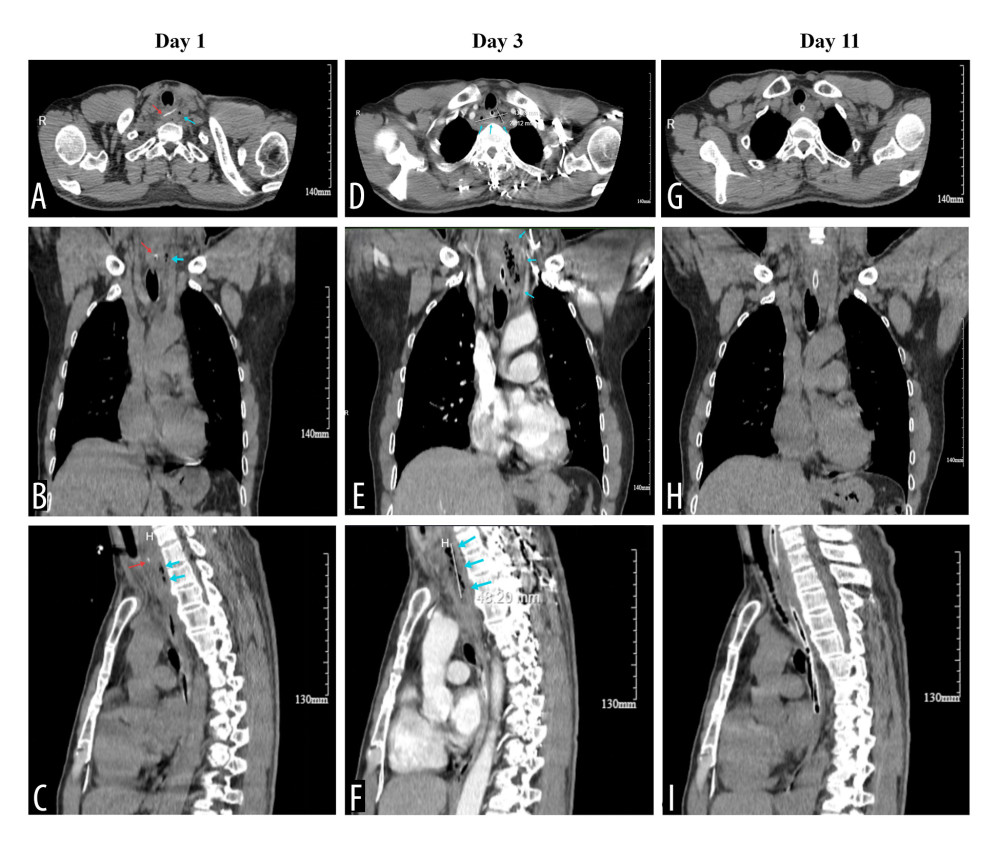 Figure 1. Serial computed tomography (CT) images demonstrating the evolution of cervical esophageal perforation with abscess formation. (A–C) Pre-admission CT images showing the fish bone (red arrows) impacted in the cervical esophagus with periesophageal air (blue arrows). (D–F) CT images on day 3 of hospitalization (48 hours after admission) showing abscess expansion. (D) Axial view demonstrating the abscess measuring 43 × 20 mm at its maximal cross-section. (F) Sagittal view showing a craniocaudal length of approximately 48 mm; the blue arrows indicate the abscess cavity. (G–I) CT images on day 11 of hospitalization (8 days after initiation of simplified negative-pressure nasoesophageal drainage) showing resolution of the abscess with residual esophageal wall edema.
 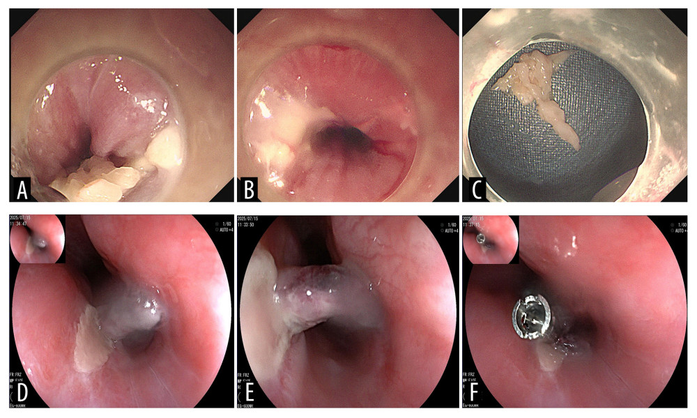 Figure 2. Endoscopic images of the patient. (A–C) Initial endoscopy was performed on day 1 using an Olympus GIF-290 gastroscope with a transparent cap attached to the distal tip. (A) Fish bone was impacted at the esophageal entrance (cervical esophagus) and penetrated the left esophageal wall. (B) A penetrating ulcer was found on the left esophageal wall (20 cm from the incisors) after foreign body removal. (C) T-shaped fish bone (transverse diameter approximately 2 cm) covered with food debris. (D–F) Follow-up endoscopy performed on Day 11 (8 days after initiation of simplified negative-pressure nasoesophageal drainage) using a Fujifilm EG-600WR gastroscope, showing the left upper esophageal wall at 18–20 cm from the incisors. (D) Healing mucosa with superficial ulceration. (E) Necrotic eschar at the ulcer base on the left esophageal wall. (F) Residual superficial ulcer with purplish skin tag at the distal edge, closed with a titanium clip.
 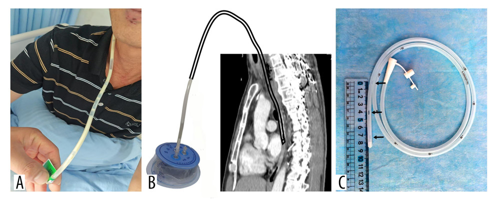 Figure 3. The simplified negative-pressure nasoesophageal drainage (SNPND) device and procedural demonstration. (A) Clinical photograph showing yellow purulent effluent drained through the nasogastric tube. (B) Composite schematic illustration combining the disposable negative pressure drainage ball and sagittal computed tomography imaging, demonstrating the SNPND procedure. The end of the nasogastric tube is positioned in the mid-esophagus, and it is connected to the external negative pressure ball. (C) Close-up photograph of the distal end of the 16-Fr nasogastric tube, showing 3 factory-manufactured side holes (each 3 mm in diameter) spaced 3 cm apart at different orientations along the distal segment.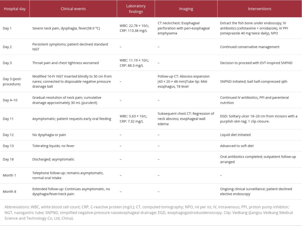
References
1. Chirica M, Kelly MD, Siboni S, Esophageal emergencies: WSES guidelines: World J Emerg Surg, 2019; 14; 26
2. Shaheem S, Panikkaveettil H, Aetiology, Clinical manifestations, diagnosis, and treatment of oesophageal perforation: A review: Cureus, 2024; 16(2); e55041
3. Khaitan PG, Famiglietti A, Watson TJ, The etiology, diagnosis, and management of esophageal perforation: J Gastrointest Surg, 2022; 26(12); 2606-15
4. Newton NJ, Sharrock A, Rickard R, Mughal M, Systematic review of the use of endo-luminal topical negative pressure in oesophageal leaks and perforations: Dis Esophagus, 2017; 30(3); 1-5
5. Sendino O, Loras C, Mata A, Safety and efficacy of endoscopic vacuum therapy for the treatment of perforations and anastomotic leaks of the upper gastrointestinal tract: Gastroenterol Hepatol, 2020; 43(8); 431-38
6. Luttikhold J, Pattynama LMD, Seewald S, Endoscopic vacuum therapy for esophageal perforation: A multicenter retrospective cohort study: Endoscopy, 2023; 55(9); 859-64
7. Vohra I, Gopakumar H, Sharma NR, Puli SR, Efficacy of endoscopic vacuum therapy in esophageal luminal defects: A systematic review and meta-analysis: Clin Endosc, 2025; 58(1); 53-62
8. Eroglu A, Can Kurkcuoglu I, Karaoganoglu N, Tekinbas C, Yimaz O, Basoglu M, Esophageal perforation: The importance of early diagnosis and primary repair: Dis Esophagus, 2004; 17(1); 91-94
9. Sdralis EIK, Petousis S, Rashid F, Epidemiology, diagnosis, and management of esophageal perforations: Systematic review: Dis Esophagus, 2017; 30(8); 1-6
10. Yan X, Dai G, Esophageal foreign body missed diagnosis; An analysis of 12 cases: Arch Acad Emerg Med, 2023; 11(1); e65
11. Xu J, Chen B, Liu Y, Clinical outcomes of sharp esophageal foreign bodies in elderly patients: A retrospective study from Wuhan, China: Front Med (Lausanne), 2025; 12; 1653609
12. Liao F, Zhu Z, Pan X, Safety and efficacy of nonoperative treatment in esophageal perforation caused by foreign bodies: Clin Transl Gastroenterol, 2022; 13(1); e00451
13. Paspatis GA, Arvanitakis M, Dumonceau JM, Diagnosis and management of iatrogenic endoscopic perforations: European Society of Gastrointestinal Endoscopy (ESGE) Position Statement – Update 2020: Endoscopy, 2020; 52(9); 792-810
14. Kouladouros K, Applications of endoscopic vacuum therapy in the upper gastrointestinal tract: World J Gastrointest Endosc, 2023; 15(6); 420-33
15. Livingstone I, Pollock L, Sgromo B, Mastoridis S, Current status of endoscopic vacuum therapy in the management of esophageal perforations and post-operative leaks: Clin Endosc, 2021; 54(6); 787-97
16. Ooi G, Burton P, Packiyanathan A, Indications and efficacy of endoscopic vacuum-assisted closure therapy for upper gastrointestinal perforations: ANZ J Surg, 2018; 88(4); E257-E63
17. Loske G, Scharsack E, Gobrecht O, Emergency endoscopic negative pressure therapy of a long oesophageal perforation in eosinophilic esophagitis with a single-lumen nasogastric tube-like open-pore film drain: Endoscopy, 2022; 54(5); E247-E48
18. Yoon BW, Yi KI, Kang JH, Negative pressure wound therapy for cervical esophageal perforation with abscess: Auris Nasus Larynx, 2015; 42(3); 254-57
19. Trocchia C, Shieh HF, Dolan I, Pulling instead of pushing: A case report of gastrostomy-assisted pull technique as an alternative method for endoluminal sponge placement in EVAC therapy: JPGN Rep, 2024; 5(1); 74-78
20. de Moura DTH, Hirsch BS, Ribas P, Endoscopic vacuum therapy: Pitfalls, tips and tricks, insights, and perspectives: Transl Gastroenterol Hepatol, 2024; 9; 50
Figures
Figure 1. Serial computed tomography (CT) images demonstrating the evolution of cervical esophageal perforation with abscess formation. (A–C) Pre-admission CT images showing the fish bone (red arrows) impacted in the cervical esophagus with periesophageal air (blue arrows). (D–F) CT images on day 3 of hospitalization (48 hours after admission) showing abscess expansion. (D) Axial view demonstrating the abscess measuring 43 × 20 mm at its maximal cross-section. (F) Sagittal view showing a craniocaudal length of approximately 48 mm; the blue arrows indicate the abscess cavity. (G–I) CT images on day 11 of hospitalization (8 days after initiation of simplified negative-pressure nasoesophageal drainage) showing resolution of the abscess with residual esophageal wall edema.Figure 2. Endoscopic images of the patient. (A–C) Initial endoscopy was performed on day 1 using an Olympus GIF-290 gastroscope with a transparent cap attached to the distal tip. (A) Fish bone was impacted at the esophageal entrance (cervical esophagus) and penetrated the left esophageal wall. (B) A penetrating ulcer was found on the left esophageal wall (20 cm from the incisors) after foreign body removal. (C) T-shaped fish bone (transverse diameter approximately 2 cm) covered with food debris. (D–F) Follow-up endoscopy performed on Day 11 (8 days after initiation of simplified negative-pressure nasoesophageal drainage) using a Fujifilm EG-600WR gastroscope, showing the left upper esophageal wall at 18–20 cm from the incisors. (D) Healing mucosa with superficial ulceration. (E) Necrotic eschar at the ulcer base on the left esophageal wall. (F) Residual superficial ulcer with purplish skin tag at the distal edge, closed with a titanium clip.Figure 3. The simplified negative-pressure nasoesophageal drainage (SNPND) device and procedural demonstration. (A) Clinical photograph showing yellow purulent effluent drained through the nasogastric tube. (B) Composite schematic illustration combining the disposable negative pressure drainage ball and sagittal computed tomography imaging, demonstrating the SNPND procedure. The end of the nasogastric tube is positioned in the mid-esophagus, and it is connected to the external negative pressure ball. (C) Close-up photograph of the distal end of the 16-Fr nasogastric tube, showing 3 factory-manufactured side holes (each 3 mm in diameter) spaced 3 cm apart at different orientations along the distal segment. In Press
Case report
Am J Case Rep In Press; DOI: 10.12659/AJCR.953317
Case report 
Am J Case Rep In Press; DOI: 10.12659/AJCR.954643
Case report 
Am J Case Rep In Press; DOI: 10.12659/AJCR.954074
Case report 
Am J Case Rep In Press; DOI: 10.12659/AJCR.953592
Most Viewed Current Articles
07 Dec 2021 : Case report
28,712,429
DOI :10.12659/AJCR.934347
Am J Case Rep 2021; 22:e934347
06 Dec 2021 : Case report  180,530
DOI :10.12659/AJCR.934406
Am J Case Rep 2021; 22:e934406
21 Jun 2024 : Case report
124,276
DOI :10.12659/AJCR.944371
Am J Case Rep 2024; 25:e944371
08 May 2026 : Case report
95,045
DOI :10.12659/AJCR.952158
Am J Case Rep 2026; 27:e952158