


21 September 2026
: Case report 
EVT-Inspired Simplified Negative-Pressure Nasoesophageal Drainage for Cervical Abscess Secondary to Esophageal Perforation
Management of emergency care
Lisha Yi AEF 1*, Linquan Zheng BDE 2, Zelong Huang BC 2, Yuling Yang DE 3DOI: 10.12659/AJCR.953266
Am J Case Rep 2026; 27:e953266


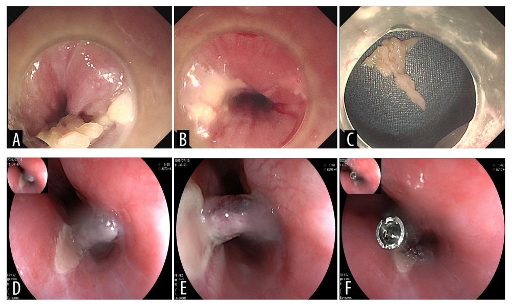
Figure 2 Endoscopic images of the patient. (A–C) Initial endoscopy was performed on day 1 using an Olympus GIF-290 gastroscope with a transparent cap attached to the distal tip. (A) Fish bone was impacted at the esophageal entrance (cervical esophagus) and penetrated the left esophageal wall. (B) A penetrating ulcer was found on the left esophageal wall (20 cm from the incisors) after foreign body removal. (C) T-shaped fish bone (transverse diameter approximately 2 cm) covered with food debris. (D–F) Follow-up endoscopy performed on Day 11 (8 days after initiation of simplified negative-pressure nasoesophageal drainage) using a Fujifilm EG-600WR gastroscope, showing the left upper esophageal wall at 18–20 cm from the incisors. (D) Healing mucosa with superficial ulceration. (E) Necrotic eschar at the ulcer base on the left esophageal wall. (F) Residual superficial ulcer with purplish skin tag at the distal edge, closed with a titanium clip.