


12 August 2026
: Case report 
Solitary Peritoneal Metastasis of AFP-Negative Hepatocellular Carcinoma in a 65-Year-Old Man with a History of Lung Adenocarcinoma: A Case Report
Unusual clinical course, Challenging differential diagnosis, Educational Purpose (only if useful for a systematic review or synthesis)
Zhineng Xie ABE 1, Jie Huang G 1*, Kailong Fu F 1DOI: 10.12659/AJCR.953374
Am J Case Rep 2026; 27:e953374


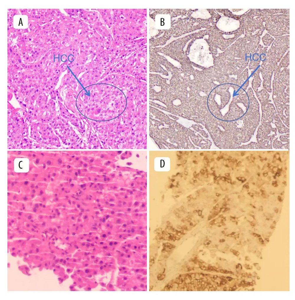
Figure 2 Pathological findings revealed moderately differentiated hepatocellular carcinoma (HCC). (A) Hematoxylin and eosin staining; observation mode: 2-dimensional planar observation; internal scale: 1 unit = 100–200 μm (low power), showing a typical trabecular arrangement of tumor cells with pronounced cellular atypia, an increased nuclear-to-cytoplasmic ratio, and no obvious cholestasis. (B) Immunohistochemical (IHC) staining; observation mode: 2-dimensional planar observation; internal scale: 1 unit = 100–200 μm (low power), showing negative expression of alpha-fetoprotein (AFP) in tumor cells, confirming the diagnosis of AFP-negative HCC. (C) Hematoxylin and eosin staining; observation mode: 2-dimensional planar observation; internal scale: 1 unit = 25–50 μm (high power), showing largely preserved hepatic lobular architecture, a regular arrangement of hepatocytes, and no clinically significant inflammation or fibrosis. (D) IHC staining; observation mode: 2-dimensional planar observation; internal scale: 1 unit = 25–50 μm (high power), showing diffuse cytoplasmic expression of Ki-67 in HCC cells and supporting the pathological diagnosis of HCC.