


12 August 2026: Articles 
Solitary Peritoneal Metastasis of AFP-Negative Hepatocellular Carcinoma in a 65-Year-Old Man with a History of Lung Adenocarcinoma: A Case Report
Unusual clinical course, Challenging differential diagnosis, Educational Purpose (only if useful for a systematic review or synthesis)
Zhineng Xie ABE 1, Jie Huang G 1*, Kailong Fu F 1DOI: 10.12659/AJCR.953374
Am J Case Rep 2026; 27:e953374


Abstract
BACKGROUND: Hepatocellular carcinoma (HCC) is an aggressive malignancy. Peritoneal metastasis is uncommon but carries a poor prognosis. Diagnosis of HCC with peritoneal metastasis is particularly challenging when alpha-fetoprotein (AFP) levels are normal. This report highlights diagnostic difficulties and the critical role of histopathological confirmation in such atypical presentations.
CASE REPORT: A 65-year-old man with chronic hepatitis B and a history of lung adenocarcinoma, who had previously undergone radiofrequency ablation and partial hepatectomy for HCC (the primary tumor was histologically confirmed as moderately differentiated HCC with negative AFP immunostaining), presented with recurrent left lower abdominal pain. Laboratory tests showed normal serum AFP but elevated carbohydrate antigen 125 (CA125). Imaging revealed a solitary peritoneal mass without evidence of intrahepatic recurrence. The patient underwent surgical resection. Immunohistochemistry confirmed hepatocellular origin (hepatocyte paraffin 1-positive, arginase-1-positive) and excluded lung adenocarcinoma (thyroid transcription factor-1-negative, napsin A-negative). Importantly, AFP immunohistochemistry findings were negative in the metastatic tumor, consistent with the primary tumor. The Ki-67 proliferation index was 50%. Postoperative recovery was uneventful, and no recurrence was observed at the 3-month follow-up.
CONCLUSIONS: Normal AFP levels do not exclude recurrent or metastatic HCC. In patients with multiple primary malignancies, diagnosis of new lesions requires careful integration of clinical history, imaging findings, and histopathological evaluation. Surgery may offer both diagnostic and therapeutic value in selected patients, although its long-term benefit remains uncertain.
Keywords: Carcinoma, Hepatocellular, Peritoneal Neoplasms, Surgical Procedures, Operative
Introduction
Hepatocellular carcinoma (HCC) is among the most common primary liver malignancies and a leading cause of cancer-related mortality worldwide. Extrahepatic metastases are associated with poor outcomes and most often involve the lungs, bones, lymph nodes, and adrenal glands [1]. Peritoneal metastasis (PM), although relatively rare, with an incidence of approximately 3% to 16% reported in laparoscopic and autopsy studies, generally indicates advanced disease and an unfavorable prognosis [2].
The diagnosis of HCC-PM can be particularly challenging in patients with atypical clinical or biochemical presentations. Although alpha-fetoprotein (AFP) is widely used as a tumor marker for HCC, it remains within the normal range in a substantial proportion of patients. Additionally, nonspecific markers such as cancer antigen 125 (CA125) may be elevated, further complicating diagnostic interpretation [3,4].
In patients with a history of multiple malignancies, determining the origin of a newly identified abdominal lesion can be especially difficult. This report describes a rare case of solitary peritoneal metastasis in a patient with HCC and normal AFP levels, highlighting diagnostic challenges and emphasizing the critical role of histopathological confirmation—including AFP immunohistochemistry (IHC)—in such atypical presentations.
Case Report
A 65-year-old man presented with recurrent left lower abdominal pain that had persisted for several weeks. His medical history was notable for more than 10 years of chronic hepatitis B infection, for which he had been receiving continuous antiviral therapy with tenofovir. He had undergone right upper lobectomy for lung adenocarcinoma 7 years earlier, followed by adjuvant therapy. Additionally, he had previously undergone radiofrequency ablation for HCC 3 years earlier and partial hepatectomy 2 years earlier, followed by 1 year of postoperative therapy with tislelizumab plus lenvatinib.
Laboratory investigations revealed normal AFP levels, elevated aminotransferase levels, and a substantially increased CA125 level (102.01 U/mL). Contrast-enhanced abdominal computed tomography (CT) demonstrated a poorly defined soft-tissue mass in the left lower abdomen (Figure 1). Positron emission tomography/CT identified hypermetabolic lesions in the same region, without evidence of intrahepatic recurrence.
Given the patient’s history of both HCC and lung adenocarcinoma, metastatic disease was strongly suspected. However, the primary origin of the lesion remained uncertain. The solitary and potentially resectable nature of the mass supported surgical exploration and resection to establish a definitive diagnosis and achieve local disease control.
Intraoperatively, the mass was confined to the left lower abdomen without invasion of adjacent bowel structures, and complete resection was achieved. The postoperative course was uneventful. Histopathological examination confirmed moderately differentiated hepatocellular carcinoma. Immunohistochemical analysis demonstrated positive staining for cytokeratin 8, cytokeratin 18, arginase-1 (Arg-1), and hepatocyte paraffin 1 (Hepa); it showed negative staining for cytokeratin 7, AFP, thyroid transcription factor-1 (TTF-1), and napsin A aspartic peptidase (Napsin A), effectively excluding metastatic lung adenocarcinoma. The Ki-67 proliferation index was approximately 50% in hotspot areas, indicating high proliferative activity (Figure 2). Based on these findings, a definitive diagnosis of HCC-PM was established.
Following surgery, the patient’s abdominal pain resolved, and CA125 levels substantially decreased (Table 1). Subsequently, he received 2 cycles of systemic therapy comprising bevacizumab plus sintilimab. At the 3-month follow-up, the patient remained clinically stable, without evidence of recurrence on imaging.
Discussion
From a clinical perspective, this case provides several important insights. First, normal AFP levels do not exclude recurrent or metastatic HCC [5]. Second, HCC-PM should be considered in the differential diagnosis of new abdominal lesions among patients with a history of hepatic malignancy, even in the presence of other primary cancers. Third, although CA125 is a nonspecific marker, its elevation may be associated with peritoneal involvement [6]. Finally, a multidisciplinary approach integrating clinical data, imaging findings, and pathology is essential for accurate diagnosis. Although no standardized treatment strategy exists for HCC-PM, surgical resection in carefully selected patients with a limited metastatic burden may provide both diagnostic clarification and potential clinical benefit [7]. In the present case, the patient achieved a favorable short-term outcome after surgery plus systemic therapy. However, the long-term efficacy of this approach remains uncertain and warrants further investigation.
Peritoneal metastasis from HCC is uncommon but represents an aggressive disease with a poor prognosis. Approximately 30% to 40% of HCC cases are AFP-negative [5], and diagnosis is particularly challenging in such cases. Several previous case reports have described HCC with peritoneal metastasis [7,8]. However, few have documented AFP IHC negativity in both the primary and metastatic tumors, which is critical for confirming that the metastasis originated from an AFP-negative HCC rather than a tumor of another origin (eg, metastatic lung adenocarcinoma). In the present case, IHC confirmed that the primary HCC was AFP-negative; the peritoneal metastatic tumor also showed AFP IHC negativity, providing strong evidence of a common clonal origin.
The differential diagnosis of a peritoneal mass in a patient with both HCC and lung adenocarcinoma includes metastatic HCC, metastatic lung adenocarcinoma, primary peritoneal malignancy, and hepatoid adenocarcinoma of the lung. Hepatoid adenocarcinoma of the lung is a rare subtype that can produce AFP and exhibit hepatoid differentiation, but it typically lacks expression of TTF-1 and Napsin A. In the present case, negative TTF-1 and Napsin A staining results excluded typical lung adenocarcinoma, whereas positive Hepa and Arg-1 staining results supported a hepatocellular origin. The absence of AFP expression in both the primary and metastatic tumors further argued against AFP-producing hepatoid adenocarcinoma of the lung. Primary peritoneal serous carcinoma was excluded by the negative cytokeratin 7 and positive Arg-1/Hepa immunophenotype. The elevated Ki-67 index (50%) indicated aggressive biological behavior, consistent with metastatic disease.
These findings highlight the indispensable role of pathological evaluation as the diagnostic gold standard, particularly in cases with atypical tumor marker profiles or multiple potential primary sources.
Conclusions
This case demonstrates that solitary peritoneal metastasis can occur several years after apparently successful treatment of HCC. Importantly, normal AFP levels do not exclude recurrence or metastasis. Accurate diagnosis of such atypical presentations requires a comprehensive multidisciplinary approach; histopathological confirmation plays a decisive role. In selected patients with limited metastatic disease, surgical resection plus systemic therapy may be considered; however, its long-term benefit remains to be established.
Figures
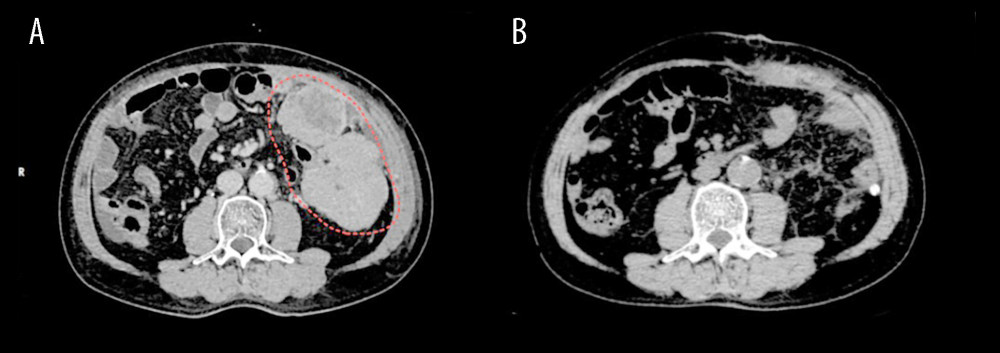 Figure 1. Abdominal computed tomography (CT) images of the patient with peritoneal metastasis from hepatocellular carcinoma. (A) Pretreatment CT scan showing a solitary soft-tissue mass in the left abdominal cavity (outlined by the red dashed line) with heterogeneous enhancement. (B) Posttreatment CT scan demonstrating disappearance of the previous peritoneal lesion, indicating a favorable treatment response.
 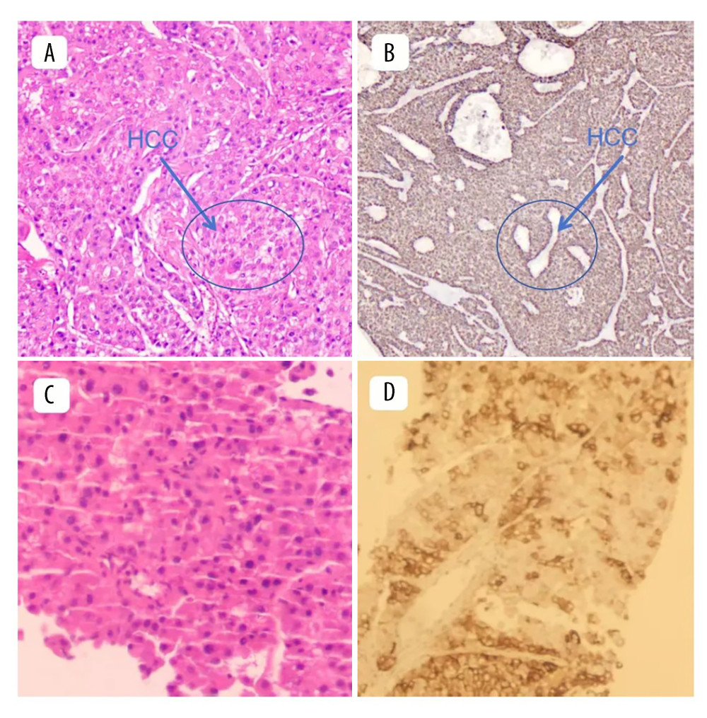 Figure 2. Pathological findings revealed moderately differentiated hepatocellular carcinoma (HCC). (A) Hematoxylin and eosin staining; observation mode: 2-dimensional planar observation; internal scale: 1 unit = 100–200 μm (low power), showing a typical trabecular arrangement of tumor cells with pronounced cellular atypia, an increased nuclear-to-cytoplasmic ratio, and no obvious cholestasis. (B) Immunohistochemical (IHC) staining; observation mode: 2-dimensional planar observation; internal scale: 1 unit = 100–200 μm (low power), showing negative expression of alpha-fetoprotein (AFP) in tumor cells, confirming the diagnosis of AFP-negative HCC. (C) Hematoxylin and eosin staining; observation mode: 2-dimensional planar observation; internal scale: 1 unit = 25–50 μm (high power), showing largely preserved hepatic lobular architecture, a regular arrangement of hepatocytes, and no clinically significant inflammation or fibrosis. (D) IHC staining; observation mode: 2-dimensional planar observation; internal scale: 1 unit = 25–50 μm (high power), showing diffuse cytoplasmic expression of Ki-67 in HCC cells and supporting the pathological diagnosis of HCC.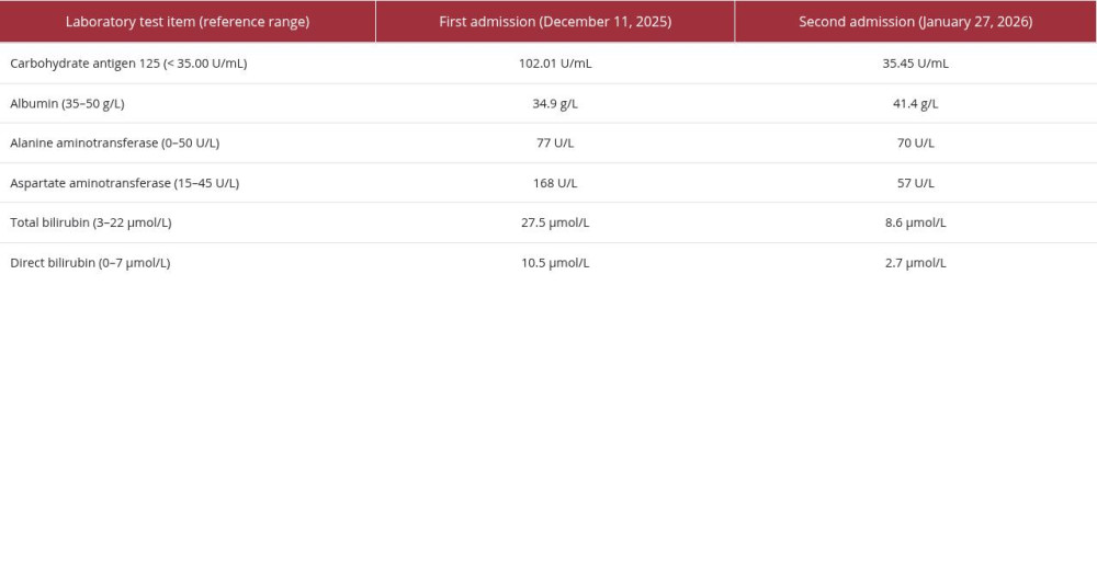
References
1. Chaudhry H, Sohal A, Iqbal H, Unusual sites of hepatocellular carcinoma metastasis: Case report: SAGE Open Med Case Rep, 2023; 11; 2050313X231211709
2. Lu YX, Zhao JP, Yuan GD, Prevalence, patterns, risk factors and outcomes of peritoneal metastases after laparoscopic hepatectomy for hepatocellular carcinoma: A multicenter study from China: Hepatobiliary Surg Nutr, 2024; 13(1); 3-15
3. Huang Y, Zeng J, Liu T, Prognostic significance of elevated preoperative serum CA125 levels after curative hepatectomy for hepatocellular carcinoma: Onco Targets Ther, 2020; 13; 4559-67
4. Devarbhavi H, Kaese D, Williams AW, Cancer antigen 125 in patients with chronic liver disease: Mayo Clin Proc, 2002; 77(6); 538-41
5. Agopian VG, Harlander-Locke MP, Markovic D, Evaluation of patients with hepatocellular carcinomas that do not produce α-fetoprotein: JAMA Surg, 2017; 152(1); 55-64
6. Ueda A, Yuki S, Ando T, CA125 kinetics as a potential biomarker for peritoneal metastasis progression following taxane-plus-ramucirumab administration in patients with advanced gastric cancer: Cancers (Basel), 2024; 16(5); 871
7. Sasaki N, Seyama Y, Ome Y, A resected case of ruptured peritoneal metastasis of hepatocellular carcinoma: Clin J Gastroenterol, 2021; 14(4); 1240-43
8. Otsuki Y, Shimizu S, Hosoda Y, Hepatocellular carcinoma with mesothelioma-like dissemination: Pathol Int, 2005; 55(11); 738-44
Figures
Figure 1. Abdominal computed tomography (CT) images of the patient with peritoneal metastasis from hepatocellular carcinoma. (A) Pretreatment CT scan showing a solitary soft-tissue mass in the left abdominal cavity (outlined by the red dashed line) with heterogeneous enhancement. (B) Posttreatment CT scan demonstrating disappearance of the previous peritoneal lesion, indicating a favorable treatment response.Figure 2. Pathological findings revealed moderately differentiated hepatocellular carcinoma (HCC). (A) Hematoxylin and eosin staining; observation mode: 2-dimensional planar observation; internal scale: 1 unit = 100–200 μm (low power), showing a typical trabecular arrangement of tumor cells with pronounced cellular atypia, an increased nuclear-to-cytoplasmic ratio, and no obvious cholestasis. (B) Immunohistochemical (IHC) staining; observation mode: 2-dimensional planar observation; internal scale: 1 unit = 100–200 μm (low power), showing negative expression of alpha-fetoprotein (AFP) in tumor cells, confirming the diagnosis of AFP-negative HCC. (C) Hematoxylin and eosin staining; observation mode: 2-dimensional planar observation; internal scale: 1 unit = 25–50 μm (high power), showing largely preserved hepatic lobular architecture, a regular arrangement of hepatocytes, and no clinically significant inflammation or fibrosis. (D) IHC staining; observation mode: 2-dimensional planar observation; internal scale: 1 unit = 25–50 μm (high power), showing diffuse cytoplasmic expression of Ki-67 in HCC cells and supporting the pathological diagnosis of HCC. In Press
Case report 
Am J Case Rep In Press; DOI: 10.12659/AJCR.953543
Case report 
Am J Case Rep In Press; DOI: 10.12659/AJCR.954216
Case report
Am J Case Rep In Press; DOI: 10.12659/AJCR.954932
Case report 
Am J Case Rep In Press; DOI: 10.12659/AJCR.954146
Most Viewed Current Articles
07 Dec 2021 : Case report
28,712,695
DOI :10.12659/AJCR.934347
Am J Case Rep 2021; 22:e934347
06 Dec 2021 : Case report  180,721
DOI :10.12659/AJCR.934406
Am J Case Rep 2021; 22:e934406
21 Jun 2024 : Case report
124,419
DOI :10.12659/AJCR.944371
Am J Case Rep 2024; 25:e944371
08 May 2026 : Case report
95,462
DOI :10.12659/AJCR.952158
Am J Case Rep 2026; 27:e952158