


27 September 2026
: Case report 
Pulmonary Actinomycosis Diagnosed via BALF mNGS in a 53-Year-Old Nonsmoking Man With Pneumoconiosis and Penicillin-Induced Thrombocytopenia: A Case Report
Unusual clinical course, Challenging differential diagnosis, Unusual or unexpected effect of treatment, Adverse events of drug therapy, Educational Purpose (only if useful for a systematic review or synthesis), Rare coexistence of disease or pathology
Xin Feng BE 1, Lin-yu Wang CE 2, Ni-jiao Li DE 1, Ze-qiang Lin F 1, He-ming Huang F 1, Yi-ling Xie F 1, Jie Liang F 1, Lunkai Yao F 1, Xin-yi Zhuo F 1, Yan Li DF 2*, Ye Qiu AG 1
DOI: 10.12659/AJCR.953438
Am J Case Rep 2026; 27:e953438


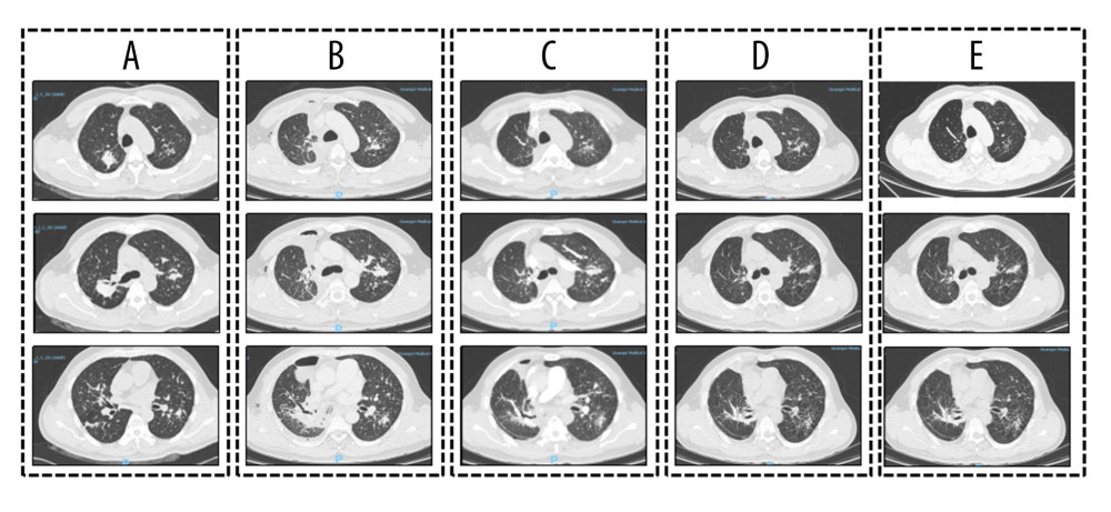
Figure 1 Chest computed tomography (CT) findings at various clinical stages(A) Before surgery: CT images show irregular contrast-enhanced soft-tissue masses in the bilateral upper lungs, multiple bilateral solid nodules, and enlarged lymph nodes in the right supraclavicular fossa and mediastinum. (B) After surgery and 2 weeks of antituberculosis therapy (isoniazid, rifampin, ethambutol, and pyrazinamide): CT images show postoperative distortion of the right lung architecture, right hydropneumothorax, persistent bilateral solid nodules without appreciable interval resolution, and persistent lymphadenopathy in the right supraclavicular fossa and mediastinum. (C) After 12 days of treatment with penicillin sodium: CT images show prominent resolution of the pulmonary lesions compared with previous images. (D) After 2 weeks of omadacycline treatment: CT images show further interval resolution of the pulmonary lesions. (E) After 4 weeks of omadacycline followed by 6 months of doxycycline treatment: CT images show continued resolution and improvement of the pulmonary lesions.