


12 September 2026
: Case report 
Recurrent Intravenous Contrast-Induced Submandibular Sialadenitis: A Case Report and Literature Review
Unknown etiology, Challenging differential diagnosis, Diagnostic / therapeutic accidents, Unusual setting of medical care, Unexpected drug reaction, Educational Purpose (only if useful for a systematic review or synthesis)
Narisara Chobaroon ABCDEF 1, Thitiporn Suwatanapongched ACDEF 1*DOI: 10.12659/AJCR.953439
Am J Case Rep 2026; 27:e953439


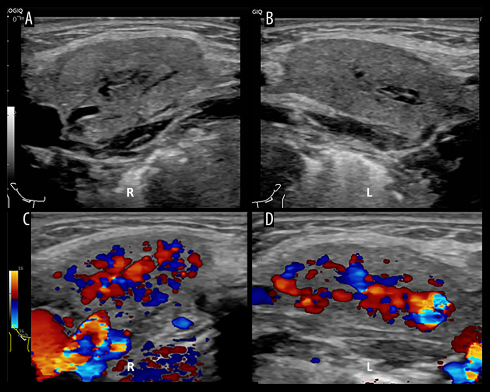
Figure 2 Ultrasonographic images of the submandibular glands obtained 18 hours after contrast administration. (A, B) Gray-scale images of the right (A) and left (B) submandibular glands demonstrate mild heterogeneous parenchymal echotexture with mild intraglandular ductal dilatation. (C, D) Color Doppler images demonstrate increased parenchymal vascularity within the right (C) and left (D) submandibular glands.