


19 August 2026
: Case report 
Malignant Soft Tissue Tumor Mimicking Chronic Expanding Hematoma After a Long Clinical Course: A Case Report
Rare disease
Itaru Ogawa ABDEF 1, Yoichi Kaneuchi ABDEF 1, Takeo Suzuki ABDF 1, Fumiya Ara BF 1, Shoki Yamada BDE 2, Osamu Hasegawa BDE 3, Michiyuki Hakozaki ABDEF 1,4*, Yoshihiro Matsumoto ADE 1
DOI: 10.12659/AJCR.953515
Am J Case Rep 2026; 27:e953515


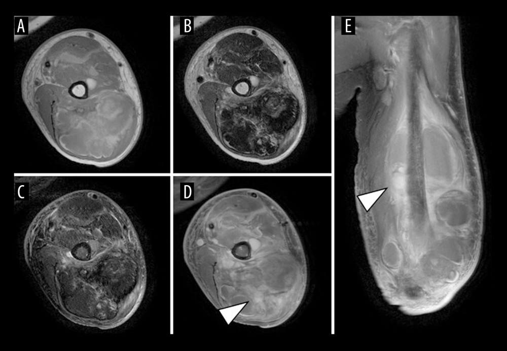
Figure 1 Magnetic resonance imaging (MRI) findings at the initial presentationMRI shows a multilocular lesion measuring 16.1 × 8.1 × 5.3 cm, mainly within the left triceps brachii muscle, extending to the flexor and extensor aspects of the upper arm. The lesion has iso- to high-signal intensity on T1-weighted images (A, axial) and peripheral high-signal intensity with internal low-signal intensity on T2-weighted images (B, axial). On short tau inversion recovery (STIR) images, the internal low-signal components are more clearly delineated (C, axial). Gadolinium-enhanced MRI shows heterogeneous enhancement in part of the lesion (arrowheads) (D, axial; E, coronal).