


23 March 2026: Articles 
Malignant Soft Tissue Tumor Mimicking Chronic Expanding Hematoma After a Long Clinical Course: A Case Report
Rare disease
Itaru Ogawa ABDEF 1, Yoichi Kaneuchi ABDEF 1, Takeo Suzuki ABDF 1, Fumiya Ara BF 1, Shoki Yamada BDE 2, Osamu Hasegawa BDE 3, Michiyuki Hakozaki ABDEF 1,4*, Yoshihiro Matsumoto ADE 1
DOI: 10.12659/AJCR.953515
Am J Case Rep 2026; 27:e953515


Abstract
BACKGROUND: Chronic expanding hematoma (CEH) is a benign lesion characterized by slow enlargement over a prolonged period, and it can closely mimic soft tissue tumors. Differentiating CEH from malignant tumors is particularly challenging in patients with a long clinical course.
CASE REPORT: A 66-year-old man presented with a mass in the distal left upper arm that had been present for >10 years. He was referred to the Orthopaedic Department for suspected malignancy. Initial contrast-enhanced magnetic resonance imaging revealed a cystic lesion, and positron emission tomography/computed tomography revealed fluorodeoxyglucose (FDG) uptake in the septal components of the lesion and in the ipsilateral axillary lymph nodes. Incisional biopsy identified only hematoma components, and the lesion was diagnosed as consistent with CEH. Although conservative treatment, including embolization, drainage, and sclerotherapy, was applied, the mass rapidly enlarged 10 months later. Follow-up imaging revealed a markedly enlarged multilocular cystic lesion containing newly developed solid components with markedly increased FDG uptake. Left shoulder disarticulation with axillary lymph node dissection was subsequently performed. Histopathological examination of the resected tissue sample revealed an undifferentiated malignant tumor that could not be distinguished as a sarcoma or a carcinoma.
CONCLUSIONS: This case shows that lesions initially considered consistent with CEH can later reveal an underlying malignant tumor or possible malignant transformation. Careful interpretation of histopathology results, awareness of potential sampling error, and meticulous radiological evaluation with close follow-up are essential.
Keywords: Hematoma, Magnetic Resonance Imaging, Soft Tissue Neoplasms, Positron-Emission Tomography, Sarcoma, Carcinoma
Introduction
Chronic expanding hematoma (CEH) is a benign condition characterized by gradual enlargement over time [1]. CEH is thought to gradually enlarge due to repeated minor bleeding and chronic inflammation within a fibrous capsule formed after the initial hemorrhage [1]. Clinically, it is often detected as a slowly enlarging mass over months to years, which is typically painless or associated with mild pain [1]. It can arise in various anatomical locations, including the extremities and trunk. Although trauma, prior surgery, and anticoagulant therapy have been reported as risk factors, no obvious predisposing factor is identified in many cases [1,2]. The prognosis is generally favorable; however, its clinical and radiological features can mimic those of malignant tumors, which can lead to diagnostic delay or overtreatment, representing an important clinical concern [3]. Herein, we report the case of a patient in whom a malignant tumor became apparent after a long clinical course of > 10 years, followed by rapid progression over a 12-month period.
Case Report
CLINICAL COURSE:
The patient was a 66-year-old left-handed Japanese man with no relevant medical history. He presented to a local clinic with a mass in the distal portion of the left upper arm that had been present for 10 years and it had gradually enlarged over the preceding 2 years. Plain magnetic resonance imaging (MRI) performed at a local clinic raised suspicion of a malignant tumor, and the patient was referred to our Orthopaedic Department.
At initial presentation, physical examination revealed a mass measuring 17 × 11 cm on the dorsal aspect of the distal left upper arm, accompanied by local warmth and tenderness. There were no apparent signs of vascular compromise, neurological deficits, or limitation in the range of motion. Laboratory examination revealed anemia, with a hemoglobin level of 10.9 g/dL (normal range: 13.7–16.8 g/dL). Coagulation studies showed a prothrombin time–international normalized ratio of 1.05 (normal range: 0.9–1.1), an activated partial thromboplastin time of 30.8 s (normal range: 24–34 s), and an elevated fibrinogen level of 516 mg/dL (normal range: 200–400 mg/dL). The patient had no history of trauma, surgery, or anticoagulant therapy.
Contrast-enhanced MRI revealed a well-circumscribed multilocular lesion measuring 16.1 × 8.1 × 5.3 cm within the triceps brachii muscle, extending to the flexor and extensor compartments of the upper arm without apparent invasion of adjacent bone or major neurovascular structures (Figure 1). The lesion had iso- to high-signal intensity on T1-weighted images, peripheral high-signal intensity with internal low-signal intensity on T2-weighted and short tau inversion recovery images, and heterogeneous gadolinium enhancement.
Positron emission tomography/computed tomography (PET/CT) revealed fluorodeoxyglucose (FDG) uptake in the septal structures of the lesion (maximum standardized uptake value [SUVmax], 5.3) and in the left axillary lymph nodes (Figure 2). An incisional biopsy was performed from the cyst wall at the site of FDG uptake, and 1 tissue sample was obtained; however, histopathological examination revealed only hematoma components, with no evidence of viable tumor cells. Based on these findings, chronic expanding hematoma (CEH) was suspected, although malignancy, particularly a malignant soft tissue tumor, could not be definitively excluded. As the patient declined further invasive examinations, conservative treatment, including transcatheter arterial embolization, percutaneous drainage, and sclerotherapy, were applied at another hospital.
Ten months after the initial biopsy, the lesion rapidly enlarged, and cytological examination at that hospital revealed a malignancy. The patient was then re-referred to our department.
Compared to the initial MRI findings, contrast-enhanced MRI at re-presentation revealed a markedly enlarged multilocular cystic lesion (15.0 × 17.6 × 10.0 cm), encircling the left humerus and containing newly developed nodular solid components (Figure 3). The lesion had relatively well-defined margins, with no clear evidence of bone invasion, but had expanded to involve the major neurovascular structures of the left upper arm. PET/CT revealed intense FDG uptake in the solid components (SUVmax, 15.0) and in the left axillary lymph nodes (SUVmax, 10.5) (Figure 4). Incisional biopsies of the newly developed solid nodules and axillary lymph nodes confirmed malignancy.
Although amputation of the left upper arm was proposed, the patient initially refused because his dominant hand was the left hand. One course of chemotherapy with doxorubicin and ifosfamide was administered; however, severe myelosuppression and septic shock developed, accompanied by rapid progression of the disease. Ultimately, left shoulder disarticulation and axillary lymph node dissection were performed 2 months after re-presentation. Immediately following the surgery, his general condition improved rapidly; in particular, he recovered from myelosuppression, and the severe inflammation that had been diagnosed as sepsis has subsided. Postoperatively, radiotherapy (60 Gy/30 Fr) was administered to the residual metastatic subclavian and axillary lymph nodes. No significant worsening of treatment-related adverse events was observed during the postoperative course. At 12 months postoperatively, the patient remained in good general condition, with reduced size of the irradiated subclavian and axillary lymph node metastases and no evidence of distant metastasis.
HISTOPATHOLOGICAL FINDINGS:
Histopathological examination of the initial incisional biopsy revealed fibrous connective tissue consistent with a cyst wall, with no definitive atypical cells suggestive of malignancy (Figure 5A).
There were numerous hemosiderin-laden macrophages on the surface of, and within, the fibrous connective tissue, supporting a diagnosis of a chronic expanding hematoma (Figure 5B).
In contrast, examination of the subsequent surgically resected specimen revealed multiple multilocular cysts containing hemorrhagic components. In addition, there were areas with a hemangioma-like architecture and regions with solid proliferation of tumor cells, which were not present in the initial biopsy (Figure 5C).
At the periphery of the hemangioma-like structures, markedly atypical tumor cells lined the vascular-like spaces. (Figure 5D). These tumor cells had enlarged hyperchromatic nuclei with marked pleomorphism and prominent nucleoli. (Figure 6A)
Immunohistochemically, the tumor cells showed diffuse positivity for vimentin (Figure 6B) and CD99 (Figure 6C), and focal positivity for epithelial membrane antigen (EMA) (Figure 6D). In contrast, markers of epithelial (AE1/AE3, Figure 6E), vascular (CD34, CD31, D2–40, ERG), myogenic (desmin, Figure 6F; alpha-smooth muscle actin, caldesmon, myogenin, myoD1), neural (S-100 protein, SOX10), melanocytic (melan A), and hematolymphoid differentiation (CD45) were negative.
In addition, amplification-associated markers, including MDM2 and CDK4, were negative, and there was no SS18–SSX fusion. Expression of SMARCB1, SMARCA4, and H3K27me3 was preserved.
Based on these findings, the tumor was diagnosed as a high-grade undifferentiated malignant tumor, not otherwise specified. It could not be distinguished as a sarcoma or a carcinoma.
Discussion
This case involved a patient with a soft tissue mass and a clinical history spanning > 10 years from initial onset. At the first presentation to our institution, CEH was suspected based on imaging and histopathological findings. However, the mass subsequently had rapid growth over a relatively short period of 10 months, accompanied by marked deterioration in imaging and histopathological features. Ultimately, the lesion was diagnosed as a malignant tumor, and a left shoulder disarticulation was performed.
Although no established treatment guidelines exist for CEH, complete surgical excision, including the hematoma and pseudocapsule, is generally recommended to prevent recurrence [1,3]. Alternatively, adjunctive treatments such as transcatheter arterial embolization have been reported; however, their indications remain limited [4]. In the present case, the patient declined further invasive procedures after the initial biopsy; therefore, palliative treatments were performed at another hospital.
Several cases in which differentiation between CEH and soft tissue sarcoma proved clinically challenging have been reported [2,5,6]. Factors contributing to this diagnostic difficulty include heterogeneous contrast enhancement within CEH on contrast-enhanced MRI, and increased FDG uptake on PET/CT [3,7]. In the present case, the contrast-enhanced MRI findings at the initial presentation were largely consistent with the characteristic imaging features of CEH [2,3]. Specifically, the lesion demonstrated iso- to slightly high-signal intensity on T1-weighted images and a heterogeneous structure with peripheral high-signal intensity and internal low-signal intensity on T2-weighted and short tau inversion recovery (STIR) images, with partially heterogeneous enhancement on contrast-enhanced T1-weighted images [2,3]. These findings reflect a mixture of blood products at different stages and pseudocapsule formation, and were not inconsistent with CEH at the initial presentation. In contrast, follow-up MRI demonstrated a marked increase in size and development of enhancing solid components, raising strong suspicion for malignancy. In addition, soft tissue sarcomas are occasionally complicated by intra-tumoral hemorrhage. When the resulting hematoma-like lesion is regarded as the primary lesion, there is a risk of misdiagnosing the underlying sarcoma as CEH [8].
On the other hand, the possibility of malignant transformation of CEH has also been suggested. To the best of our knowledge, 10 cases of suspected malignant transformation of CEH have been reported in the English literature to date [9–13]. Notably, all these cases were histopathologically diagnosed as angiosarcoma, which is inconsistent with the histopathological diagnosis in the present case. In previously reported cases, the interval from the onset of CEH to malignant transformation ranged from 2 to 38 years, indicating that even long-standing CEH can harbor a potential risk of malignant transformation and it should therefore be managed with caution.
In the present case, immunohistochemical analysis of the surgical specimen revealed no markers indicative of specific differentiation. Therefore, based on the histopathological findings and immunohistochemical results, soft tissue sarcomas with specific differentiation (pleomorphic leiomyosarcoma, pleomorphic rhabdomyosarcoma, pleomorphic liposarcoma, dedifferentiated liposarcoma, malignant peripheral nerve sheath tumor, high-grade myxofibrosarcoma, and angiosarcoma) were ruled out from the differential diagnosis, leaving undifferentiated pleomorphic sarcoma and sarcomatoid carcinoma as the most likely diagnoses. Ultimately, the tumor was considered a high-grade undifferentiated malignant tumor.
Treatment was conducted accordingly. Systemic chemotherapy with doxorubicin and ifosfamide was administered [14]; however, treatment was complicated by severe myelosuppression and septic shock, and limb-sparing surgery was deemed unfeasible. Therefore, left shoulder disarticulation and axillary lymph node dissection followed by additional radiotherapy for the residual metastatic subclavian and axillary lymph nodes were performed, in accordance with previously reported treatment strategies for high-grade soft tissue sarcomas [14]. Although the overall management was generally consistent with existing recommendations, the eventual diagnosis of malignancy and the inability to preserve the limb highlight the diagnostic challenges associated with CEH-like lesions.
Even with retrospective analysis, it remains unclear whether the lesion in the present case was malignant from initial onset 10 years earlier or whether a pre-existing CEH underwent rapid malignant transformation during follow-up remains unclear. However, the possibility that an underlying malignant tumor, insufficiently sampled in the early stages, which gradually became clinically apparent over time, cannot be excluded. In lesions predominantly composed of hematoma components, clinicians should be aware of the potential for false-negative biopsy results due to sampling error, depending on the biopsy site and timing. To reduce this risk, it is important to obtain sufficient tissue samples from multiple sites, particularly targeting areas showing abnormalities on FDG-PET or contrast-enhanced MRI, such as enhancing solid components or mural nodules. Furthermore, when discrepancies exist among clinical course, imaging findings, and pathological results, early consideration of repeat biopsy or surgical resection is warranted.
In clinical practice, when CEH is considered as a definitive diagnosis, a thorough evaluation using PET imaging, contrast-enhanced MRI, and biopsy is essential. In addition, even after a diagnosis of CEH has been established, careful radiological follow-up and consideration of early surgical intervention, when appropriate, are crucial for optimal patient management.
Conclusions
This case demonstrates that lesions initially considered consistent with CEH can later reveal an underlying malignant tumor or possible malignant transformation. Therefore, a definitive diagnosis of CEH should be made with caution, and careful long-term follow-up with appropriate re-evaluation is warranted.
Figures
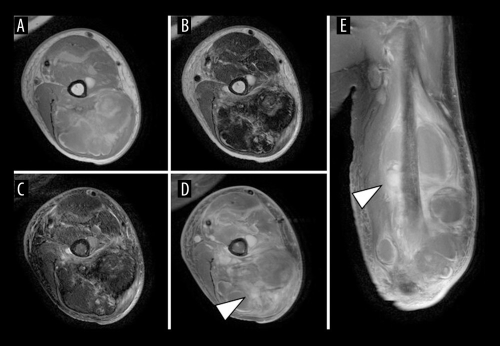 Figure 1. Magnetic resonance imaging (MRI) findings at the initial presentationMRI shows a multilocular lesion measuring 16.1 × 8.1 × 5.3 cm, mainly within the left triceps brachii muscle, extending to the flexor and extensor aspects of the upper arm. The lesion has iso- to high-signal intensity on T1-weighted images (A, axial) and peripheral high-signal intensity with internal low-signal intensity on T2-weighted images (B, axial). On short tau inversion recovery (STIR) images, the internal low-signal components are more clearly delineated (C, axial). Gadolinium-enhanced MRI shows heterogeneous enhancement in part of the lesion (arrowheads) (D, axial; E, coronal).
 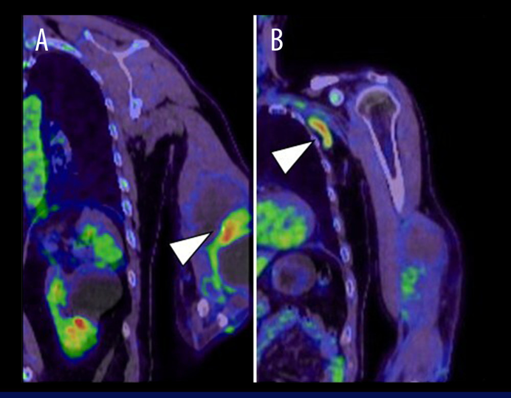 Figure 2. Fluorodeoxyglucose positron emission tomography/computed tomography (FDG-PET/CT) findings at the initial presentationFDG-PET/CT shows FDG uptake with a maximum standardized uptake value (SUVmax) of 5.3 in the septal components of the lesion (arrowhead) (A, coronal). In addition, there is FDG uptake in the left axillary lymph node (arrowhead) (B, coronal).
 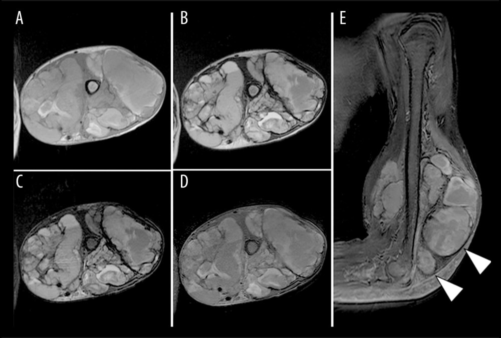 Figure 3. Magnetic resonance imaging (MRI) findings at the re-presentationMRI shows a multilocular cystic lesion measuring approximately 15.0 × 17.6 × 10.0 cm in the distal portion of the left upper arm. The lesion has iso-to-high-signal intensity on T1-weighted images (A, axial), and slightly low-signal intensity on T2-weighted images (B, axial) and short tau inversion recovery images (C, axial). Gadolinium-enhanced MRI shows solid components with contrast enhancement adjacent to the cystic lesion (arrowheads) (D, axial; E, sagittal).
 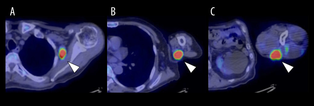 Figure 4. Fluorodeoxyglucose (FDG) positron emission tomography/computed tomography (PET/CT) findings at the re-presentationThere is FDG uptake with a maximum standardized uptake value (SUVmax) of 10.5 in the left axillary lymph node (arrowhead) (A, axial). In addition, there is FDG uptake with an SUVmax of 15.0 in a proximal dorsal nodule of the left upper arm (arrowhead) (B, axial), and FDG uptake with an SUVmax of 15.2 in a distal dorsal nodule (arrowhead) (C, axial).
 ![Histopathological findingsThe biopsy specimen had a structure consistent with a cyst wall composed of thick fibrous connective tissue, with no definitive atypical tumor cells (A, hematoxylin and eosin [H&E] stain, × 100) (bar, 200 μm). There are numerous hemosiderin-laden macrophages on the surface of, and within, the fibrous connective tissue, which are presumed to represent the cyst wall (B, H&E stain, × 200) (bar, 100 μm). Surgical specimens reveal multiple multilocular cysts containing hemorrhagic components. In addition, areas with hemangioma-like architecture and regions have solid proliferation of tumor cells (C, H&E staining, × 40) (bar, 500 μm). There are markedly atypical tumor cells lining the vascular-like spaces are at the periphery of the hemangioma-like structures (D, H&E stain, × 200) (bar, 100 μm).](https://jours.isi-science.com/imageXml.php?i=amjcaserep-27-e953515-g005.jpg&idArt=953515&w=1000) Figure 5. Histopathological findingsThe biopsy specimen had a structure consistent with a cyst wall composed of thick fibrous connective tissue, with no definitive atypical tumor cells (A, hematoxylin and eosin [H&E] stain, × 100) (bar, 200 μm). There are numerous hemosiderin-laden macrophages on the surface of, and within, the fibrous connective tissue, which are presumed to represent the cyst wall (B, H&E stain, × 200) (bar, 100 μm). Surgical specimens reveal multiple multilocular cysts containing hemorrhagic components. In addition, areas with hemangioma-like architecture and regions have solid proliferation of tumor cells (C, H&E staining, × 40) (bar, 500 μm). There are markedly atypical tumor cells lining the vascular-like spaces are at the periphery of the hemangioma-like structures (D, H&E stain, × 200) (bar, 100 μm).
Figure 5. Histopathological findingsThe biopsy specimen had a structure consistent with a cyst wall composed of thick fibrous connective tissue, with no definitive atypical tumor cells (A, hematoxylin and eosin [H&E] stain, × 100) (bar, 200 μm). There are numerous hemosiderin-laden macrophages on the surface of, and within, the fibrous connective tissue, which are presumed to represent the cyst wall (B, H&E stain, × 200) (bar, 100 μm). Surgical specimens reveal multiple multilocular cysts containing hemorrhagic components. In addition, areas with hemangioma-like architecture and regions have solid proliferation of tumor cells (C, H&E staining, × 40) (bar, 500 μm). There are markedly atypical tumor cells lining the vascular-like spaces are at the periphery of the hemangioma-like structures (D, H&E stain, × 200) (bar, 100 μm). 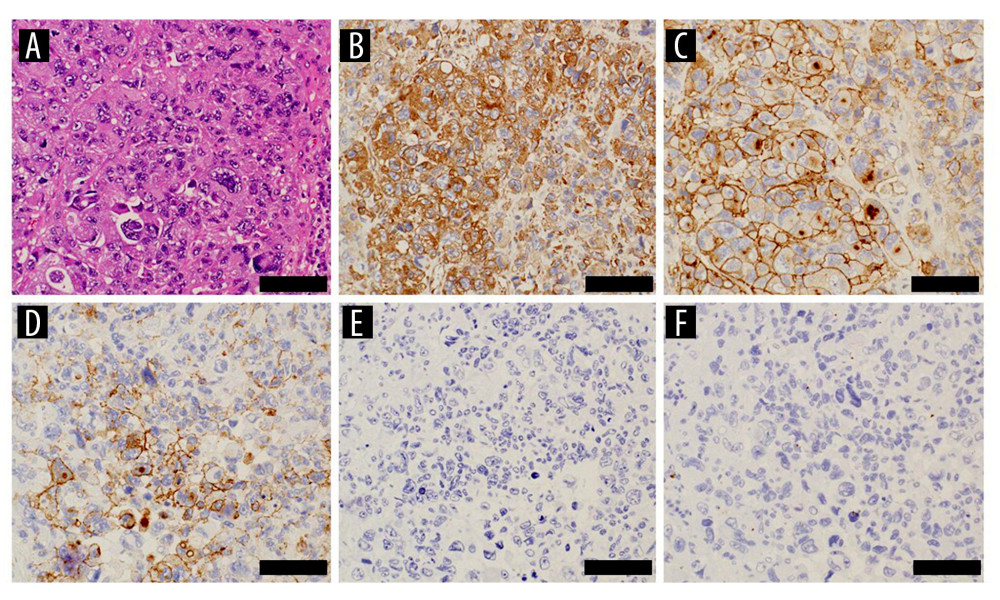 Figure 6. High-power views of H&E staining and immunohistochemical stainings of the surgical specimenSolid proliferation of the tumor cells is observed (A, × 200) (bar, 100 μm). The tumor cells show diffuse positivity for vimentin (B, × 200) and CD99 (C, × 200), and focal positivity for EMA (D, × 200), but negativity for AE1/AE3 (E, × 200) and desmin (F, × 200) (bars, 100 μm).
 References
1. Reid JD, Kommareddi S, Lankerani M, Park MC, Chronic expanding hematomas. A clinicopathologic entity: JAMA, 1980; 244; 2441-42
2. Sakamoto A, Okamoto T, Tsuboyama T, Matsuda S, Chronic expanding hematoma in the thigh: A late complication 32 years after treatment of synovial sarcoma: A case report: Am J Case Rep, 2019; 20; 1449-53
3. Nishida Y, Kobayashi E, Kubota D: J Med Case Rep, 2014; 8; 349
4. Higuchi T, Inaki S, Baba A, A case of chronic expanding hematoma in the thoracic cavity treated with transcatheter arterial embolization: Ann Vasc Dis, 2026; 19; 25-00148
5. Gupta A, Menon A, Agarwal M, Agashe VM, Soft tissue sarcoma masquerading as chronic hematoma: A case report and literature review: J Orthop Case Rep, 2025; 15; 156-60
6. Wells ME, Qiao J, Decker KE, A masquerading hematoma resulting in the delayed diagnosis of a soft tissue sarcoma: A case report: Cureus, 2023; 15; e44822
7. Lee J, Lee T, Oh E, Yoon YC, Temporal evolution of a chronic expanding organizing hematoma on MRI, including functional MR imaging techniques: A case report: Investig Magn Reson Imaging, 2017; 21; 43-50
8. Hoshi M, Oebisu N, Ieguchi M, Clinical features of soft tissue sarcoma presenting intra-tumour haematoma: Case series and review of the literature: Int Orthop, 2017; 41; 203-9
9. Burgert-Lon CE, Riddle ND, Lackman RD, Angiosarcoma arising in chronic expanding hematoma: Five cases of an underrecognized association: Am J Surg Pathol, 2015; 39; 1540-47
10. Alshalfan R, Almalki S, Alnasser M, Alhaqbani A, Case report: Chronic expanding hematoma becomes angiosarcoma: J Surg Case Rep, 2025; 2025; rjaf063
11. Meis-Kindblom JM, Kindblom LG, Angiosarcoma of soft tissue: A study of 80 cases: Am J Surg Pathol, 1998; 22; 683-97
12. Lăzureanu C, Baderca F, Burlacu O, Nicodin A, Soft tissue epithelioid angiosarcoma: Rom J Morphol Embryol, 2010; 51; 787-92
13. Miyazaki H, Goto A, Hino R, Pleural cavity angiosarcoma arising in chronic expanding hematoma after pneumonectomy: Hum Pathol, 2011; 42; 1576-79
14. National Comprehensive Cancer Network: NCCN Clinical Practice Guidelines in Oncology: Soft Tissue Sarcoma. Version 2.2024 Available from: https://www.nccn.org
Figures
Figure 1. Magnetic resonance imaging (MRI) findings at the initial presentationMRI shows a multilocular lesion measuring 16.1 × 8.1 × 5.3 cm, mainly within the left triceps brachii muscle, extending to the flexor and extensor aspects of the upper arm. The lesion has iso- to high-signal intensity on T1-weighted images (A, axial) and peripheral high-signal intensity with internal low-signal intensity on T2-weighted images (B, axial). On short tau inversion recovery (STIR) images, the internal low-signal components are more clearly delineated (C, axial). Gadolinium-enhanced MRI shows heterogeneous enhancement in part of the lesion (arrowheads) (D, axial; E, coronal).Figure 2. Fluorodeoxyglucose positron emission tomography/computed tomography (FDG-PET/CT) findings at the initial presentationFDG-PET/CT shows FDG uptake with a maximum standardized uptake value (SUVmax) of 5.3 in the septal components of the lesion (arrowhead) (A, coronal). In addition, there is FDG uptake in the left axillary lymph node (arrowhead) (B, coronal).Figure 3. Magnetic resonance imaging (MRI) findings at the re-presentationMRI shows a multilocular cystic lesion measuring approximately 15.0 × 17.6 × 10.0 cm in the distal portion of the left upper arm. The lesion has iso-to-high-signal intensity on T1-weighted images (A, axial), and slightly low-signal intensity on T2-weighted images (B, axial) and short tau inversion recovery images (C, axial). Gadolinium-enhanced MRI shows solid components with contrast enhancement adjacent to the cystic lesion (arrowheads) (D, axial; E, sagittal).Figure 4. Fluorodeoxyglucose (FDG) positron emission tomography/computed tomography (PET/CT) findings at the re-presentationThere is FDG uptake with a maximum standardized uptake value (SUVmax) of 10.5 in the left axillary lymph node (arrowhead) (A, axial). In addition, there is FDG uptake with an SUVmax of 15.0 in a proximal dorsal nodule of the left upper arm (arrowhead) (B, axial), and FDG uptake with an SUVmax of 15.2 in a distal dorsal nodule (arrowhead) (C, axial).Figure 5. Histopathological findingsThe biopsy specimen had a structure consistent with a cyst wall composed of thick fibrous connective tissue, with no definitive atypical tumor cells (A, hematoxylin and eosin [H&E] stain, × 100) (bar, 200 μm). There are numerous hemosiderin-laden macrophages on the surface of, and within, the fibrous connective tissue, which are presumed to represent the cyst wall (B, H&E stain, × 200) (bar, 100 μm). Surgical specimens reveal multiple multilocular cysts containing hemorrhagic components. In addition, areas with hemangioma-like architecture and regions have solid proliferation of tumor cells (C, H&E staining, × 40) (bar, 500 μm). There are markedly atypical tumor cells lining the vascular-like spaces are at the periphery of the hemangioma-like structures (D, H&E stain, × 200) (bar, 100 μm).Figure 6. High-power views of H&E staining and immunohistochemical stainings of the surgical specimenSolid proliferation of the tumor cells is observed (A, × 200) (bar, 100 μm). The tumor cells show diffuse positivity for vimentin (B, × 200) and CD99 (C, × 200), and focal positivity for EMA (D, × 200), but negativity for AE1/AE3 (E, × 200) and desmin (F, × 200) (bars, 100 μm). In Press
Case report 
Am J Case Rep In Press; DOI: 10.12659/AJCR.953543
Case report
Am J Case Rep In Press; DOI: 10.12659/AJCR.954216
Case report 
Am J Case Rep In Press; DOI: 10.12659/AJCR.954932
Case report 
Am J Case Rep In Press; DOI: 10.12659/AJCR.954146
Most Viewed Current Articles
07 Dec 2021 : Case report
28,712,695
DOI :10.12659/AJCR.934347
Am J Case Rep 2021; 22:e934347
06 Dec 2021 : Case report  180,721
DOI :10.12659/AJCR.934406
Am J Case Rep 2021; 22:e934406
21 Jun 2024 : Case report
124,419
DOI :10.12659/AJCR.944371
Am J Case Rep 2024; 25:e944371
08 May 2026 : Case report
95,462
DOI :10.12659/AJCR.952158
Am J Case Rep 2026; 27:e952158