


19 August 2026
: Case report 
Malignant Soft Tissue Tumor Mimicking Chronic Expanding Hematoma After a Long Clinical Course: A Case Report
Rare disease
Itaru Ogawa ABDEF 1, Yoichi Kaneuchi ABDEF 1, Takeo Suzuki ABDF 1, Fumiya Ara BF 1, Shoki Yamada BDE 2, Osamu Hasegawa BDE 3, Michiyuki Hakozaki ABDEF 1,4*, Yoshihiro Matsumoto ADE 1
DOI: 10.12659/AJCR.953515
Am J Case Rep 2026; 27:e953515


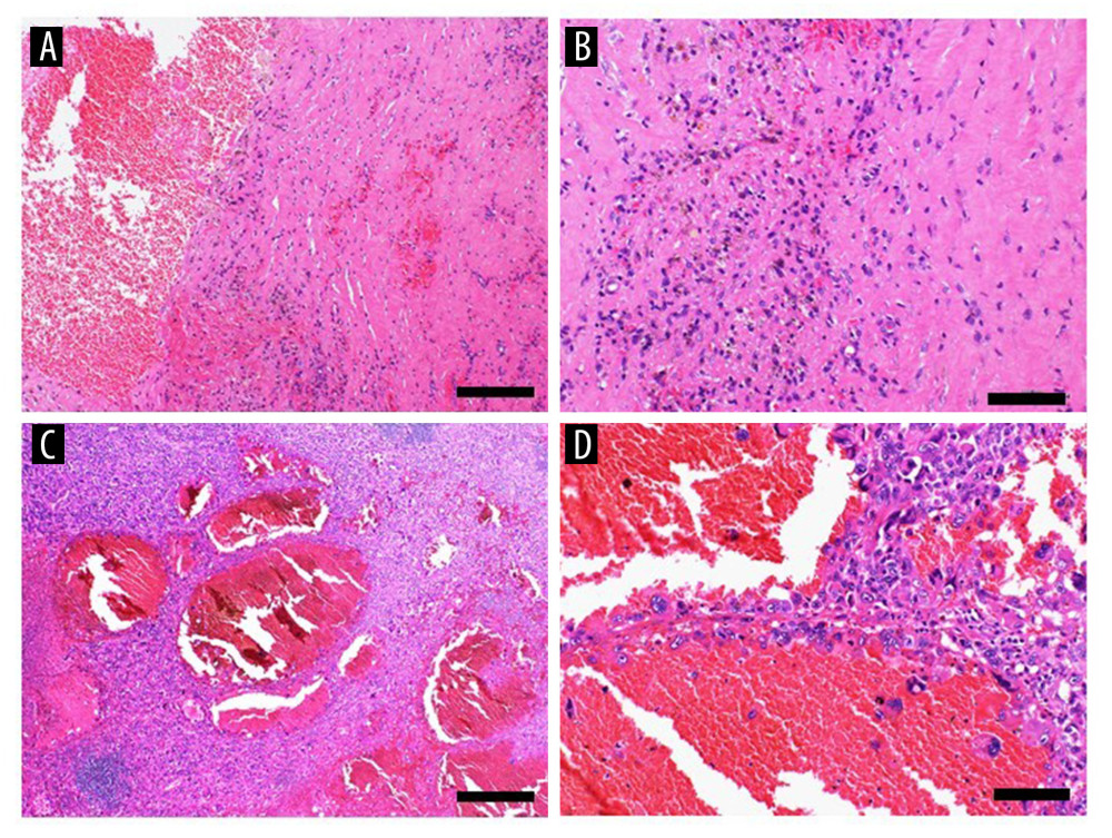
Figure 5 Histopathological findingsThe biopsy specimen had a structure consistent with a cyst wall composed of thick fibrous connective tissue, with no definitive atypical tumor cells (A, hematoxylin and eosin [H&E] stain, × 100) (bar, 200 μm). There are numerous hemosiderin-laden macrophages on the surface of, and within, the fibrous connective tissue, which are presumed to represent the cyst wall (B, H&E stain, × 200) (bar, 100 μm). Surgical specimens reveal multiple multilocular cysts containing hemorrhagic components. In addition, areas with hemangioma-like architecture and regions have solid proliferation of tumor cells (C, H&E staining, × 40) (bar, 500 μm). There are markedly atypical tumor cells lining the vascular-like spaces are at the periphery of the hemangioma-like structures (D, H&E stain, × 200) (bar, 100 μm).