


05 September 2026
: Case report 
Isolated Psychiatric Presentation of Cerebral Venous Thrombosis in a Patient Without Risk Factors: A Diagnostic Challenge
Unusual clinical course, Challenging differential diagnosis, Management of emergency care
Mahdi Abdulrahman Kanjo A 1,2*, Hanin Abdulbast AboTaleb DEF 1, Asim Musaad Almalawi AB 1,3, Reham Jamal Balbaid AB 1
DOI: 10.12659/AJCR.953812
Am J Case Rep 2026; 27:e953812


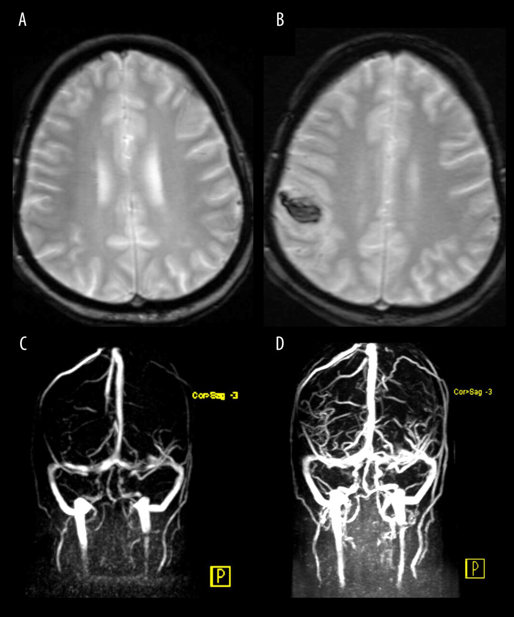
Figure 1 Magnetic resonance imaging (MRI) studies of the brain performed during hospitalization.(A) Contrast-enhanced MRI of the brain on admission demonstrates small, nonspecific foci of altered signal intensity within the bilateral deep and subcortical white matter. No areas of abnormal restricted diffusion are identified. There is no evidence of hyperacute, acute, or early subacute ischemic insult. No extra-axial or intracerebral hemorrhage is seen. (B) Contrast-enhanced MRI of the brain on day 6 of admission reveals a newly developed intra-axial acute hematoma in the right high frontoparietal subcortical region. (C, D) MR venography of the brain performed on day 6 demonstrates occlusion of the right superior anastomotic vein (vein of Trolard), findings consistent with venous hemorrhage.