


18 April 2026: Articles 
Isolated Psychiatric Presentation of Cerebral Venous Thrombosis in a Patient Without Risk Factors: A Diagnostic Challenge
Unusual clinical course, Challenging differential diagnosis, Management of emergency care
Mahdi Abdulrahman Kanjo A 1,2*, Hanin Abdulbast AboTaleb DEF 1, Asim Musaad Almalawi AB 1,3, Reham Jamal Balbaid AB 1
DOI: 10.12659/AJCR.953812
Am J Case Rep 2026; 27:e953812


Abstract
BACKGROUND: Cerebral venous thrombosis (CVT) is an uncommon form of stroke with highly variable clinical manifestations. Although headache and focal neurological deficits are typical presenting features, isolated psychiatric symptoms as an initial manifestation are exceptionally rare and may substantially delay diagnosis. CVT most commonly affects young adults and women with identifiable prothrombotic risk factors.
CASE REPORT: We report the case of a 37-year-old previously healthy woman with no identifiable thrombotic risk factors who presented with acute behavioral disturbances characterized by insomnia, agitation, emotional lability, and aggressive behavior. There was no history of prior psychiatric illness. Initial investigations, including brain magnetic resonance imaging (MRI), cerebrospinal fluid analysis, and infectious workup, were unremarkable, with no evidence of structural abnormalities on early neuroimaging. She was admitted with a working diagnosis of acute polymorphic psychotic disorder and started on psychiatric treatment. Six days later, she developed sudden loss of consciousness followed by generalized tonic-clonic seizures, prompting urgent neuroimaging. Imaging revealed a right high-parietal intracerebral hemorrhage, and subsequent venous imaging confirmed CVT involving the superficial superior cerebral vein. The patient was treated with antiepileptic therapy and anticoagulation, leading to gradual neurological and psychiatric improvement.
CONCLUSIONS: This case highlights an unusual presentation of CVT with isolated psychiatric manifestations, absence of classical risk factors, and initially normal neuroimaging findings, all of which contributed to delayed diagnosis. Abrupt neurological deterioration can occur despite non-specific early investigations. Early consideration of cerebral venous imaging may be warranted in atypical acute psychiatric presentations to avoid delayed diagnosis and potentially life-threatening complications.
Keywords: Thrombosis, Seizures, Stroke
Introduction
Cerebral venous thrombosis (CVT) is an uncommon cerebrovascular disease that accounts for approximately 0.5% to 3% of cerebrovascular disease cases [1]. Unlike arterial stroke, which is typically associated with atherosclerosis and older age, CVT frequently affects young and middle-aged adults and has a higher prevalence in women [1,2]. This female predominance, often reported at ratios of 3: 1 or higher, is partly due to sex-specific risk factors such as oral contraceptive use, pregnancy, puerperium, and hormone replacement therapy, which are present in approximately 65% of affected women [3].
CVT results from thrombus formation in the cerebral venous sinuses or cortical veins, causing impaired venous drainage and increased intracranial pressure. Because venous occlusion often develops gradually, the clinical presentation is typically subacute and variable, making diagnosis challenging [4]. Headache is the most frequently reported symptom, occurring in up to 90% of affected patients [5]. Other common manifestations include seizures, focal neurological deficits, papilledema, visual disturbances, altered level of consciousness, and symptoms of increased intracranial pressure [1]. In nearly one-third of cases, intracranial hemorrhage occurs because of venous congestion and rupture of thin-cortical veins due to increased pressure. This can lead to significant complications and increased morbidity if not promptly addressed [6].
Indeed, CVT can occasionally present with atypical clinical features, including isolated psychiatric or behavioral abnormalities [7–9]. Although these are limited and can mimic primary psychiatric disorders, patients are at risk of initial misdiagnosis and delayed neuroimaging. Early diagnosis relies on high clinical suspicion and appropriate neurovascular imaging, particularly magnetic resonance (MR) venography or computed tomography (CT) venography, as routine brain imaging may appear nonspecific in the early stages [1].
The present report describes a rare and diagnostically challenging case of CVT that initially presented with isolated psychiatric manifestations before progressing to complications, including seizures. In this case, we sought to raise awareness of atypical neuropsychiatric presentations of CVT and reinforce the need for early consideration of venous thrombosis in the differential diagnosis of acute psychosis, even in patients without identifiable prothrombotic risk factors.
Case Report
A 37-year-old woman with no known chronic medical conditions and no prior psychiatric or neurological disease presented to the emergency department with acute behavioral changes. According to her family, she developed severe insomnia over several days, accompanied by marked agitation, anxiety, excessive talkativeness, and episodes of aggressive behavior. There was no history of fever, head trauma, substance use, or recent infection, and the patient did not report headache or other neurological symptoms. Her body mass index was 32.8 kg/m2. She was not taking any regular medications, including hormonal or emergency contraceptive pills, and there was no history of recent menstrual irregularities or use of agents such as tranexamic acid.
On initial evaluation (day 1), she was conscious and oriented to person, place, and time but demonstrated significant agitation and lack of cooperation. Her vital signs were stable except for an elevated blood pressure of 150/90 mm Hg. Neurological examination was partially limited due to poor patient cooperation; however, no gross focal neurological deficits were identified on motor, sensory, or cranial nerve assessment.
Given the acute onset of behavioral disturbances, encephalitis was suspected. She was admitted for further evaluation and started on ceftriaxone, vancomycin, and acyclovir empirically.
Laboratory investigations, including complete blood count, renal and liver function tests, and inflammatory marker levels, were within normal limits. Cerebrospinal fluid analysis revealed normal glucose and protein levels with no pleocytosis, and cultures were negative. Viral and tuberculosis panels were negative.
Magnetic resonance imaging (MRI) of the brain with contrast, performed on the first day of admission, showed some small nonspecific foci of altered signal intensity without clear evidence of encephalitis (Figure 1A). Considering the unremarkable cerebrospinal fluid findings and the absence of structural abnormalities explaining her symptoms, an infectious etiology was considered unlikely.
Over the following days (days 2 and 3), her behavioral symptoms persisted; she became increasingly agitated and refused investigations, necessitating sedation with benzodiazepines and haloperidol. She was transferred to a psychiatric service with a diagnosis of acute polymorphic psychotic disorder, without symptoms of schizophrenia. She received treatment with the antipsychotics olanzapine and risperidone, along with the antidepressant desvenlafaxine.
On day 6 of hospitalization, the patient suddenly collapsed and developed generalized tonic-clonic seizures, associated with a decreased level of consciousness. An urgent CT scan of the brain revealed a new right frontoparietal subcortical intraparenchymal hematoma with hyperdense cortical veins (Figure 2A).
Subsequent magnetic resonance angiography and MR venography demonstrated occlusion of the right superior anastomotic vein (vein of Trolard) and confirmed CVT with venous hemorrhage (Figure 1B–1D).
She was immediately transferred to the intensive care unit (ICU) and started on therapeutic low-molecular-weight heparin in accordance with established management guidelines for CVT. She received a loading dose of intravenous levetiracetam and midazolam for seizure control, followed by maintenance therapy with twice daily levetiracetam. Antipsychotic medications were continued with close neurological and hemodynamic monitoring.
Clinically, she remained awake and oriented, without the need for sedation. Anticoagulation and antipsychotics were then continued. Neurological examination revealed normal cranial nerve function and full motor strength. Electroencephalography showed focal epileptiform activity consistent with focal cerebral disturbances. Seizure prophylaxis was intensified with lacosamide and valproic acid. Her hemoglobin level dropped to 8.5 g/dL, prompting evaluation for anemia and work-up for thrombophilic disorders (factor V assay, fibrinogen, anti-SSA/SSB antibodies, antiphospholipid antibodies, and lipoprotein a), which were mostly unremarkable.
Over the subsequent days, the patient’s condition stabilized clinically, her neurological status improved, and she was subsequently transferred from the ICU to the neurology ward (day 11). Follow-up imaging demonstrated an interval reduction in the size of the hematoma without evidence of a new hemorrhage (Figure 2B). She was switched from low-molecular-weight heparin to apixaban 5 mg twice daily and maintained on levetiracetam, lacosamide, and valproic acid. Her seizures were controlled and her mental status progressively stabilized. No additional seizures or neurological deficits were noted.
Thrombophilia screening returned negative results, except for slightly above the typical upper limit of lupus anticoagulant (LA1; 42.1 s) and mildly positive antinuclear antibody (48.9). Laboratory test results revealed microcytic anemia associated with folic acid and vitamin B12 deficiencies, and appropriate supplementation was initiated. Additionally, her plasma homocysteine level was elevated to 12.5 μmol/L, consistent with mild hyperhomocysteinemia.
The diagnosis was revised, concluding that her psychosis was secondary to the underlying venous thrombosis. No new abnormalities were observed on imaging.
She was discharged in a stable condition on day 13 on oral anticoagulation and antiepileptic therapy, with outpatient follow-up. At subsequent follow-up visits (day 27), the patient remained clinically stable with no recurrence of psychiatric symptoms or seizures. The follow-up CT brain imaging results are shown in Figure 2C. The patient’s clinical course is summarized in Figure 3.
Discussion
Recent evidence highlights the heterogeneous and atypical clinical presentations of CVT, including isolated headache [10], recurrent falls and weakness [11], sudden loss of consciousness [12], dizziness and visual disturbances [13], generalized seizure-like activity consistent with status epilepticus [14], and psychosis [7], all of which can contribute to delayed diagnosis. The present case further illustrates the diverse clinical spectrum of CVT and the diagnostic challenges associated with its atypical presentation, as the patient initially presented with isolated behavioral and psychiatric symptoms despite nonspecific early imaging findings, while subsequent neurological deterioration, including seizures and hemorrhage, ultimately prompted definitive diagnosis by MR venography. It highlights the importance of considering early venous imaging in patients with acute psychiatric presentations when clinical evolution is atypical.
Psychiatric or neuropsychiatric presentations of CVT, including acute agitation, mood disturbance, or acute psychosis, are uncommon but are increasingly recognized in case reports and small case series [7–9]. However, the interval from symptom onset to the final diagnosis of CVT varies in reported cases, ranging from less than 2 hours to 4 weeks [15]. In this case, psychiatric manifestations of CVT preceded the development of typical neurological symptoms by approximately 1 week, culminating in the onset of seizures. The exact mechanisms underlying these psychiatric manifestations remain incompletely understood but are hypothesized to involve frontal lobe venous congestion, microhemorrhages, and subcortical dysfunction, as well as the effects of diffuse inflammatory responses on neural networks involved in mood and behavior regulation [16]. Neuroimaging plays a central role in the diagnosis of CVT. However, early diagnosis remains challenging, and conventional CT or MRI may fail to detect venous thrombosis in some cases [17]. Previous studies have reported that the sensitivity of conventional imaging modalities is lower than that of dedicated venographic techniques, such as computed tomography venography or MR venography, which have demonstrated very high sensitivity in several studies [18]. Furthermore, the diagnostic yield of MRI depends on the imaging sequence used, with some sequences demonstrating limited sensitivity during the first week of symptom onset [1]. These factors may explain why the initial MRI in our patient was negative, whereas MR venography performed after clinical deterioration revealed the presence of CVT.
Notably, our patient lacked the most commonly recognized risk factors for CVT, oral contraceptive use and pregnancy, with obesity as the only identifiable predisposing factor. Obesity (body mass index ≥ 30) has been increasingly recognized as a significant and independent risk factor for CVT [19]. Although psychiatric manifestations of CVT are relatively uncommon, this case highlights the importance of maintaining a high index of suspicion, particularly in patients with underlying risk factors, such as obesity.
In the absence of focal neurological deficits or radiological evidence suggestive of CVT at presentation, extensive laboratory investigations of the underlying prothrombotic conditions were not initially performed. However, following the development of intracranial hemorrhage and subsequent seizure, a more comprehensive diagnostic evaluation was undertaken, including laboratory testing for potential prothrombotic and metabolic risk factors. These investigations revealed several findings that may represent possible prothrombotic signals rather than definitive etiological factors. Specifically, the patient demonstrated mild hyperhomocysteinemia (homocysteine, 12.5 μmol/L) and autoimmune seropositivity, including a mildly positive antinuclear antibody and a slightly elevated lupus anticoagulant (LA1). While hyperhomocysteinemia has been reported as a risk factor for CVT [20], the degree of homocysteine elevation in this case was modest, and its clinical significance remains uncertain. It may be related to nutritional factors such as folate or vitamin B12 deficiency, both of which are involved in homocysteine metabolism [21].
Similarly, the presence of antinuclear antibodies and lupus anticoagulant may suggest an underlying autoimmune tendency [22,23]; however, these findings were identified at a single time point and were not supported by confirmatory repeating within this report. Therefore, their contribution to thrombus formation cannot be established. Prior studies have described associations between antiphospholipid antibodies and CVT, but such relationships typically rely on persistent positivity and compatible clinical criteria [17]. Overall, autoimmune evaluation may be helpful in selected patients with CVT, particularly in atypical presentations or in the absence of clear provoking factors; however, autoimmune markers may be absent despite clinically significant disease.
As described earlier, no traditional risk factors for CVT, including oral contraceptive use, pregnancy, or the puerperium, were identified in this patient. In contrast, the observed factors in this case—obesity, possible vitamin deficiencies with mild hyperhomocysteinemia, and limited autoimmune seropositivity—should be interpreted cautiously as potential contributors. Other thrombophilia markers, including protein C, protein S, and factor V Leiden, were within normal ranges.
Overall, this case highlights the multifactorial and often uncertain nature of CVT risk assessment. The laboratory findings in this patient represent possible associations identified during workup rather than definitive etiological factors. While targeted laboratory evaluation may provide supportive information in selected patients with atypical neuropsychiatric presentations, it should be interpreted cautiously and cannot replace definitive imaging. This may be particularly relevant in settings where advanced imaging such as MR venography is not readily available, in which case clinical suspicion and adjunctive investigations may help guide the need for further diagnostic evaluation.
Lastly, the patient experienced a good clinical outcome, with resolution of the hematoma and stabilization of neurological and psychiatric symptoms, which is consistent with evidence showing that most patients with CVT achieve good functional recovery when treated promptly and comprehensively with anticoagulation and multidisciplinary supportive care including neurological monitoring, seizure control, and treatment of underlying risk factors [24]. Nevertheless, diagnostic delays remain a major barrier to optimal care. Clinicians across disciplines, including emergency medicine, psychiatry, and neurology, should be aware of this potential diagnostic mimic and incorporate venous imaging earlier in the evaluation of atypical behavioral presentations.
Conclusions
This case highlights that CVT can present with atypical psychiatric and neurological symptoms even in the absence of classic risk factors, such as oral contraceptive use or pregnancy. Obesity and mild laboratory abnormalities, including hyperhomocysteinemia and limited autoimmune seropositivity, may represent potential contributing factors, although these require confirmation on repeat testing. Diagnosis was established by neuroimaging. Early recognition, prompt imaging, and multidisciplinary management, including anticoagulation and symptomatic treatment, resulted in complete recovery. Clinicians should maintain a high index of suspicion for CVT in patients with acute or atypical psychiatric presentations, and MR venography remains the preferred diagnostic modality when CVT is suspected.
Figures
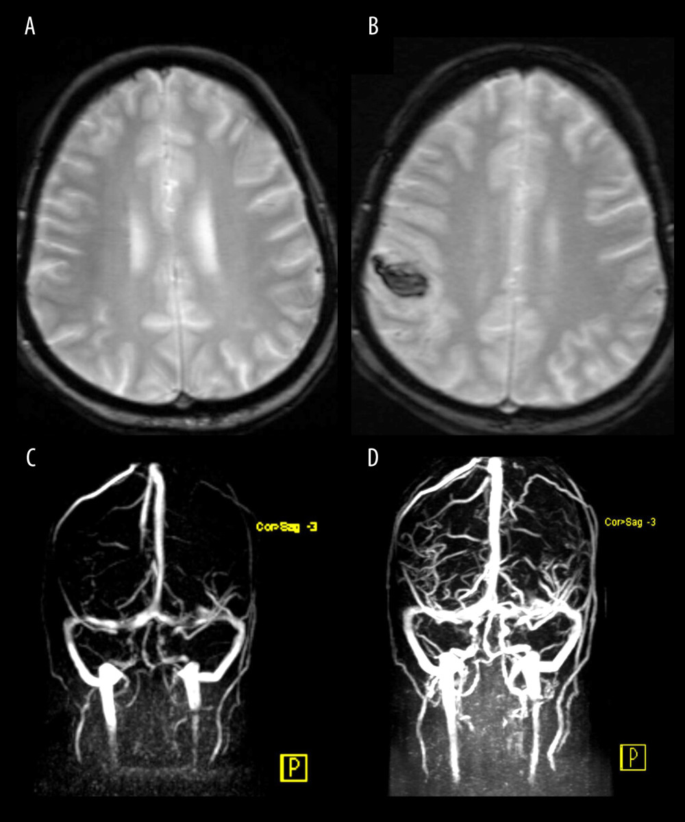 Figure 1. Magnetic resonance imaging (MRI) studies of the brain performed during hospitalization.(A) Contrast-enhanced MRI of the brain on admission demonstrates small, nonspecific foci of altered signal intensity within the bilateral deep and subcortical white matter. No areas of abnormal restricted diffusion are identified. There is no evidence of hyperacute, acute, or early subacute ischemic insult. No extra-axial or intracerebral hemorrhage is seen. (B) Contrast-enhanced MRI of the brain on day 6 of admission reveals a newly developed intra-axial acute hematoma in the right high frontoparietal subcortical region. (C, D) MR venography of the brain performed on day 6 demonstrates occlusion of the right superior anastomotic vein (vein of Trolard), findings consistent with venous hemorrhage.
 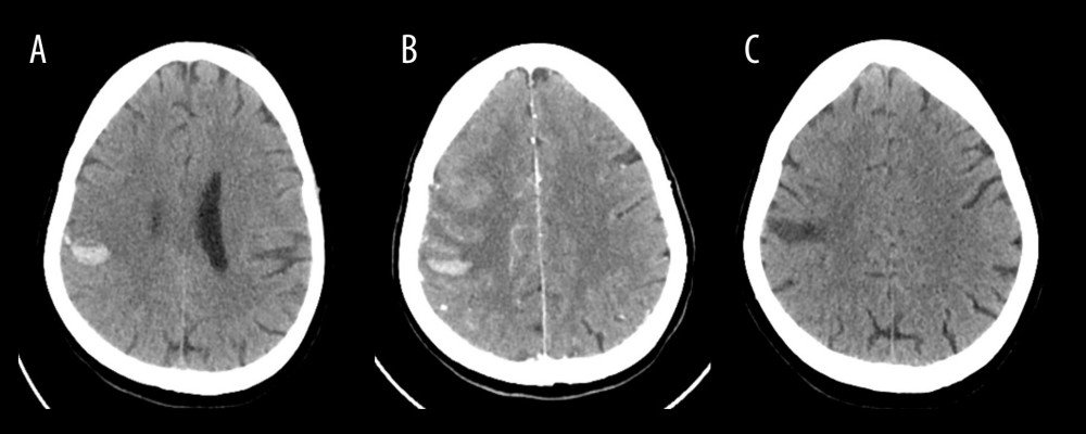 Figure 2. Computed tomography (CT) brain imaging studies performed during hospitalization.(A) Non–contrast-enhanced CT of the brain performed on day 6 following the onset of a generalized tonic-clonic seizure reveals a newly developed intra-axial acute hematoma in the right high frontoparietal subcortical region. Adjacent hyperdense cortical veins are noted, suggestive of thrombosed cortical veins. (B) Follow-up CT angiography of the brain and neck on day 11 demonstrates unchanged right-sided peripherally located hyperdense/thrombosed superficial superior cerebral veins draining to the superior sagittal sinus. Otherwise; no filling defect in dural venous sinuses. Plain CT images show stable multiple foci of intra-axial acute hematoma noted at the right frontoparietal lobe. (C) Follow-up non–contrast-enhanced CT of the brain performed 2 weeks after discharge (day 23) shows interval resolution of the previously noted right frontoparietal intraparenchymal hyperdense hematoma, now appearing hypodense, with resolution of the associated vasogenic edema.
 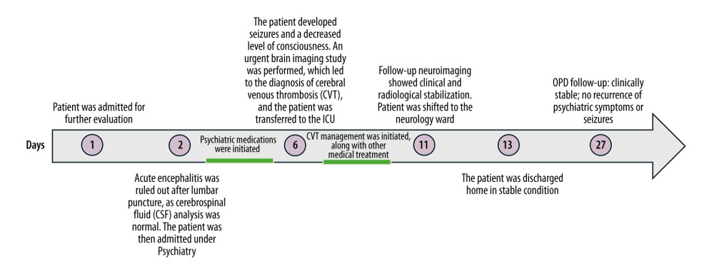 Figure 3. Timeline of the patient’s clinical course from initial presentation to outpatient follow-up.
 References
1. Saposnik G, Barinagarrementeria F, Brown RD, Diagnosis and management of cerebral venous thrombosis: Stroke, 2011; 42(4); 1158-92
2. Murphy SJX, Werring DJ, Stroke: Causes and clinical features: Medicine (Baltimore), 2020; 48(9); 561-66
3. Coutinho JM, Ferro JM, Canhão P, Cerebral venous and sinus thrombosis in women: Stroke, 2009; 40(7); 2356-61
4. Ranjan R, Ken-Dror G, Sharma P, Pathophysiology, diagnosis and management of cerebral venous thrombosis: A comprehensive review: Medicine (Baltimore), 2023; 102(48); e36366
5. Wasay M, Kojan S, Dai AI, Headache in cerebral venous thrombosis: Incidence, pattern and location in 200 consecutive patients: J Headache Pain, 2010; 11(2); 137-39
6. Afifi K, Bellanger G, Buyck PJ, Features of intracranial hemorrhage in cerebral venous thrombosis: J Neurol, 2020; 267(11); 3292-98
7. Calva-González M, Tello-Gerez TJ, Serrano-Arias F, The neuropsychiatric side of cerebral venous thrombosis: A case of delirium and catatonia: J Psychiatr Pract, 2023; 29(6); 493
8. Ozdilek B, Zincir SB, Domac FM, Consideration of cerebral venous thrombosis as a cause of delirium in psychiatry clinics: Psychiatry Clin Psychopharmacol, 2015; 25(2); 190-92
9. von Mering M, Stiefel M, Brockmann K, Nau R, Deep cerebral venous sinus thrombosis often presents with neuropsychologic symptoms: J Clin Neurosci, 2003; 10(3); 310-12
10. Piwowarski E, Mantha K, Wegman M, The commonly uncommon presentation of cerebral venous thrombosis: A case report: HCA Healthc J Med, 2025; 6(3); 287-91
11. Nandy A, Banerjee S, Gupta R, A case report on atypical presentation of cerebral venous sinus thrombosis, a young adult with recurrent fall: A clinical quandary: Open J Emerg Med, 2021; 9(4); 171-78
12. Katwal S, Suwal S, Lamichhane S, Cerebral venous sinus thrombosis with hemorrhagic infarct: A rare presentation in a risk-defying male patient: Radiol Case Rep, 2023; 19(1); 153-57
13. Gaba W, Mahdi A, Atypical presentation of isolated cortical vein thrombosis: J Neurol Sci, 2019; 405; 163 Abstracts from the World Congress of Neurology (WCN 2019)
14. Malone M, Ritchie D, Okonoboh P, Ebrahim A, An atypical presentation of cerebral venous sinus thrombosis in a 19-year-old woman: Consultant360 [Internet], 2023 Available from: [cited 2026 May 21]https://www.consultant360.com/photoclinic/atypical-presentation-cerebral-venous-sinus-thrombosis-19-year-old-woman
15. Ferro JM, Canhão P, Stam J, Prognosis of cerebral vein and dural sinus thrombosis: Stroke, 2004; 35(3); 664-70
16. Li AY, Tong E, Yedavalli VS, A case-based review of cerebral venous infarcts with perfusion imaging and comparison to arterial ischemic stroke: Front Radiol, 2021; 1; 687045
17. Ferro JM, Aguiar de Sousa D, Cerebral venous thrombosis: An update: Curr Neurol Neurosci Rep, 2019; 19(10); 74
18. van Dam LF, van Walderveen MAA, Kroft LJM, Current imaging modalities for diagnosing cerebral vein thrombosis – A critical review: Thromb Res, 2020; 189; 132-39
19. Zuurbier SM, Arnold M, Middeldorp S, Risk of cerebral venous thrombosis in obese women: JAMA Neurol, 2016; 73(5); 579-84
20. Boncoraglio G, Carriero MR, Chiapparini L, Hyperhomocysteinemia and other thrombophilic risk factors in 26 patients with cerebral venous thrombosis: Eur J Neurol, 2004; 11(6); 405-9
21. Kalita J, Singh VK, Misra UK, A study of hyperhomocysteinemia in cerebral venous sinus thrombosis: Indian J Med Res, 2020; 152(6); 584-94
22. Natorska J, Celińska-Lowenhoff M, Undas AI, High prevalence of antinuclear antibodies in patients following venous thromboembolism: Adv Clin Exp Med, 2018; 27(6); 827-32
23. Jerez-Lienas A, Mathian A, Aboab J, Cerebral vein thrombosis in the antiphospholipid syndrome: Analysis of a series of 27 patients and review of the literature: Brain Sci, 2021; 11(12); 1641
24. Liao S, Wang R, Li Z, Long-term outcome and predictors in cerebral venous sinus thrombosis: A prospective cohort study: BMC Neurol, 2026; 26; 113
Figures
Figure 1. Magnetic resonance imaging (MRI) studies of the brain performed during hospitalization.(A) Contrast-enhanced MRI of the brain on admission demonstrates small, nonspecific foci of altered signal intensity within the bilateral deep and subcortical white matter. No areas of abnormal restricted diffusion are identified. There is no evidence of hyperacute, acute, or early subacute ischemic insult. No extra-axial or intracerebral hemorrhage is seen. (B) Contrast-enhanced MRI of the brain on day 6 of admission reveals a newly developed intra-axial acute hematoma in the right high frontoparietal subcortical region. (C, D) MR venography of the brain performed on day 6 demonstrates occlusion of the right superior anastomotic vein (vein of Trolard), findings consistent with venous hemorrhage.Figure 2. Computed tomography (CT) brain imaging studies performed during hospitalization.(A) Non–contrast-enhanced CT of the brain performed on day 6 following the onset of a generalized tonic-clonic seizure reveals a newly developed intra-axial acute hematoma in the right high frontoparietal subcortical region. Adjacent hyperdense cortical veins are noted, suggestive of thrombosed cortical veins. (B) Follow-up CT angiography of the brain and neck on day 11 demonstrates unchanged right-sided peripherally located hyperdense/thrombosed superficial superior cerebral veins draining to the superior sagittal sinus. Otherwise; no filling defect in dural venous sinuses. Plain CT images show stable multiple foci of intra-axial acute hematoma noted at the right frontoparietal lobe. (C) Follow-up non–contrast-enhanced CT of the brain performed 2 weeks after discharge (day 23) shows interval resolution of the previously noted right frontoparietal intraparenchymal hyperdense hematoma, now appearing hypodense, with resolution of the associated vasogenic edema.Figure 3. Timeline of the patient’s clinical course from initial presentation to outpatient follow-up. In Press
Case report 
Am J Case Rep In Press; DOI: 10.12659/AJCR.953543
Case report 
Am J Case Rep In Press; DOI: 10.12659/AJCR.954216
Case report 
Am J Case Rep In Press; DOI: 10.12659/AJCR.954932
Case report 
Am J Case Rep In Press; DOI: 10.12659/AJCR.954146
Most Viewed Current Articles
07 Dec 2021 : Case report
28,712,695
DOI :10.12659/AJCR.934347
Am J Case Rep 2021; 22:e934347
06 Dec 2021 : Case report  180,721
DOI :10.12659/AJCR.934406
Am J Case Rep 2021; 22:e934406
21 Jun 2024 : Case report
124,419
DOI :10.12659/AJCR.944371
Am J Case Rep 2024; 25:e944371
08 May 2026 : Case report
95,462
DOI :10.12659/AJCR.952158
Am J Case Rep 2026; 27:e952158