


05 September 2026
: Case report 
Isolated Psychiatric Presentation of Cerebral Venous Thrombosis in a Patient Without Risk Factors: A Diagnostic Challenge
Unusual clinical course, Challenging differential diagnosis, Management of emergency care
Mahdi Abdulrahman Kanjo A 1,2*, Hanin Abdulbast AboTaleb DEF 1, Asim Musaad Almalawi AB 1,3, Reham Jamal Balbaid AB 1
DOI: 10.12659/AJCR.953812
Am J Case Rep 2026; 27:e953812


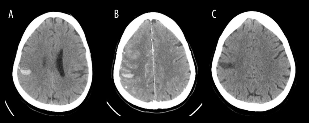
Figure 2 Computed tomography (CT) brain imaging studies performed during hospitalization.(A) Non–contrast-enhanced CT of the brain performed on day 6 following the onset of a generalized tonic-clonic seizure reveals a newly developed intra-axial acute hematoma in the right high frontoparietal subcortical region. Adjacent hyperdense cortical veins are noted, suggestive of thrombosed cortical veins. (B) Follow-up CT angiography of the brain and neck on day 11 demonstrates unchanged right-sided peripherally located hyperdense/thrombosed superficial superior cerebral veins draining to the superior sagittal sinus. Otherwise; no filling defect in dural venous sinuses. Plain CT images show stable multiple foci of intra-axial acute hematoma noted at the right frontoparietal lobe. (C) Follow-up non–contrast-enhanced CT of the brain performed 2 weeks after discharge (day 23) shows interval resolution of the previously noted right frontoparietal intraparenchymal hyperdense hematoma, now appearing hypodense, with resolution of the associated vasogenic edema.