


14 September 2026
: Case report 
Retrieval of a Displaced Mandibular Tooth Fragment From the Floor of the Mouth Using a Combined Navigation System and Endoscope: A Case Report
Unusual setting of medical care
Taifu Hirano ABCDEF 1*, Tadashi Kawai AE 1, Shu Suzuki BD 1, Noriaki Takahashi BD 2, Mitsuru Izumisawa BD 2, Hiroyuki Yamada AE 1
DOI: 10.12659/AJCR.953836
Am J Case Rep 2026; 27:e953836


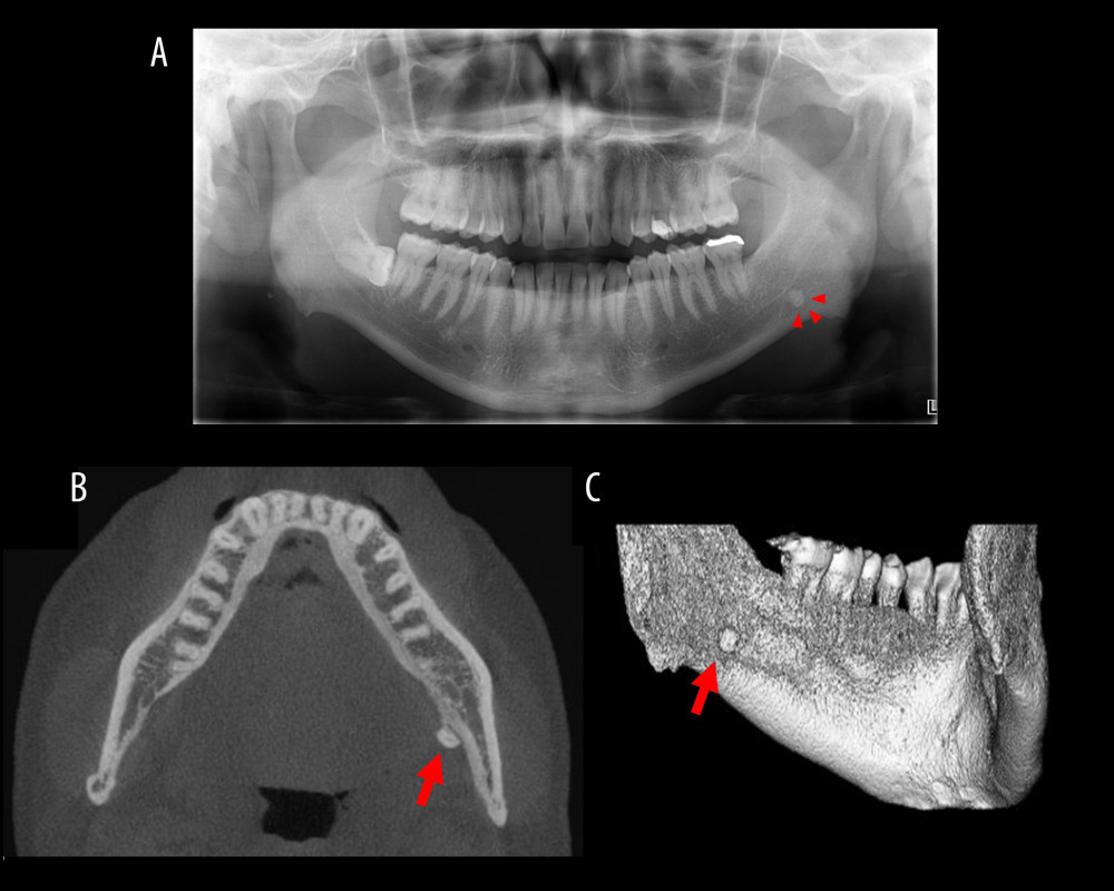
Figure 2 Panoramic radiograph and CBCT images. (A) Panoramic radiograph showing hard tissue on the left side of the mandible. (B) Axial CBCT imaging showing hard tissue on the lingual side of the left mandible. (C) Three-dimensional CBCT image showing hard tissue under the left mylohyoid line. Arrows indicate the displaced tooth fragment.