


14 September 2026: Articles 
Retrieval of a Displaced Mandibular Tooth Fragment From the Floor of the Mouth Using a Combined Navigation System and Endoscope: A Case Report
Unusual setting of medical care
Taifu Hirano ABCDEF 1*, Tadashi Kawai AE 1, Shu Suzuki BD 1, Noriaki Takahashi BD 2, Mitsuru Izumisawa BD 2, Hiroyuki Yamada AE 1
DOI: 10.12659/AJCR.953836
Am J Case Rep 2026; 27:e953836


Abstract
BACKGROUND: If a tooth fragment is displaced into the floor of the mouth during mandibular third molar extraction, surgical retrieval can be challenging because the fragment cannot be directly visualized in the operative field. Several studies have described the use of navigation systems or endoscopic techniques individually. However, to the best of our knowledge, no reports have described their combined use for this condition.
CASE REPORT: A 37-year-old man underwent extraction of a left mandibular third molar at a dental clinic in May 2021, during which a root fragment was displaced into the floor of the mouth. An attempt to retrieve the fragment under general anesthesia at another hospital was unsuccessful, and the patient was subsequently referred to our department. Because direct retrieval was considered technically difficult, surgical retrieval was performed by using a combination of electromagnetic navigation and endoscopic guidance. The fragment was successfully removed via an intraoral approach with minimal surgical invasion. Postoperatively, the wound healed uneventfully and no lingual nerve injury was observed.
CONCLUSIONS: Electromagnetic navigation systems provide real-time spatial localization based on preoperative imaging, and are particularly useful when the target is difficult to palpate or directly visualize. Endoscopic guidance provides enhanced illumination and direct visualization within confined anatomical spaces, thereby facilitating minimally invasive surgery. In the present case, the combined use of electromagnetic navigation and endoscopic guidance can facilitate accurate intraoperative localization and minimally invasive retrieval of a displaced mandibular root fragment on the floor of the mouth.
Keywords: Surgery, Oral, Tooth, Endoscopes, Mouth Floor, Molar, Third
Introduction
Mandibular third molar extraction is the most common surgical procedure performed by oral and maxillofacial surgeons. Wisdom tooth extraction surgery can result in accidents and complications, such as damage to adjacent teeth, soft tissue damage, bone damage, dislocation of the TMJ, aspiration of extracted teeth, subcutaneous emphysema, and tooth displacement into the floor of the mouth [1,2]. If the tooth root is displaced to the floor of the mouth, it should be extracted as soon as possible to prevent inflammation [3]. However, removal can be difficult within a confined surgical field. A displaced tooth should not be extracted in the absence of symptoms [4].
The causes of tooth displacement include anatomical factors such as lingual inclination of the tooth and perforation of the lingual cortical bone [5]. Excessive force, improper treatment, and inadequate clinical and radiographic examinations and evaluations are significant factors that contribute to tooth displacement [6].
There is no uniformly applicable surgical technique because of differences in the direction and size of the root, time after extraction, and tissue reaction [7].
Several authors have reported the use of surgical navigation systems and endoscopic techniques to manage displaced tooth fragments. Navigation systems provide real-time spatial localization, while endoscopic techniques provide enhanced visualization in confined surgical fields. Although navigation systems and endoscopic techniques have been reported separately, we were unable to identify any reports describing their combined use for the retrieval of displaced tooth fragments from the floor of the mouth.
The aim of this case report is to demonstrate the technical feasibility of localizing and retrieving a displaced mandibular tooth root fragment in the floor of the mouth using a combination of a navigation system and endoscopic guidance in a case where direct localization was difficult.
Case Report
The patient was a 37-year-old man. In May 2021, during the extraction of his left mandibular third molar at a dental clinic, the tooth root was displaced into the floor of the mouth. In November 2022, an attempt to remove the tooth under general anesthesia at another hospital was unsuccessful because the displaced tooth could not be identified intraoperatively. The patient was referred to our department in December 2023. An extraoral examination revealed no abnormal findings (Figure 1A). An intraoral examination revealed no signs of inflammation, and the displaced tooth was not palpable. A faint postoperative scar was observed on the floor of the mouth. There were no signs of lingual nerve injury. The patient reported occasionally having foreign body sensation on the floor of the mouth (Figure 1B).
Panoramic radiography showed hard tissue on the left side of the mandible (Figure. 2A). Cone-beam computed tomography (CBCT) image showed a hard tissue at the lingual side of the left mandible (Figure 2B). The 3-dimensional CBCT imaging showed hard tissue under the left mylohyoid line (Figure 2C). The clinical diagnosis was a fragment of the lower left wisdom tooth displaced into the floor of the mouth, and extraction of the fragment was planned under general anesthesia.
A navigation system (ENT Navigation System; Stryker Japan, Tokyo, Japan) and a 0° rigid endoscope (VISERA ELITE II OLYMPUS OTV-S300; Olympus Medical Systems, Inc., Tokyo, Japan) with a diameter of 4 mm were used. This navigation system can arbitrarily set a reference point using a magnetic field and perform navigation based on the preoperative image data obtained before the surgery. Before surgery, the navigation registration error was confirmed to be within the clinically acceptable range. The navigation system detects changes in the magnetic field through a tracker or pointer and displays their position on the image.
Initially, a pointer was set up to locate the displaced tooth in the CBCT image. Pointers were registered on the mucosal raspatory and endoscope (Figure 3A). Subsequently, a tracker was placed to set the reference point in the surgical field (Figure 3B). Reference points were established on the bilateral mandibular canines and molars (Figure 3C). To minimize mandibular movement during navigation, the mandible was stabilized using an occlusal fixation device made of autopolymerized resin, which allowed secure tracker placement (Figure 3B). After setup was completed, the operator moved the pointer while viewing the endoscopic image, and the system displayed the pointer position on the CBCT image. This allowed precise localization of the tooth fragment.
Actual use in an operating room is shown. A field generator that received the magnetic field was placed near the surgical field (Figure 4). The intraoperative images obtained during navigation are shown in Figure 5A. The image on the right shows the estimated position of the displaced fragment based on the CBCT image and the registered reference points. The fan-shaped area represents the endoscopic field of view, and the corresponding endoscopic image showed the displaced root fragment. The endoscopic image and fan-shaped field of view on the CBCT image matched perfectly, and the displaced tooth root fragment was located. The clearly identified displaced tooth was extracted using mosquito forceps (Figure 5B). The operative time was 68 minutes, and blood loss was 10 mL.
Two weeks after surgery, there were no obvious abnormal extraoral findings (Figure 6A), and the intraoral findings were also good. There were no wound abnormalities or lingual neurological symptoms, and the patient was doing well (Figure 6B).
Postoperative panoramic radiography showed that the tooth fragment on the left side of the mandible was completely extracted (Figure 7A). Postoperative axial and 3D CBCT image showed that the tooth fragment near the left mylohyoid line was extracted (Figure 7B, 7C).
Discussion
During the extraction of impacted mandibular third molars, the tooth may occasionally be displaced into the lingual side because of factors such as thinness or perforation of the lingual cortical bone associated with the lingual inclination of the root apex [8]. Panoramic radiography alone cannot identify thinning or the absence of the lingual cortical bone; therefore, preoperative CT evaluation is important to confirm mandibular morphology and root orientation [8].
Displacement of a tooth fragment into the floor of the mouth is an occasional complication of mandibular third molar extraction. Excessive force during surgery and inadequate preoperative evaluation are considered major contributing factors. Displacement into the submandibular space is the most common [9], and retrieval is particularly difficult when the fragment is located below the mylohyoid line [7].
There are 2 surgical approaches: the direct method and the lingual mucoperiosteal flap method [10]. The direct method is indicated for palpable fragments in the sublingual or mylohyoid regions, whereas the lingual mucoperiosteal flap method is often used for non-palpable fragments in the submandibular or sublingual spaces [10]. In the present case, the fragment was successfully removed using an intraoral incision alone; however, a combined intraoral and extraoral approach may be required [11].
The displacement of a tooth fragment into the floor of the mouth can lead to infection involving the sublingual, submandibular, or deeper cervical spaces [12]. If the displaced fragment is left untreated, infection can spread to adjacent anatomical spaces and become life-threatening [12]. Therefore, surgical retrieval is generally recommended, particularly when the fragment is deeply displaced or associated with symptoms [7,13]. In the present case, extraction was performed due to the risk of infection.
In recent years, navigation-assisted surgery has been used in various fields. In the oral and maxillofacial regions, it has been used for implant placement and other surgical procedures[3,14–16]. The navigation system used in this study is frequently used in otorhinolaryngology, particularly in sinus surgery. Previous reports have described its use in the retrieval of displaced tooth fragment in the maxillary sinus [3,16]. Navigation systems provide real-time spatial localization based on preoperative imaging, and are particularly useful when the target is difficult to palpate or directly visualize. However, navigation alone may not provide sufficient direct visualization within a narrow surgical field.
Endoscopic techniques for the retrieval of displaced mandibular third molars and tooth fragments have also been reported [17,18]. Endoscopy provides enhanced illumination and direct visualization of confined anatomical spaces, thereby facilitating minimally invasive surgery. However, endoscopic visualization alone may be insufficient for the precise localization of deeply displaced or non-palpable fragments.
In the present case, the tooth fragment was small and non-palpable and located deep in the floor of the mouth, making retrieval particularly challenging. Therefore, a combined approach involving navigation and endoscopic guidance was selected. The navigation system enabled accurate spatial localization of the fragment, whereas the endoscope allowed for real-time visual confirmation during dissection and retrieval. This combined approach facilitates minimally invasive intraoral removal while avoiding blind exploration of the floor of the mouth.
If the surgical site is in the maxilla, the position of the target usually remains stable during surgery, making the exact location easier to determine. However, in the mandible, mouth opening and closing can change the position of the displaced fragment during surgery. Because the tooth fragment in this case had shifted toward the floor of the mouth, it was necessary to place a tracker on the mandible. Therefore, we developed a special device to secure a dental arch tracker.
This approach may be particularly useful in cases where the fragment is non-palpable, located below the mylohyoid muscle, or when previous retrieval attempts have failed. However, the generalizability of this technique remains limited because this is a single-case report.
Conclusions
This case report demonstrates the successful retrieval of a non-palpable displaced mandibular root fragment using a combined electromagnetic navigation and endoscopic approach. The integration of real-time three-dimensional navigation guidance with direct endoscopic visualization enabled accurate intraoral localization and minimally invasive retrieval. This technique may be a valuable option for managing displaced tooth fragments in the anatomically challenging regions of the oral cavity. However, further studies are required to evaluate its broader clinical applicability because this report describes a single case.
Figures
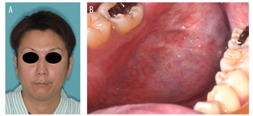 Figure 1. Patient photographs. (A) Extraoral photograph showing no abnormalities. (B) Intraoral photograph showing no inflammatory findings. A faint postoperative scar is observed in the floor of the mouth; the roots were not palpable.
 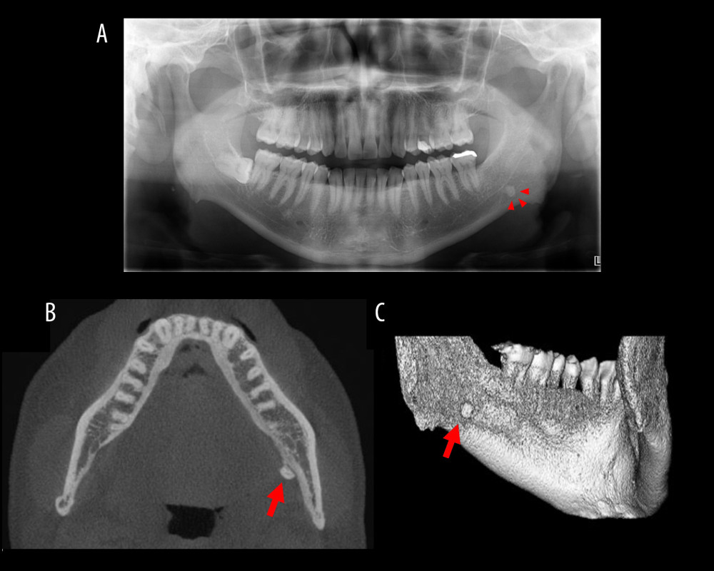 Figure 2. Panoramic radiograph and CBCT images. (A) Panoramic radiograph showing hard tissue on the left side of the mandible. (B) Axial CBCT imaging showing hard tissue on the lingual side of the left mandible. (C) Three-dimensional CBCT image showing hard tissue under the left mylohyoid line. Arrows indicate the displaced tooth fragment.
 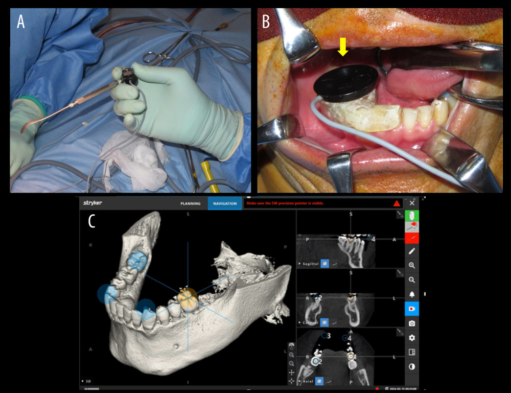 Figure 3. Photograph of the device used for navigation and the screen where the reference point was set. (A) Photograph of a mucosal raspatory set up as a pointer. (B) Photograph of a tracker placed with resin in the intraoral surgical field. (C) Monitor screen when a reference point is set on the teeth.
 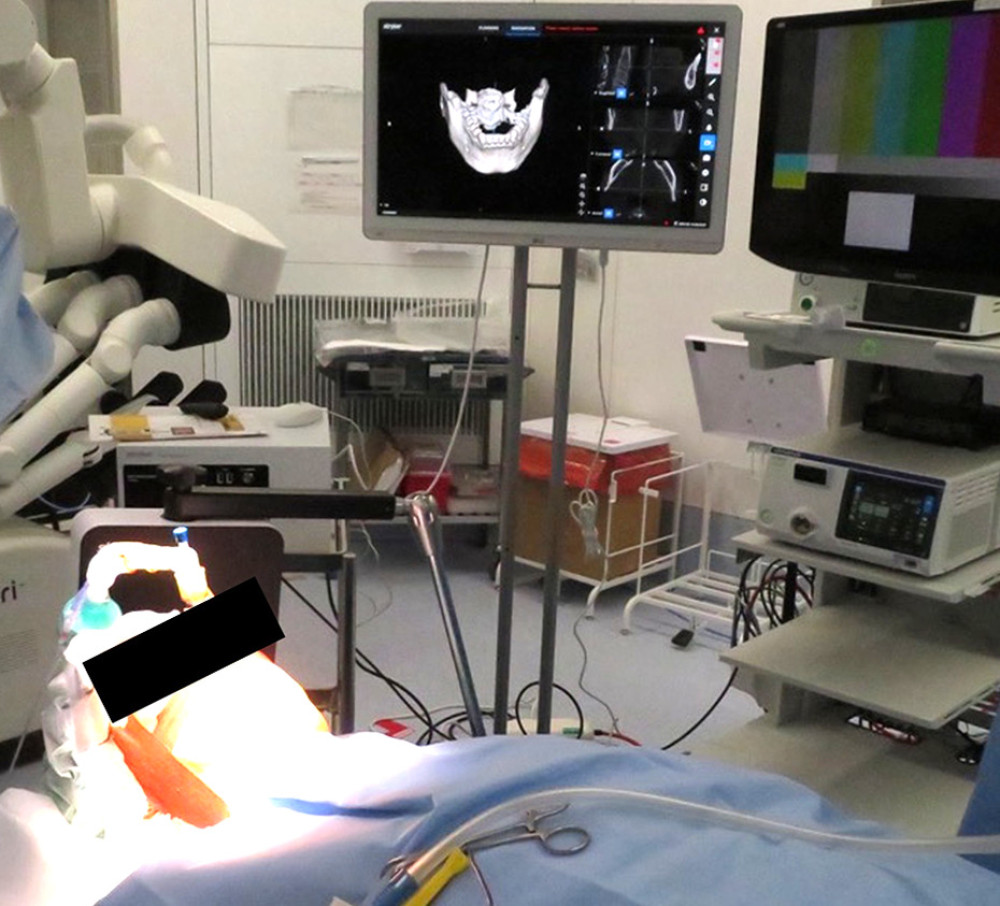 Figure 4. Field generator and navigation system in the operating room.
 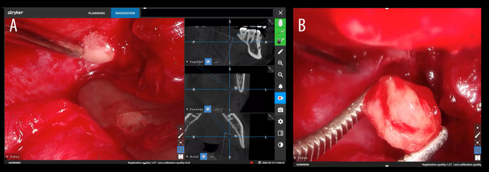 Figure 5. Intraoperative images under navigation. (A) Monitor image showing alignment between the endoscopic view and the fan-shaped field on the CBCT image. (B) Endoscopic view of the extraction of the tooth fragment.
 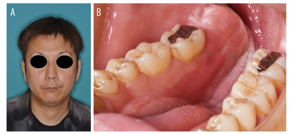 Figure 6. Photographs at 2 weeks after the surgery. (A) Extraoral findings were normal. (B) Intraoral findings were normal, with no wound abnormalities or lingual neurological symptoms.
 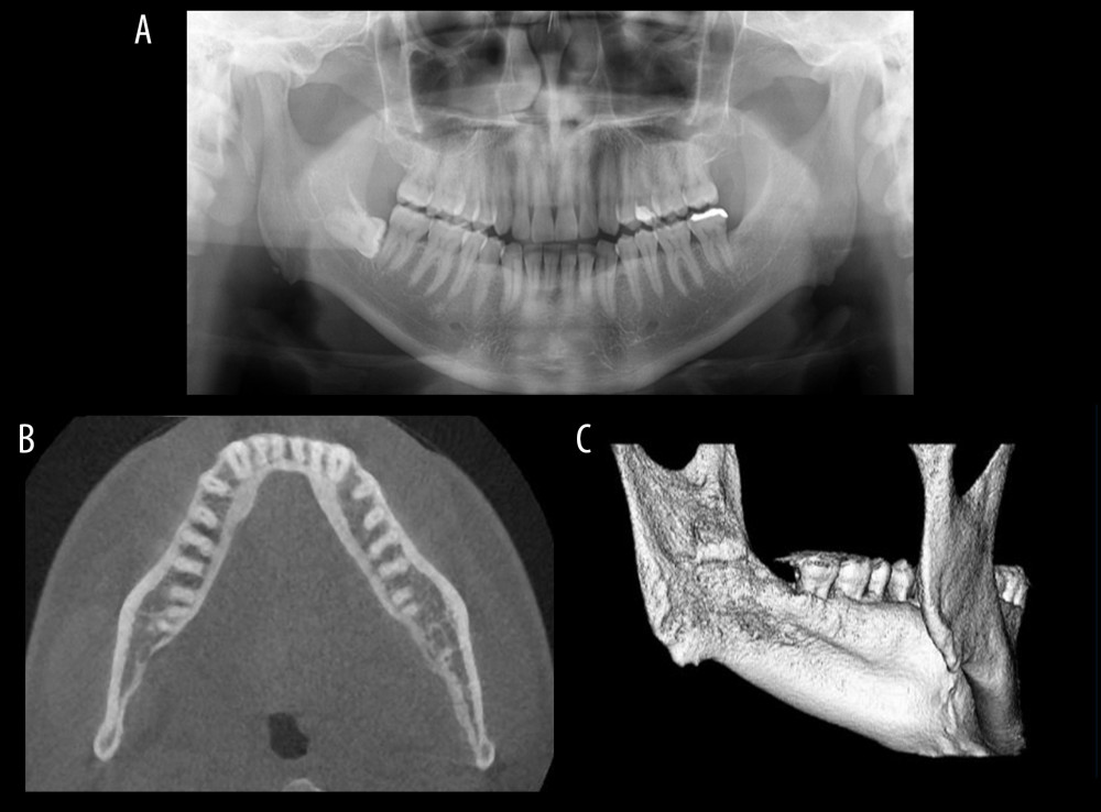 Figure 7. Postoperative imaging. (A) Panoramic radiograph showing complete removal of the tooth fragment. (B) Axial CBCT image. (C) Three-dimensional CBCT image.
 References
1. Sayed N, Bakathir A, Pasha M, Al-Sudairy S, Complications of third molar extraction A retrospective study from a tertiary healthcare centre in Oman: Sultan Qaboos Univ Med J, 2019; 19; 230-35
2. Zhao S, Huang Z, Geng T, Huang L, Case report intraoral management of iatrogenically displaced lower third molar roots in the sublingual space: A report of 2 cases: Int J Clin Exp Med, 2015; 8; 19591-95
3. Wang H, Yang CY, Li Z, Traumatic displacement of teeth into maxillary sinus and the retrieval assisted by computer-assisted navigation: A case report: Medicine (Baltimore), 2018; 97(51); e13865
4. Aznar-Arasa L, Figueiredo R, Gay-Escoda C, Iatrogenic displacement of lower third molar roots into the sublingual space: Report of 6 cases: J Oral Maxillofac Surg, 2012; 70; e107-15
5. Solanki R, Khangwal M, Kumar D, Goel M, Retrieval of mandibular third molar tooth accidentally displaced in submandibular space: Series of two cases: Indian J Dent, 2016; 7; 105
6. Goldberg MH, Nemarich AN, Marco WP, The impacted third molar: Referral patterns, patient compliance, and surgical requirements: J Am Dent Assoc, 1983; 107; 439-41
7. Huang IY, Wu CW, Worthington P, The displaced lower third molar: A literature review and suggestions for management: J Oral Maxillofac Surg, 2007; 65; 1186-90
8. Nusrath MA, Banks RJ, Unrecognised displacement of mandibular molar root into the submandibular space: Br Dent J, 2010; 209(6); 279-80
9. Cheung LK, Leung YY, Chow LK, Incidence of neurosensory deficits and recovery after lower third molar surgery: A prospective clinical study of 4338 cases: Int J Oral Maxillofac Surg, 2010; 39; 320-26
10. Masui M, Ibaragi S, Takeshita Y, Displacement of lower third molar into floor of mouth: A single institutional experience: Oral Maxillofac Surg, 2022; 26; 447-53
11. Yeh C-J, A simple retrieval technique for accidentally displaced mandibular third molars: J Oral Maxillofac Surg, 2002; 60; 836-37
12. Iwanaga J, Watanabe K, Kumar Anand M, Shane Tubbs AR, Air dissection of the spaces of the head and neck: A new teaching and dissection method: Clin Anat, 2020; 33(2); 207-13
13. Medeiros N, Gaffrée G, Accidental displacement of inferior third molar into the lateral pharyngeal space: Case report: J Oral Maxillofac Surg, 2008; 66; 578-80
14. Wang J, Cui NH, Guo YJ, Zhang W, Navigation-guided extraction of impacted supernumerary teeth: A case report: J Oral Maxillofac Surg, 2017; 75; 1136e1-e5
15. Block M, Emery R, Lank K, Ryan J, Implant placement accuracy using dynamic navigation: Int J Oral Maxillofac Implants, 2017; 32; 92-99
16. Campbell A, Costello BJ, Retrieval of a displaced third molar using navigation and active image guidance: J Oral Maxillofac Surg, 2010; 68; 480-85
17. Taylor H, Alsabbagh AY, Downie I, Endoscope-assisted surgical removal of an ectopic wisdom tooth below lower border of the mandible – A case report: Ann Maxillofac Surg, 2022; 12; 117-20
18. Şimşek MS, Üstündağ İ, Compound odontoma associated with calcifying odontogenic cyst in the maxillary sinus of a pediatric patient: A case report: J Maxillofac Oral Surg, 2026 s12663-026-03019-3
Figures
Figure 1. Patient photographs. (A) Extraoral photograph showing no abnormalities. (B) Intraoral photograph showing no inflammatory findings. A faint postoperative scar is observed in the floor of the mouth; the roots were not palpable.Figure 2. Panoramic radiograph and CBCT images. (A) Panoramic radiograph showing hard tissue on the left side of the mandible. (B) Axial CBCT imaging showing hard tissue on the lingual side of the left mandible. (C) Three-dimensional CBCT image showing hard tissue under the left mylohyoid line. Arrows indicate the displaced tooth fragment.Figure 3. Photograph of the device used for navigation and the screen where the reference point was set. (A) Photograph of a mucosal raspatory set up as a pointer. (B) Photograph of a tracker placed with resin in the intraoral surgical field. (C) Monitor screen when a reference point is set on the teeth.Figure 4. Field generator and navigation system in the operating room.Figure 5. Intraoperative images under navigation. (A) Monitor image showing alignment between the endoscopic view and the fan-shaped field on the CBCT image. (B) Endoscopic view of the extraction of the tooth fragment.Figure 6. Photographs at 2 weeks after the surgery. (A) Extraoral findings were normal. (B) Intraoral findings were normal, with no wound abnormalities or lingual neurological symptoms.Figure 7. Postoperative imaging. (A) Panoramic radiograph showing complete removal of the tooth fragment. (B) Axial CBCT image. (C) Three-dimensional CBCT image. In Press
Case report 
Am J Case Rep In Press; DOI: 10.12659/AJCR.953317
Case report 
Am J Case Rep In Press; DOI: 10.12659/AJCR.954643
Case report 
Am J Case Rep In Press; DOI: 10.12659/AJCR.954074
Case report 
Am J Case Rep In Press; DOI: 10.12659/AJCR.953592
Most Viewed Current Articles
07 Dec 2021 : Case report
28,712,429
DOI :10.12659/AJCR.934347
Am J Case Rep 2021; 22:e934347
06 Dec 2021 : Case report  180,530
DOI :10.12659/AJCR.934406
Am J Case Rep 2021; 22:e934406
21 Jun 2024 : Case report
124,276
DOI :10.12659/AJCR.944371
Am J Case Rep 2024; 25:e944371
08 May 2026 : Case report
95,045
DOI :10.12659/AJCR.952158
Am J Case Rep 2026; 27:e952158