


22 September 2026
: Case report 
Dual-Site Postsurgical Pyoderma Gangrenosum of the Breast and Back With Delayed Diagnosis: A Case Report
Unusual clinical course, Challenging differential diagnosis
Tristan Packard ABCDEF 1*, Grant Dolan ABCD 2, Cathryn Chance BEF 1, Mark Jones ABDE 3DOI: 10.12659/AJCR.954079
Am J Case Rep 2026; 27:e954079


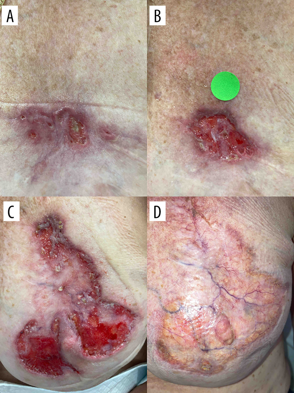
Figure 1 Clinical progression of the left breast lesion. (A) Initial presentation of the nonhealing left superior breast ulcer, measuring approximately 1.0 × 3.0 cm at the time of initial wound clinic evaluation. (B) Interval enlargement following serial debridement, with the lesion increasing to approximately 4.5 × 3.5 cm. (C) Peak lesion size prior to initiation of immunosuppressive therapy, measuring approximately 10.0 × 10.5 cm. (D) Complete resolution after initiation of immunosuppressive therapy. Images were obtained during routine clinical care; therefore, acquisition distance, angle, lighting, and scale were not fully standardized.