


09 September 2026
: Case report 
Cone-Beam Computed Tomography Imaging of Primary Non-Hodgkin Lymphoma of the Jaws: A 6-Case Series
Challenging differential diagnosis, Rare disease
Emmanouil Chatzipetros ABCDEFG 1*, Konstantina-Eleni Alexiou BDEG 1, Kostas Tsiklakis ABEG 1, Christos Angelopoulos AFG 1
DOI: 10.12659/AJCR.954314
Am J Case Rep 2026; 27:e954314


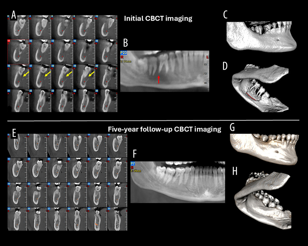
Figure 3 Case 3. (A) Cone-beam computed tomography (CBCT) cross-sectional images showing moth-eaten bone destruction (yellow arrows) and involvement of the mandibular canal (red dots). (B) CBCT panoramic reconstruction demonstrating the floating teeth appearance and spiked-tooth effect (red arrow). (C, D) CBCT three-dimensional (3D) reconstructions (buccal and lingual views) demonstrating lingual cortical perforation and destruction of the supporting alveolar bone of the molars. (E) Five-year follow-up CBCT cross-sectional images showing complete healing of the osteolytic lesion and restoration of the lingual cortical plate. (F) Five-year follow-up CBCT panoramic reconstruction showing complete osseous support of the affected teeth. (G, H) Five-year follow-up CBCT 3D reconstructions showing complete healing of the osteolytic lesion and restoration of the lingual plate.