


09 September 2026: Articles 
Cone-Beam Computed Tomography Imaging of Primary Non-Hodgkin Lymphoma of the Jaws: A 6-Case Series
Challenging differential diagnosis, Rare disease
Emmanouil Chatzipetros ABCDEFG 1*, Konstantina-Eleni Alexiou BDEG 1, Kostas Tsiklakis ABEG 1, Christos Angelopoulos AFG 1
DOI: 10.12659/AJCR.954314
Am J Case Rep 2026; 27:e954314


Abstract
BACKGROUND: Primary non-Hodgkin lymphoma (NHL) of the jaws is a rare malignancy often clinically and radiographically mimicking odontogenic or inflammatory conditions, leading to delayed diagnosis. Because imaging findings are frequently nonspecific, recognition of characteristic radiographic patterns may facilitate earlier diagnostic suspicion and appropriate biopsy-based investigation. This study evaluated cone-beam computed tomography (CBCT) imaging characteristics of primary NHL of the maxilla and mandible in patients with histopathologically confirmed disease and no initial suspicion of malignancy.
CASE REPORT: Six patients with histopathologically confirmed primary NHL of the jaws were retrospectively analyzed. Clinical findings and medical histories were reviewed. CBCT images were evaluated for lesion location, border definition, internal architecture, and effects on adjacent anatomical regions. Diagnosis was established by biopsy, with histopathological and/or immunohistochemical confirmation. Most patients had unremarkable medical history. Common findings included paresthesia, tooth mobility, and facial swelling. Mandible was more frequently affected than maxilla. All lesions appeared as osteolytic areas with ill-defined and/or moth-eaten borders, causing cortical destruction and infiltration into adjacent structures. Involvement of the mandibular canal was observed in mandibular cases, whereas maxillary cases demonstrated extension into the maxillary sinus or nasal cavity. Floating teeth and/or spiked-tooth appearance were identified in several cases, reflecting aggressive disease.
CONCLUSIONS: Primary NHL of the jaws demonstrates nonspecific but aggressive radiographic features on CBCT that may overlap with odontogenic and inflammatory conditions. However, a recurring pattern characterized by ill-defined osteolysis, cortical involvement, and floating-teeth or spiked-tooth appearance should raise suspicion for malignancy. Recognizing these imaging features may support timely biopsy-oriented evaluation and histopathological confirmation, particularly when clinical and radiographic findings are atypical for routine odontogenic disease.
Keywords: Cone-Beam Computed Tomography, Jaw, Lymphoma, Non-Hodgkin
Introduction
Hematolymphoid neoplasms are a heterogeneous group of malignancies arising from lymphoid cells at various stages of differentiation, encompassing a broad spectrum of subtypes. Malignant lymphomas are broadly classified into Hodgkin lymphoma and non-Hodgkin lymphoma (NHL). These neoplasms originate from B- or T-lymphocyte lineages and exhibit diverse biological and clinical behaviors depending on their stage of differentiation [1–3]. NHL encompasses a wide range of B- and T-cell subtypes, with B-cell lymphomas accounting for most cases (approximately 85% to 90%), while T-cell lymphomas represent the remaining 10% to 15% [1–6]. Among these, diffuse large B-cell lymphoma (DLBCL) is the most common and aggressive subtype, whereas follicular lymphoma typically follows a more indolent course. DLBCL is the most frequently encountered subtype in the maxillofacial region [5].
Lymphomas arising outside the lymph nodes are classified as extranodal lymphomas [4]. They may be primary, originating within extranodal tissues, or secondary, representing dissemination from an initial nodal or extranodal site [5]. In the head and neck region, lymphomas are among the most common malignancies following squamous cell carcinomas [2]. The most frequent primary extranodal sites in this region include the Waldeyer’s ring; however, other locations such as the buccal mucosa, salivary glands, floor of the mouth, paranasal sinuses, and facial bones have also been reported [4,5]. Primary osseous NHL is a rare malignancy that arises within the skeletal system in the absence of nodal or visceral involvement [2]. When it arises within the medullary cavity in the absence of systemic disease, it is referred to as primary intraosseous lymphoma, a distinction important for diagnostic and therapeutic considerations [3].
The radiological appearance of NHL of the jaws exhibits significant variability and is frequently mistaken for common inflammatory or infectious odontogenic conditions [6,7]. While computed tomography (CT) and magnetic resonance imaging (MRI) are often indispensable for assessing systemic disease burden, cone-beam computed tomography (CBCT) provides high-resolution visualization of early intraosseous changes within the jaws [4,5,8]. Early recognition of radiographic features, including osteolytic lesions, ill-defined or moth-eaten borders, cortical erosion, infiltration, and destruction, as well as the floating teeth appearance or the spiked tooth effect, is critical, as delayed diagnosis remains a common challenge with significant impact on patient prognosis [1,6–8].
The aim of this retrospective case series was to describe recurring CBCT features of histopathologically confirmed primary NHL of the jaws in patients in whom malignancy was not initially suspected.
Case Reports
STUDY DESIGN AND ETHICS APPROVAL:
Records from the database of a private oral and maxillofacial radiology practice (Athens, Greece) between 2006 and 2019 were retrospectively reviewed to identify histopathologically confirmed cases of NHL involving the jaws with available clinical and CBCT imaging data. Cases were considered primary jaw NHL when the initial clinical and radiographic presentation involved the maxilla or mandible and no prior diagnosis of lymphoma was documented in the available records. Based on referral documentation and clinical notes, none of the included patients had a documented clinical suspicion of lymphoma at the time of CBCT examination.
This study was approved by the Ethics, Regulation and Research Committee of the Athens Dental Association (Athens, Greece; protocol number 215/01-31-2024). The study was conducted in accordance with the principles of the Declaration of Helsinki. Informed consent was obtained from all patients.
IMAGING PROTOCOL:
CBCT examinations were performed using NewTom 3G and VGi units (QR, Cefla, Verona, Italy). The NewTom 3G operated at 110 kV, 1.5 to 6 mA, with field of views of 6, 9, and 12 inches and an exposure time of 7.2 seconds. The NewTom VGi utilized variable acquisition parameters of 110 kV, 2.5 to 4.1 mA, and exposure time of 3.6 seconds, with fields of view of 12 × 8 cm or 15 × 12 cm, depending on the clinical requirements and lesion extent. Image reconstruction was performed using a fixed isotropic voxel size of 0.30 mm. All scans were analyzed using NNT software (version 13.1).
CLINICAL AND IMAGING CHARACTERISTICS:
Demographic and clinical data, including age, sex, chief concern, and medical history, were recorded for all patients. In addition, referral information and provisional clinical diagnoses were reviewed to document the clinical context in which patients were referred for CBCT examination. The radiological parameters analyzed are shown in Table 1 and include lesion location (anterior or posterior maxilla or mandible); number of lesions (single or multiple); internal architecture (osteolytic, sclerotic, or mixed); lesion extent [limited (≤ 1 cm), moderate (> 1 cm and ≤ 2 cm), or extensive (> 2 cm)]; lesion borders (well-defined, ill-defined, or moth-eaten); effects on the bony cortex (erosion, infiltration, or destruction); effects on adjacent teeth (floating teeth appearance or spiked-tooth effect); periosteal reaction; involvement of the mandibular canal; involvement of the maxillary sinus and/or nasal cavity; and presence of pathological fracture.
HISTOPATHOLOGICAL FINDINGS:
Histopathological and immunohistochemical analysis following biopsy confirmed the diagnosis of NHL in all cases. Only histopathologically confirmed cases were included in the final study sample. Five patients were diagnosed with diffuse large B-cell lymphoma, not otherwise specified (DLBCL NOS), while 1 patient was diagnosed with plasmablastic lymphoma (PBL) and was subsequently found to be positive for HIV.
CASE 1:
A 45-year-old woman presented with paresthesia and mild discomfort in the left posterior mandible. Clinical examination showed no obvious soft tissue swelling, and her medical history was unremarkable. CBCT demonstrated a limited osteolytic lesion in the left posterior mandible with ill-defined/moth-eaten borders, cortical plate erosion and infiltration, disruption of the trabecular architecture and involvement of the mandibular canal (Figure 1A–1D). Three-dimensional (3D) reconstruction highlighted irregular cortical destruction (Figure 1D). Based on these imaging features, particularly the infiltrative borders and neurosensory deficit, malignancy was suspected. Histopathologic examination confirmed the diagnosis of primary NHL of the mandible. Following treatment, the patient was monitored with CBCT imaging.
After treatment, annual follow-up was performed using CBCT. At the latest follow-up, 5 years later, complete healing of the lesion was observed (Figure 1E–1H). The previously described ill-defined osteolytic lesion was no longer evident. The region showed substantial bone regeneration with re-established trabecular architecture. The cortical plates appeared fully reconstituted, with no cortical infiltration or ongoing destruction. The mandibular canal was clearly visible and free of infiltrative changes, correlating with complete resolution of paresthesia. Overall, the findings indicated complete radiographic remission and successful bone healing, with no evidence of recurrence.
This case highlights the remarkable regenerative capacity of mandibular bone following treatment of primary NHL, even in lesions initially presenting with aggressive, infiltrative imaging characteristics.
CASE 2:
A 66-year-old man presented with persistent paresthesia in the left posterior mandible. Clinical examination revealed no palpable masses or significant mucosal changes in the edentulous region. His medical history was unremarkable.
CBCT imaging revealed 2 distinct but closely situated osteolytic lesions within the left mandibular body (Figure 2). Both lesions exhibited an aggressive moth-eaten pattern, with ill-defined, infiltrative borders. Cross-sectional (Figure 2A) and coronal (Figure 2B) images demonstrated focal infiltration of the alveolar crest and the lingual cortical plate (yellow and red arrows, respectively), along with disruption of the trabecular architecture. Despite the infiltrative nature of the lesions, no significant bone expansion or periosteal reaction was observed.
The mandibular canal appeared to be involve the posterior lesion (red arrows) (Figure 2A, 2B). 3D reconstructions (Figure 2C, 2D) further demonstrated localized cortical erosions of the alveolar crest and the lingual cortical plate (yellow and red arrows, respectively).
The multifocal presentation further broadened the differential diagnosis to include metastatic disease, multiple myeloma, and other aggressive osteolytic conditions. Based on the imaging findings—particularly the ill-defined borders, multifocal presentation, and associated neurosensory deficit—a malignant process was strongly suspected; histopathological analysis confirmed primary NHL of the mandible.
This case highlights the importance of recognizing multifocal, ill-defined osteolytic lesions with neurosensory involvement as a potential presentation of primary NHL of the mandible.
CASE 3:
A 40-year-old man presented with a chief concern of unexplained tooth mobility in the right posterior mandible, accompanied by marked swelling of the buccal vestibule and paresthesia. His medical history was unremarkable.
CBCT imaging demonstrated an extensive, solitary, osteolytic lesion in the right mandible (Figure 3). The lesion exhibited an aggressive moth-eaten pattern with complete destruction of the alveolar process and a significant breach of the lingual cortical plate (Figure 3A–3D). This extensive cortical destruction correlated with the clinical finding of soft tissue swelling.
Key radiographic features included a pronounced floating teeth appearance of the molars and a distinct spiked-tooth effect (Figure 3B, 3D). The lesion also encompassed the mandibular canal (Figure 3A, red dots indicate the mandibular canal). These imaging features further supported an aggressive, infiltrative process.
Given the extensive bone destruction and floating teeth appearance, the differential diagnosis included metastatic disease, osteomyelitis, Langerhans cell histiocytosis, and primary malignant neoplasms. Histopathological examination confirmed the diagnosis of primary intraosseous NHL of the mandible.
After treatment, annual follow-up was performed using CBCT. At the latest follow-up, 5 years later, complete healing of the lesion was observed (Figure 3E–3H). The previously described extensive osteolytic lesion was no longer evident. The affected region demonstrated marked bone regeneration, with re-establishment of the trabecular architecture and stable integration of the involved teeth. The mandibular canal was clearly delineated. Overall, the imaging findings were consistent with complete radiographic remission and successful osseous healing, with no evidence of disease recurrence.
This case highlights the aggressive imaging presentation of primary NHL of the mandible, including floating teeth and cortical perforation, as well as the potential for remarkable bone regeneration following appropriate treatment.
CASE 4:
A 79-year-old woman was referred for preoperative evaluation prior to dental implant placement, presenting with isolated and unexplained tooth mobility in the right posterior maxilla. Clinical examination revealed no evident soft tissue swelling or palpable mass, but erythema and extensive gingival recession in the corresponding region were noted. Her medical history was unremarkable.
CBCT imaging demonstrated an extensive, aggressive osteolytic lesion involving a large portion of the right maxilla (Figure 4). Cross-sectional images (Figure 4A, yellow arrow) showed marked destruction of the cortical plate and alveolar process. The lesion exhibited an infiltrative growth pattern, extending superiorly into the right maxillary sinus and medially into the nasal cavity, as demonstrated on axial (Figure 4C, orange arrow) and coronal views (Figure 4D, blue and orange arrows).
The involved teeth demonstrated a characteristic floating teeth appearance and spiked-tooth effect (Figure 4B, red arrow). The aggressive moth-eaten pattern of bone destruction, together with cortical destruction and extension into the maxillary sinus and nasal cavity, strongly suggested an infiltrative malignant process. Although no periosteal reaction was observed, this finding was considered within the overall imaging presentation and was not interpreted as an independent indicator of malignancy, as periosteal reaction is uncommon in primary NHL of the jaws.
The radiographic differential diagnosis included metastatic disease, lymphoma, aggressive osteomyelitis, and other primary malignant neoplasms. Histopathological examination confirmed the diagnosis of primary NHL.
This case underscores the clinical challenge of primary NHL of the maxilla, which may masquerade as severe periodontal disease, and highlights the crucial role of CBCT in identifying the aggressive, infiltrative nature of such lesions.
CASE 5:
A 44-year-old woman presented with paresthesia and mild pain in the left posterior mandible, associated with a previously restored mandibular second molar. Clinical examination revealed erythema and a small ulcerative lesion resembling a sinus tract, although larger than typically observed in odontogenic infections. No significant soft tissue swelling was noted. Her medical history was unremarkable.
CBCT imaging demonstrated a moderate osteolytic lesion in the left posterior mandible (Figure 5). The lesion exhibited an ill-defined, infiltrative pattern of bone destruction, without evidence of significant cortical expansion or periosteal reaction. A spiked-tooth effect, involving the distal root of the second molar (Figure 5D, red arrow), was observed.
Cross-sectional images revealed disruption of the trabecular architecture and focal cortical thinning (Figure 5A, yellow arrows), with involvement of the mandibular canal. 3D reconstructions further highlighted the irregular cortical margins (Figure 5B, yellow arrow) and the infiltrative nature of the process.
The radiographic differential diagnosis included metastatic disease, lymphoma, osteomyelitis, and other primary malignant neoplasms. Histopathological examination confirmed the diagnosis of primary NHL of the mandible.
This case highlights the potential for primary NHL to mimic periapical pathology both clinically and radiographically, particularly in association with restored teeth, while the presence of paresthesia should raise suspicion for neural involvement and an underlying malignant process.
CASE 6:
A 35-year-old man presented with marked, rapidly progressive swelling of the right mandible, paresthesia, and significant tooth mobility and loss. His medical history was initially reported as unremarkable; however, subsequent evaluation revealed that the patient was positive for HIV.
Initial panoramic radiograph (Figure 6) demonstrated extensive osteolytic destruction of the right mandibular body, characterized by loss of the normal trabecular architecture, ill-defined margins, and an associated pathological fracture (red arrow).
CBCT imaging was subsequently performed for further evaluation and demonstrated an extensive, aggressive moth-eaten osteolytic lesion involving the right mandibular body and ramus (Figure 7A–7C). The extent of osseous destruction resulted in a pathological fracture of the inferior border (Figure 7E). The remaining teeth showed the characteristic floating teeth appearance (Figure 7D).
Histopathological and immunohistochemical analyses confirmed the diagnosis of PBL. As PBL is highly associated with HIV infection, further laboratory tests were performed and confirmed the suspected diagnosis. The patient was promptly started on systemic chemotherapy.
Follow-up panoramic imaging at 25 months revealed complete radiographic resolution of the lesion, with osseous regeneration and healing of the mandibular fracture (Figure 8).
This case highlights the highly aggressive radiographic presentation of PBL in immunocompromised patients, as well as the potential for remarkable bone regeneration following appropriate treatment, underscoring the importance of early recognition and prompt multidisciplinary management.
Discussion
EXTRA-NODAL NHL:
NHL typically originates in lymph nodes; however, a substantial proportion, approximately 24%, arises in extranodal sites [1,8]. Among these, the gastrointestinal tract, skin, bone, and Waldeyer’s ring are most frequently involved [9]. Primary extranodal NHL of the head and neck is relatively uncommon, with oral cavity involvement being particularly rare, accounting for only 0.1% to 5% of cases [10]. Primary bone NHL represents approximately 5% of all extranodal lymphomas [4]. Gomes et al reported that 59.5% of DLBCL NOS cases were located in the mandible and 40.5% in the maxilla [8]. In the present study, 5 cases involved the mandible and 1 involved the maxilla, consistent with previous reports [7]. Primary DLBCL NOS of the jaws in children and infants is extremely rare (4.8%), and most cases occurring in adults (95.2%), with a mean age of approximately 56 years [8]. A male predominance has been consistently reported in patients with NHL, with male-to-female ratios ranging from 1.5: 1 [11] to 5: 1 [12]. In the present study, 6 cases of NHL were identified (3 men and 3 women), including 5 cases of DLBCL NOS and 1 case of PBL. The patients’ ages ranged from 35 to 79 years, with a mean age of 51.5 years.
CLINICAL FINDINGS OF NHL OF THE JAWS:
Chief concerns associated with primary osseous NHL have been extensively described in the literature [13]. In a systematic review of NHL of the jaws, local swelling was reported as the most common clinical manifestation (90.7%), followed by pain (53.5%) and erythema (43.4%), whereas paresthesia was observed in 25.6% of cases [8]. Tooth mobility was identified as the most frequent dental manifestation (37.2%), followed by dental pain and/or sensitivity to percussion (31.7%) [8]. In the present study, paresthesia was the most common presenting symptom, followed by tooth mobility and facial swelling, findings that are consistent with previous reports [8,13]. These nonspecific clinical manifestations can contribute to delayed diagnosis and potential misinterpretation as odontogenic or inflammatory conditions.
CBCT FEATURES OF NHL OF THE JAWS:
NHL of the jaws most commonly presents as osteolytic lesions with ill-defined borders (85.7%) on CBCT imaging [7,8]. The most frequently reported effects on adjacent teeth include loss of the lamina dura (19%), floating teeth appearance (16.7%), and widening of the periodontal ligament space (14.3%). Other findings include bone loss (11.9%), periapical radiolucencies (7.1%), tooth displacement (7.1%), and root resorption (2.4%) [8]. Cortical bone destruction has been reported in up to 87.1% of cases, whereas periosteal reaction is rare (2.4%) [8]. Notably, the majority of jaw NHL cases (52.4%) are initially misdiagnosed as inflammatory or infectious lesions, while only 9.5% are correctly suspected to be malignant [8]. In the present study, all cases (n = 6) presented as osteolytic lesions with ill-defined and/or moth-eaten borders. Cortical bone changes ranged from erosion to infiltration and destruction. The floating teeth and/or spiked-tooth appearance were identified in 4 cases, while no periosteal reaction was observed. Overall, these radiological findings are consistent with those of previous studies [1,6–8]. In addition, involvement of the mandibular canal (5 cases) and the maxillary sinus and/or nasal cavity (1 case) was observed, while a pathological mandibular fracture was identified in 1 case. These imaging characteristics can contribute to diagnostic challenges because they overlap substantially with those of inflammatory, infectious, and odontogenic lesions. Although the findings observed in the present series are largely consistent with previously published reports, their recurrence across all 6 histopathologically confirmed cases reinforces recognizable CBCT patterns that may raise clinical suspicion of NHL and prompt timely biopsy and further investigation.
DIFFERENTIAL DIAGNOSIS OF NHL OF THE JAWS:
The differential diagnosis of NHL of the jaws is broad, as its clinical and radiographic features often resemble those of more common odontogenic and non-odontogenic conditions [6,7]. Clinically, patients may present with swelling, pain, tooth mobility, or paresthesia, findings that can be easily mistaken for odontogenic infections or inflammatory lesions such as periapical abscesses or osteomyelitis [8,13]. Radiographically, ill-defined radiolucent areas on CBCT may resemble primary bone malignancies, including osteosarcoma, as well as metastatic lesions to the jaws and squamous cell carcinoma [7,8,13]. Benign conditions, such as periodontal disease, odontogenic cysts, and odontogenic tumors, may also be considered in the early stages [1,6–8,13]. Given this wide range of possible diagnoses, a high index of suspicion is required, particularly in cases with atypical presentation or lack of response to conventional dental treatment [1,6–8]. Definitive diagnosis relies on histopathological evaluation and immunohistochemical analysis, which are essential for distinguishing NHL from other entities and guiding appropriate management [14].
ASSOCIATION OF HIV WITH NHL:
HIV infection significantly increases the risk of developing various types of lymphoma, particularly NHL, especially in advanced stages such as AIDS [15]. In this context, lymphomas tend to present as aggressive and disseminated neoplasms. DLBCL NOS represents one of the most common subtypes of HIV-associated NHL [15]. PBL is a rare and highly aggressive subtype of NHL, most commonly occurring in immunocompromised patients, particularly those with HIV infection [8,15]. In the present study, 1 patient was found to be HIV-positive, with the diagnosis established following the identification of PBL. This finding underscores the importance of considering underlying immunodeficiency in patients presenting with atypical or aggressive jaw lesions. Radiographic findings in this patient revealed an extensive, infiltrative osteolytic lesion (Figure 7). Cortical disruption and medullary involvement were also observed, along with an associated pathological fracture, reflecting the highly aggressive biological behavior of the disease. Preservation of the tooth roots, resulting in a floating teeth appearance, was also observed, representing a characteristic but nonspecific imaging feature of rapidly progressive destructive lesions of the jaws. Notably, follow-up imaging (Figure 8) demonstrated complete resolution of the lesion following systemic chemotherapy, with full osseous regeneration and restoration of mandibular integrity. Such a degree of bone healing is rarely reported in cases of mandibular PBL and highlights the potential for significant skeletal recovery following early diagnosis and appropriate oncologic management. These findings underscore the importance of including PBL in the differential diagnosis of aggressive osteolytic lesions in immunocompromised patients and illustrate the value of imaging for lesion characterization, treatment monitoring, and assessment of osseous recovery.
MANAGEMENT OF NHL:
Treatment options for NHL include chemotherapy, radiotherapy, or a combination of both modalities. The reported 5-year survival rate for patients with DLBCL NOS is approximately 83% [14]. In the present study, most patients were treated with chemotherapy. Of the 6 patients, 3 had a follow-up period of 12 to 60 months, during which substantial osseous healing was observed, and continue to undergo long-term surveillance with panoramic radiography or CBCT imaging, while no follow-up data were available for the remaining 3 patients. Although follow-up data were available for only 3 patients, the observed osseous healing following treatment illustrates the potential for substantial skeletal recovery and supports the role of imaging in long-term follow-up and disease monitoring.
The findings of the present case series should be interpreted within the context of its retrospective design and limited sample size. Nevertheless, the study provides additional evidence supporting previously reported CBCT manifestations of primary NHL of the jaws in histopathologically confirmed cases. Across the 6 cases, a recurring imaging pattern was observed, characterized by nonspecific ill-defined and/or moth-eaten osteolytic lesions, varying degrees of cortical erosion, infiltration or destruction, frequent mandibular canal involvement, and floating-teeth or spiked-tooth appearances, often accompanied by nonspecific clinical symptoms such as paresthesia, swelling, or tooth mobility. Although none of these findings is pathognomonic for NHL, their recurrent association in this series reinforces a recognizable constellation of aggressive jaw findings that may raise suspicion for malignancy when conventional odontogenic or inflammatory explanations are not fully convincing. The principal contribution of this series is not the identification of novel radiographic phenomena, but the consolidation of these recurring imaging patterns in a rare disease entity and the demonstration of their frequent resemblance to odontogenic and inflammatory conditions encountered in routine dental practice. Recognition of these patterns may assist clinicians and oral radiologists in considering NHL among the differential diagnoses of atypical osteolytic jaw lesions and may support timely biopsy-oriented evaluation.
Conclusions
In this 6-case series, primary NHL of the jaws demonstrated nonspecific but aggressive CBCT features, most commonly characterized by ill-defined and/or moth-eaten osteolytic lesions with cortical involvement and, in some cases, floating teeth or spiked-tooth appearance. Although these findings are not pathognomonic, recognition of this recurring imaging pattern may raise suspicion for malignancy and support timely biopsy and histopathological diagnosis in patients with atypical jaw lesions.
Figures
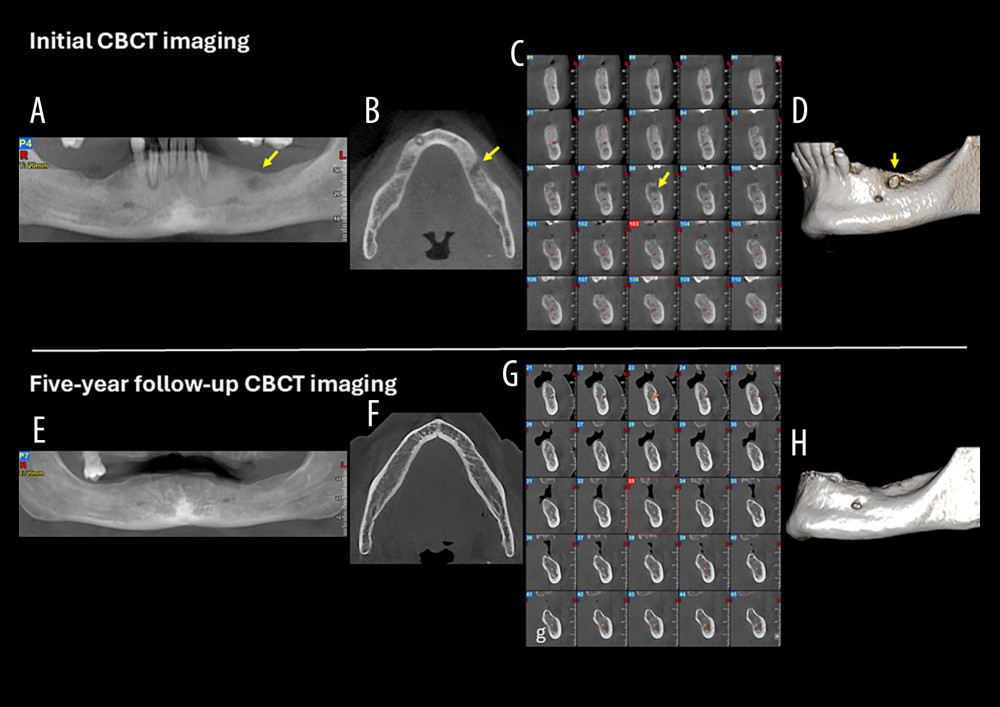 Figure 1. Case 1. (A) Cone-beam computed tomography (CBCT) panoramic reconstruction showing an ill-defined osteolytic lesion in the left edentulous mandibular body (yellow arrow). (B) CBCT axial slice demonstrating the infiltrative nature of the lesion and erosion of the buccal cortical plate (yellow arrow). (C) CBCT cross-sectional images illustrating the moth-eaten pattern of bone destruction (yellow arrow), with cortical erosion and infiltration and involvement of the mandibular canal (red dots). (D) CBCT three-dimensional (3D) reconstruction showing erosion and infiltration of the buccal cortical plate (yellow arrow). (E) Five-year follow-up CBCT panoramic reconstruction showing complete resolution of the osteolytic lesion and restoration of mandibular bone density. (F) Five-year follow-up CBCT axial slice demonstrating reconstitution of the buccal cortical plate. (G) Five-year follow-up CBCT cross-sectional images revealing bone remodeling and a clearly delineated, uninvolved mandibular canal. (H) Five-year follow-up CBCT 3D reconstruction illustrating healing of the previous cortical defect, with a smooth and continuous mandibular surface.
 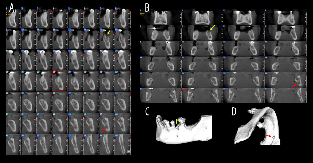 Figure 2. Case 2. (A) Cone-beam computed tomography (CBCT) cross-sectional images demonstrating 2 distinct, ill-defined osteolytic lesions in the left posterior mandible. Lesion 1 (yellow arrows) shows alveolar crest infiltration and lesion 2 (red arrows) shows lingual cortical plate infiltration. (B) CBCT coronal slices highlighting the infiltrative nature of the lesions (yellow and red arrows). (C) CBCT three-dimensional (3D) reconstruction of lesion 1 showing alveolar crest infiltration (yellow arrow). (D) 3D reconstruction of lesion 2 showing lingual infiltration (red arrow).
 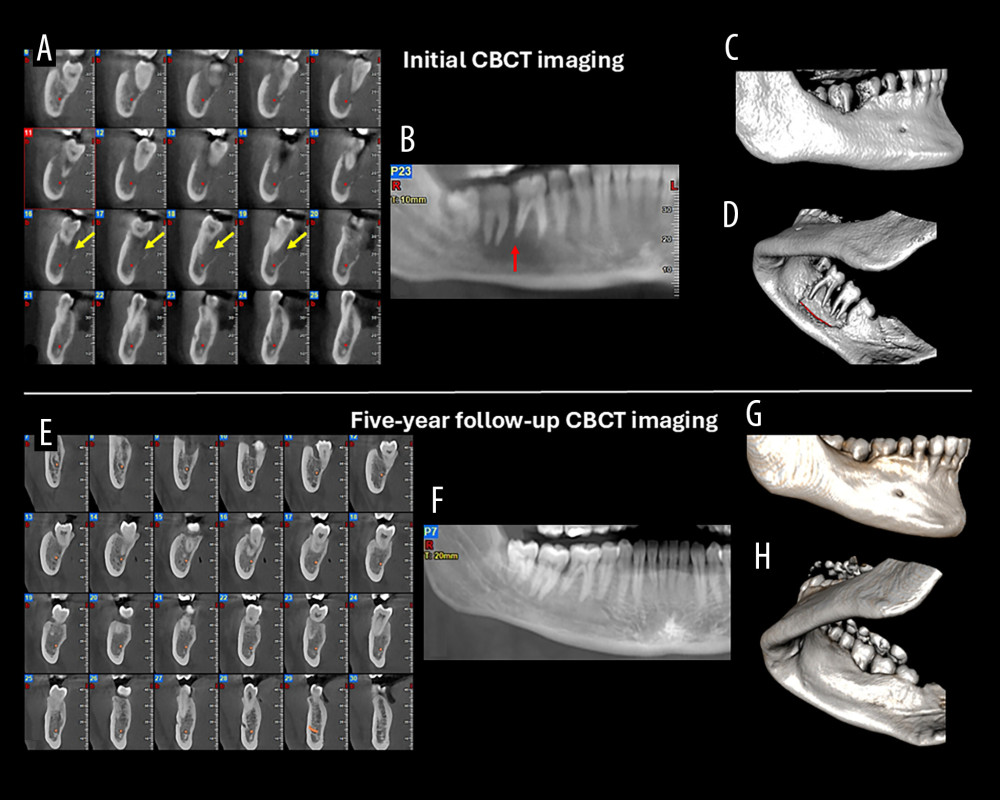 Figure 3. Case 3. (A) Cone-beam computed tomography (CBCT) cross-sectional images showing moth-eaten bone destruction (yellow arrows) and involvement of the mandibular canal (red dots). (B) CBCT panoramic reconstruction demonstrating the floating teeth appearance and spiked-tooth effect (red arrow). (C, D) CBCT three-dimensional (3D) reconstructions (buccal and lingual views) demonstrating lingual cortical perforation and destruction of the supporting alveolar bone of the molars. (E) Five-year follow-up CBCT cross-sectional images showing complete healing of the osteolytic lesion and restoration of the lingual cortical plate. (F) Five-year follow-up CBCT panoramic reconstruction showing complete osseous support of the affected teeth. (G, H) Five-year follow-up CBCT 3D reconstructions showing complete healing of the osteolytic lesion and restoration of the lingual plate.
 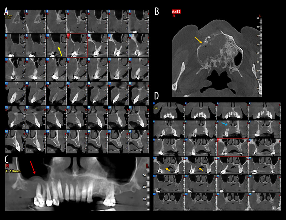 Figure 4. Case 4. (A) Cone-beam computed tomography (CBCT) cross-sectional images illustrating extensive alveolar bone destruction and cortical plate erosion (yellow arrow). (B) CBCT panoramic reconstruction showing the floating teeth appearance and spiked-tooth effect in the right posterior maxilla (red arrow). (C) CBCT axial slice demonstrating the infiltrative lesion with osseous destruction extending into the maxillary sinus and nasal cavity (orange arrow). (D) CBCT coronal slices highlighting maxillary sinus (orange arrows) and nasal cavity involvement (blue arrow).
 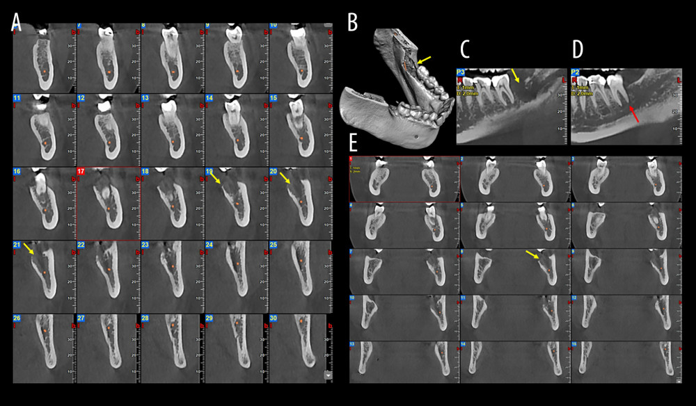 Figure 5. Case 5. (A) Cone-beam computed tomography (CBCT) cross-sectional images illustrating the ill-defined osteolytic lesion, trabecular disruption, and cortical thinning (yellow arrows). (B) CBCT three-dimensional reconstruction demonstrating localized cortical erosion (yellow arrow). (C, D) CBCT panoramic reconstructions highlighting infiltrative bone loss (yellow arrow) and the characteristic spiked-tooth effect at the distal root of the second molar (red arrow). (E) CBCT coronal slices showing the osteolytic lesion (yellow arrow).
 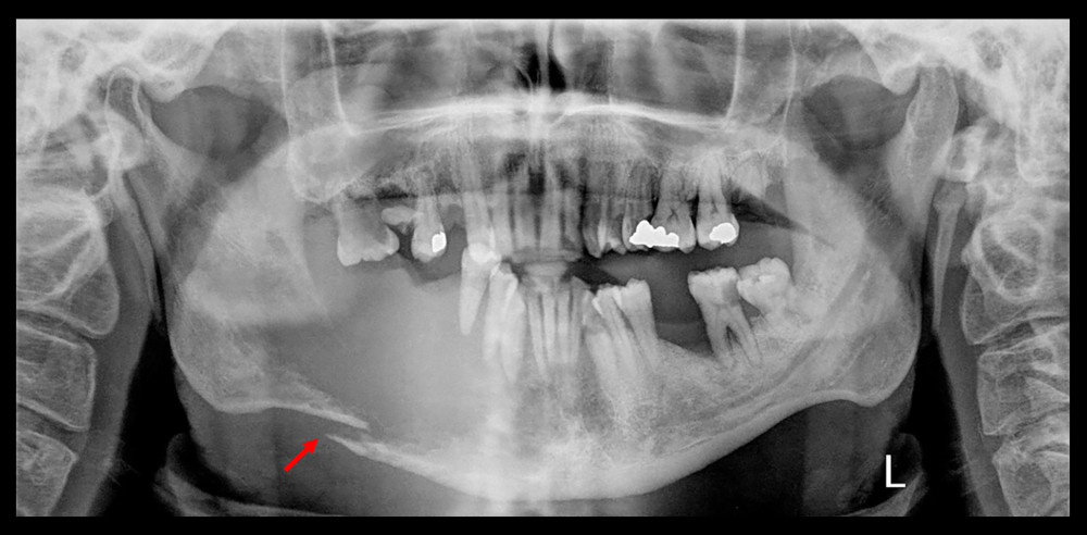 Figure 6. Initial panoramic radiograph of Case 6 demonstrating an extensive osteolytic destruction of the right mandibular body, characterized by loss of normal trabecular architecture, ill-defined margins, and pathological fracture of the lower border of the mandible (red arrow).
 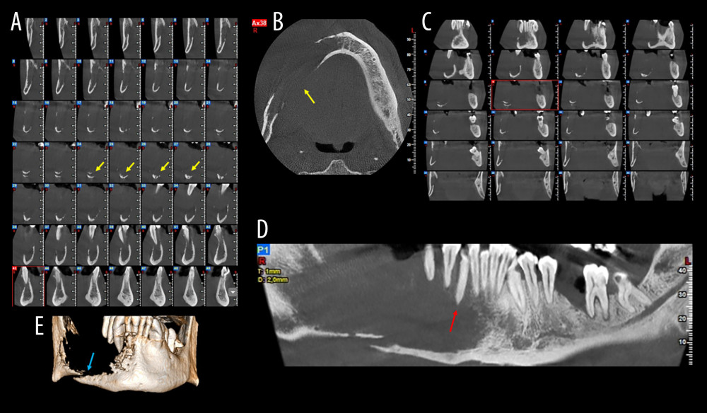 Figure 7. Case 6. (A) Cone-beam computed tomography (CBCT) cross-sectional slices demonstrating complete destruction of the right mandibular body (yellow arrows). (B) CBCT axial slice showing extensive lesion in the right mandibular body (yellow arrow). (C) CBCT coronal slices and (D) panoramic reconstruction highlighting the characteristic floating teeth appearance and spiked-tooth effect (red arrow). (E) CBCT three-dimensional reconstruction revealing a pathological fracture of the inferior border (blue arrow).
 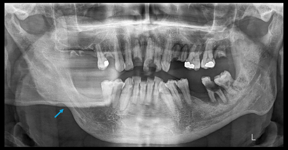 Figure 8. Twenty-five-month follow-up panoramic radiograph of case 6 showing complete healing of the lesion, with osseous regeneration and consolidation of the previous fracture site, resulting in restoration of mandibular continuity (blue arrow).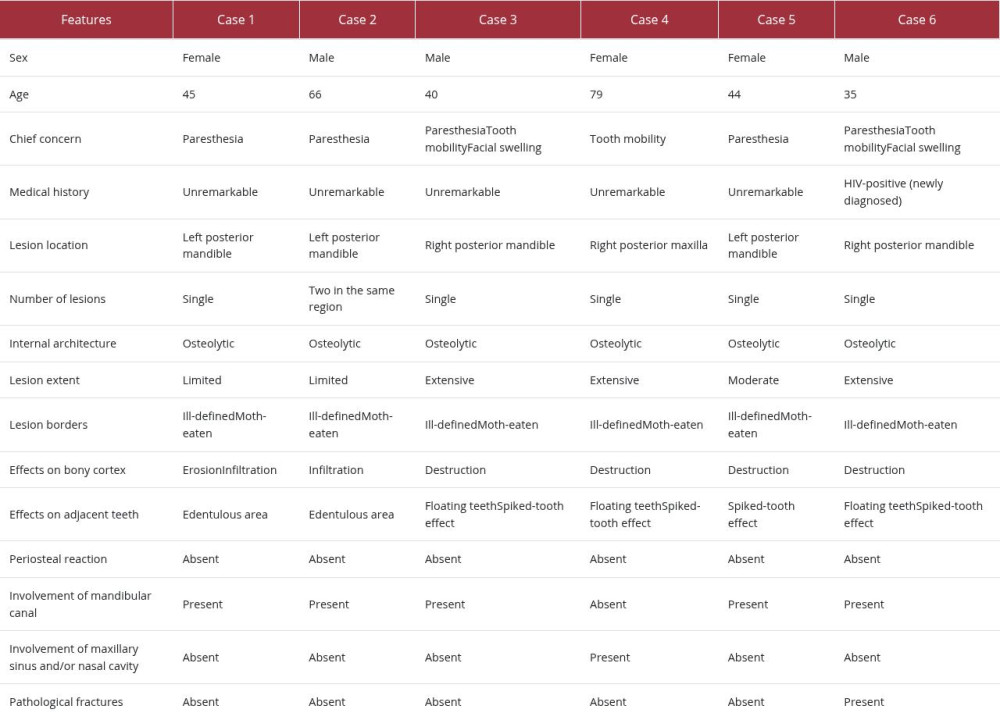
References
1. Jain T, Kumar S, Rajoria S, Silent intruders: Hematolymphoid neoplasms revealed in the jaws: A series of three cases: Indian J Pathol Microbiol, 2025; 68; 815-19
2. Sung H, Ferlay J, Siegel RL, Global cancer statistics 2020: GLOBOCAN estimates of incidence and mortality worldwide for 36 cancers in 185 countries: Cancer J Clin, 2021; 71; 209-49
3. Kemp S, Gallagher G, Kabani S, Oral non-Hodgkin’s lymphoma: Review of the literature and World Health Organization classification with reference to 40 cases: Oral Surg Oral Med Oral Pathol Oral Radiol Endod, 2008; 105; 194-201
4. MacDonald D, Lim S, Extranodal lymphoma arising within the maxillary alveolus: A systematic review: Oral Radiol, 2018; 34; 185
5. MacDonald D, Martin M, Savage K, Maxillofacial lymphomas: Br J Radiol, 2021; 94; 20191041
6. Adalja D, Tagliaferri A, Rezkalla A, Taha B, Diffuse B-cell lymphoma of the mandible disguised as acute osteomyelitis: Eur J Case Rep Intern Med, 2024; 11; 004243
7. Djavanmardi L, Oprean N, Alantar A, Malignant non-Hodgkin’s lymphoma (NHL) of the jaws: A review of 16 cases: J Craniomaxillofac Surg, 2008; 36; 410-14
8. Gomes NR, Lima LA, Morais-Perdigão AL, Radiological aspects of lymphomas and leukaemias affecting the jaws: A systematic review: J Oral Pathol Med, 2023; 52; 315-23
9. Matthews JB, Basu MK, Primary extranodal lymphoma of the oral cavity: An immunohistochemical study: Br J Oral Surg, 1983; 21; 159-70
10. Fukuda Y, Ishida T, Fujimoto M, Malignant lymphoma of the oral cavity: Clinicopathologic analysis of 20 cases: J Oral Pathol, 1987; 16; 8-12
11. Clayton F, Butler JJ, Ayala AG, Non-Hodgkin’s lymphoma in bone. Pathologic and radiologic features with clinical correlates: Cancer, 1987; 60; 2494-501
12. Potdar GG, Primary reticulum-cell sarcoma of bone in Western India: Br J Cancer, 1970; 24; 48-55
13. Nosrat A, Verma P, Glass S, Non-Hodgkin lymphoma mimicking endodontic lesion: A case report with 3-dimensional analysis, segmentation, and printing: J Endod, 2021; 47; 671-76
14. Rodrigues-Fernandes CI, de Souza LL, Santos-Costa SFD, Clinicopathological analysis of oral diffuse large B-cell lymphoma, NOS: A systematic review: J Oral Pathol Med, 2019; 48; 185-91
15. Pather S, Mohamed Z, McLeod H, Pillay K, Large cell lymphoma: Correlation of HIV status and prognosis with differentiation profiles assessed by immunophenotyping: Pathol Oncol Res, 2013; 19; 695-705
Figures
Figure 1. Case 1. (A) Cone-beam computed tomography (CBCT) panoramic reconstruction showing an ill-defined osteolytic lesion in the left edentulous mandibular body (yellow arrow). (B) CBCT axial slice demonstrating the infiltrative nature of the lesion and erosion of the buccal cortical plate (yellow arrow). (C) CBCT cross-sectional images illustrating the moth-eaten pattern of bone destruction (yellow arrow), with cortical erosion and infiltration and involvement of the mandibular canal (red dots). (D) CBCT three-dimensional (3D) reconstruction showing erosion and infiltration of the buccal cortical plate (yellow arrow). (E) Five-year follow-up CBCT panoramic reconstruction showing complete resolution of the osteolytic lesion and restoration of mandibular bone density. (F) Five-year follow-up CBCT axial slice demonstrating reconstitution of the buccal cortical plate. (G) Five-year follow-up CBCT cross-sectional images revealing bone remodeling and a clearly delineated, uninvolved mandibular canal. (H) Five-year follow-up CBCT 3D reconstruction illustrating healing of the previous cortical defect, with a smooth and continuous mandibular surface.Figure 2. Case 2. (A) Cone-beam computed tomography (CBCT) cross-sectional images demonstrating 2 distinct, ill-defined osteolytic lesions in the left posterior mandible. Lesion 1 (yellow arrows) shows alveolar crest infiltration and lesion 2 (red arrows) shows lingual cortical plate infiltration. (B) CBCT coronal slices highlighting the infiltrative nature of the lesions (yellow and red arrows). (C) CBCT three-dimensional (3D) reconstruction of lesion 1 showing alveolar crest infiltration (yellow arrow). (D) 3D reconstruction of lesion 2 showing lingual infiltration (red arrow).Figure 3. Case 3. (A) Cone-beam computed tomography (CBCT) cross-sectional images showing moth-eaten bone destruction (yellow arrows) and involvement of the mandibular canal (red dots). (B) CBCT panoramic reconstruction demonstrating the floating teeth appearance and spiked-tooth effect (red arrow). (C, D) CBCT three-dimensional (3D) reconstructions (buccal and lingual views) demonstrating lingual cortical perforation and destruction of the supporting alveolar bone of the molars. (E) Five-year follow-up CBCT cross-sectional images showing complete healing of the osteolytic lesion and restoration of the lingual cortical plate. (F) Five-year follow-up CBCT panoramic reconstruction showing complete osseous support of the affected teeth. (G, H) Five-year follow-up CBCT 3D reconstructions showing complete healing of the osteolytic lesion and restoration of the lingual plate.Figure 4. Case 4. (A) Cone-beam computed tomography (CBCT) cross-sectional images illustrating extensive alveolar bone destruction and cortical plate erosion (yellow arrow). (B) CBCT panoramic reconstruction showing the floating teeth appearance and spiked-tooth effect in the right posterior maxilla (red arrow). (C) CBCT axial slice demonstrating the infiltrative lesion with osseous destruction extending into the maxillary sinus and nasal cavity (orange arrow). (D) CBCT coronal slices highlighting maxillary sinus (orange arrows) and nasal cavity involvement (blue arrow).Figure 5. Case 5. (A) Cone-beam computed tomography (CBCT) cross-sectional images illustrating the ill-defined osteolytic lesion, trabecular disruption, and cortical thinning (yellow arrows). (B) CBCT three-dimensional reconstruction demonstrating localized cortical erosion (yellow arrow). (C, D) CBCT panoramic reconstructions highlighting infiltrative bone loss (yellow arrow) and the characteristic spiked-tooth effect at the distal root of the second molar (red arrow). (E) CBCT coronal slices showing the osteolytic lesion (yellow arrow).Figure 6. Initial panoramic radiograph of Case 6 demonstrating an extensive osteolytic destruction of the right mandibular body, characterized by loss of normal trabecular architecture, ill-defined margins, and pathological fracture of the lower border of the mandible (red arrow).Figure 7. Case 6. (A) Cone-beam computed tomography (CBCT) cross-sectional slices demonstrating complete destruction of the right mandibular body (yellow arrows). (B) CBCT axial slice showing extensive lesion in the right mandibular body (yellow arrow). (C) CBCT coronal slices and (D) panoramic reconstruction highlighting the characteristic floating teeth appearance and spiked-tooth effect (red arrow). (E) CBCT three-dimensional reconstruction revealing a pathological fracture of the inferior border (blue arrow).Figure 8. Twenty-five-month follow-up panoramic radiograph of case 6 showing complete healing of the lesion, with osseous regeneration and consolidation of the previous fracture site, resulting in restoration of mandibular continuity (blue arrow). In Press
Case report 
Am J Case Rep In Press; DOI: 10.12659/AJCR.953543
Case report 
Am J Case Rep In Press; DOI: 10.12659/AJCR.954216
Case report 
Am J Case Rep In Press; DOI: 10.12659/AJCR.954932
Case report 
Am J Case Rep In Press; DOI: 10.12659/AJCR.954146
Most Viewed Current Articles
07 Dec 2021 : Case report
28,712,695
DOI :10.12659/AJCR.934347
Am J Case Rep 2021; 22:e934347
06 Dec 2021 : Case report  180,721
DOI :10.12659/AJCR.934406
Am J Case Rep 2021; 22:e934406
21 Jun 2024 : Case report
124,419
DOI :10.12659/AJCR.944371
Am J Case Rep 2024; 25:e944371
08 May 2026 : Case report
95,462
DOI :10.12659/AJCR.952158
Am J Case Rep 2026; 27:e952158