


09 September 2026
: Case report 
Cone-Beam Computed Tomography Imaging of Primary Non-Hodgkin Lymphoma of the Jaws: A 6-Case Series
Challenging differential diagnosis, Rare disease
Emmanouil Chatzipetros ABCDEFG 1*, Konstantina-Eleni Alexiou BDEG 1, Kostas Tsiklakis ABEG 1, Christos Angelopoulos AFG 1
DOI: 10.12659/AJCR.954314
Am J Case Rep 2026; 27:e954314


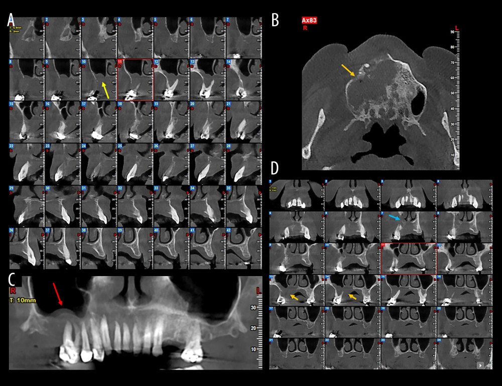
Figure 4 Case 4. (A) Cone-beam computed tomography (CBCT) cross-sectional images illustrating extensive alveolar bone destruction and cortical plate erosion (yellow arrow). (B) CBCT panoramic reconstruction showing the floating teeth appearance and spiked-tooth effect in the right posterior maxilla (red arrow). (C) CBCT axial slice demonstrating the infiltrative lesion with osseous destruction extending into the maxillary sinus and nasal cavity (orange arrow). (D) CBCT coronal slices highlighting maxillary sinus (orange arrows) and nasal cavity involvement (blue arrow).