20 December 2023: Articles 
Pigmented Poroma of the Lower Eyelid: A Case Report and Literature Review
Unusual clinical course, Challenging differential diagnosis, Educational Purpose (only if useful for a systematic review or synthesis)
Olga BogomoletsDOI: 10.12659/AJCR.941975
Am J Case Rep 2023; 24:e941975


Abstract
BACKGROUND: Eyelid tumors belong to a diverse group of neoplasms ranging from benign lesions to malignant tumors. Poromas are common, benign, mostly unpigmented tumors of the epidermal sweat duct unit, that usually grow slowly and occur in elderly people on the palms and soles. In most poroma cases some gene fusions were detected, which were caused by chromosomal aberrations.
CASE REPORT: We report the atypical case of a 30-year-old female patient suffering for more than 15 years from a solitary, polypoid, pigmented formation with a focal tuberous surface on the left lower eyelid. The lesion was not growing during the first years, but in the last 6 months before diagnosis its size more than doubled, finally reaching 12×14 mm. It was removed and histopathological analysis confirmed the diagnosis of a rare tumor – a poroma. There were no complications during healing and no recurrence was reported.
CONCLUSIONS: There have so far been only 9 reports of eyelid poromas, and the presented case significantly differed from the previous ones, as it appeared at an early age and showed rapid growth during a short time due to the war-related acute psychological stress. Moreover, it had unusual pigmentation and unpleasant smell. Reporting such untypical cases is clinically important because it is crucial to be aware of the diversity of eccrine poroma manifestation to distinguish it from malignant lesions.
Keywords: case reports, Eyelid Neoplasms, Poroma, adult, Female, Humans, Diagnosis, Differential, Eyelids, Pigmentation Disorders, Sweat Gland Neoplasms
Background
A poroma is a benign neoplasm that can be of eccrine or apocrine origin and is histogenetically associated with the intraepidermal (acrosyringial) part of the excretory ducts of the eccrine sweat glands. The disease code according to ICD-10 is D23.9. Occurrence of this tumor on the eyelid is rather rare [1]. Poroma usually occurs in older patients, without a gender predominance [2,3] and the exact pathogenesis of this lesion is unknown [4]. After radical removal, the prognosis of any form of poroma is favorable [5]. It can occur together with other skin lesions (eg, verruca plantaris and acral lentiginous melanoma), creating combined pathologies that are difficult to diagnose [6,7].
Recent research has revealed a genetic background to this disease, showing that about 85% of poroma cases are related with the following gene fusions: YAP1-MAML2, MAML2-YAP1, YAP1-NUTM1, and WWTR1-NUTM1, which are a consequence of intrachromosomal inversions and translocations [8–10]. NUT and transcriptional repressor GATA binding 1 (TRPS1) are potential molecular markers of poroma [11,12]. Ultraviolet radiation does not seem to be a key pathogenic factor in poroma development. Merkel cell polyomavirus (MCPyV) infection, existing malignancies, and organ transplantation history are important risk factors [9,13].
Our case is interesting because the neoplasm was localized on the lower eyelid at a young age (12–14 years). Other unusual characteristics of this clinical case were pigmentation, unpleasant smell, and the intense growth of the poroma, which coincided with significant acute stress because she was living in the area of military actions in Ukraine.
Case Report
TREATMENT:
The removal of the tumor was carried out with the method of radio frequency wave, followed by histopathological examination using routine staining with hematoxylin and eosin (H&E). The healing proceeded without complications, within 10 days, by secondary intension. The patient was satisfied with the treatment and the aesthetic result (Figure 1E). No recurrence was reported within 9 months after surgery.
HISTOPATHOLOGY:
Histopathological examination revealed a poroma. The main observation was the proliferation of epidermal cells with the formation of squamous bodies (Figure 2). Among the epidermal population, there were groups of cells with a small amount of eosinophilic cytoplasm and rounded hyperchromic nuclei (poroid cells). Moreover, multiple corneal pseudocysts and single mucin-filled ductal structures were observed in a series of sections. The morphological architecture was consistent with eccrine poroma (ICD-O code 8409/0) with features of traumatization. However, the subtype of poroma (eccrine/ apocrine) could not be determined. The clinical differential diagnostic was done with the seborrheic keratoma and no additional immunostaining was performed due to the lack of malignancy features.
Discussion
Poromas can arise on any skin surface, but those arising from eccrine tissue are most commonly found on the palms, soles, and scalp. That is why poroma on the eyelid is a rare pathology [1]. Other recently reported rare localizations of eccrine poroma are right thigh (lesion diagnosed in 74-year-old Chinese woman) and right lateral foot (lesion diagnosed in 82-year-old African American man) [14,15].
The development of benign poroma is based on simple hyperplasia of epidermal cells of the sweat gland, associated with exposure to an unknown trigger without visual damage to the nearby skin. The pathological process is generally slowly progressing [5].
Poroma is usually non-pigmented (17% are pigmented variants) and underdiagnosed due to confusion with other pigmented tumors. Most frequently, poroma is confused clinically with basal cell carcinoma, squamous cell carcinoma, and cutaneous melanoma [16].
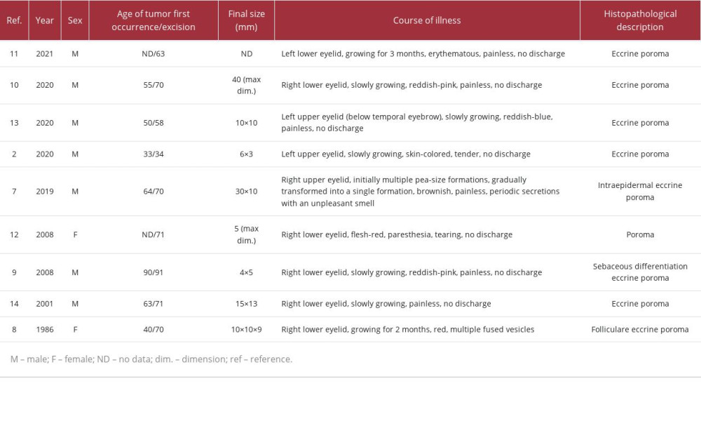
To date, there are only 9 reported cases of this tumor on the eyelids. We included the most important information about all reported clinical cases of the poroma (located in the peri-orbital zone) in Table 1, which compares the cases according to specific parameters.
We found some essential differences in our case of eyelid poroma in comparison to the generally accepted literature data, which may be important in facilitating diagnosis of this rare disease.
According to literature, all reported poromas occurred at an older age (mostly over 60 years) [2,17–24]. Thus, we show for the first time that the tumor can also appear in early adolescence (the patient was 12–14 years old). In our case, the lesion did not grow during the first 15 years and then very rapid tumor growth was observed – it doubled in size during about 6 months. This active growth coincided with the patient’s acute stress related to the war in Ukraine. Currently, the direct cause of such expansive growth is not known, but there is evidence that stress hormones promote the occurrence and development of tumors through various mechanisms [25].
Moreover, it should be noted that contrary to the information about the absence of sex predominance, our analysis (Table 1, together with our case) revealed that poroma of the eyelid was diagnosed more commonly in men than in women, with a male-to-female ratio 7: 3.
Finally, all diagnosed poromas were painless and only 2 cases (including ours) had a symptom of a bad smell. However, unlike the patient reported by Ahuja et al, in our case, there was no secretion from the tumor or the nearest eccrine ductus [17]. Thus, we hypothesized that the tumor became too big and as a result, was adhesive to the skin. Consequently, it caused difficulties in skin breathing and hygiene, resulting in bad smell.
It is also very important to differentiate benign tumors from malignant ones. The fundamental difference between these types of tumors is the absence of atypia in the structure of the benign ones. In our case, no abnormal cells or asymmetric architecture (suggestive of the malignant nature of the lesion) were observed, so there was no need to perform additional immunostaining. However, in more complex and uncertain cases, immunostaining for carcinoembryonic antigen (CAE) is crucial to differentiate between benign and malignant lesions [26].
Conclusions
Reported clinical case of eccrine poroma of the eyelid makes an important contribution to the diagnosis of skin cancer, not only due to its rare localization, but also because of its special features. Our case is the first that appeared during adolescence. It also stands out from others with its unusual pigmentation, the presence of an unpleasant smell, and extremely fast growth, which can be related to the patient’s acute war-related psychological stress. There is a need to educate doctors of various specialties (including ophthalmologists) to make them aware that poroma may occur on the eyelids of patients of all ages. Specialists should also be familiar with its treatment, especially since it can be frequently confused with other benign and malignant tumors.
Figures
References
1.. Cotton D: Neoplasms with ecrine differentiation, 1990, Philadelphia, Lea&Febiger
2.. Mencia-Gutierrez E, Navarro-Perea C, Gutierrez-Diaz E, Eyelid eccrine poroma: A case report and review of literature: Cureus, 2020; 12; e8906
3.. Shimano M, Kitamura S, Maeda T, Pigmented eccrine poroma on the palm mimicking seborrheic keratosis: Dermoscopic findings and a review of reported cases: Australas J Dermatol, 2021; 62; e477-e79
4.. Ahmed jan N, Masood S: Poroma, 2022, Treasure Island (FL), StatPearls Publishing [cited 2023 March 9]. Available from: https://www.ncbi.nlm.nih.gov/books/NBK560909/
5.. Chessa MA, Patrizi A, Baraldi C, Dermoscopic-histopathological correlation of eccrine poroma: An observational study: Dermatol Pract Concept, 2019; 9; 283-91
6.. Kneiber D, Cassarino DS, Combined poroma and verruca plantaris: Am J Dermatopathol, 2022; 44; e51-e53
7.. Sevim S, Akay BN, Kozan EN, Combined tumour of aberrant cytokeratin expressing acral lentiginous melanoma and poroma: Diagnostic challenge: Australas J Dermatol, 2023; 64; e233-e36
8.. Kervarrec T, Pissaloux D, Tirode F, Gene fusions in poroma, porocarcinoma and related adnexal skin tumours: An update: Histopathology, 2023 [Online ahead of print]
9.. Kyrmanidou E, Fotiadou C, Kemanetzi C, Eccrine poroma: Pathogenesis, new diagnostic tools and association with porocarcinoma – a review: Diagnostics (Basel), 2023; 13; 2689
10.. Sekine S, Kiyono T, Ryo E, Recurrent YAP1-MAML2 and YAP1-NUTM1 fusions in poroma and porocarcinoma: J Clin Invest, 2019; 129; 3827-32
11.. Macagno N, Kervarrec T, Sohier P, NUT is a specific immunohisto-chemical marker for the diagnosis of YAP1-NUTM1-rearranged cutaneous poroid neoplasms: Am J Surg Pathol, 2021; 45; 1221-27
12.. Taniguchi K, Goto K, Yabushita H, Transcriptional repressor GATA binding 1 (TRPS1) immunoexpression in normal skin tissues and various cutaneous tumors: J Cutan Pathol, 2023; 50(11); 1006-13
13.. Merilainen AS, Sihto H, Koljonen V, Merkel cell polyomavirus is a passenger virus in both poroma and porocarcinoma: J Cutan Pathol, 2022; 49; 49-54
14.. Li J, Ding Y, Zhang S, He W, Eccrine poroma: A case report: Asian J Surg; 874-75
15.. Mineroff J, Jagdeo J, Heilman E, Bradu S, Poroma in a patient with Fitzpatrick type V skin: J Drugs Dermatol, 2023; 22; 690-91
16.. Bombonato C, Piana S, Moscarella E, Pigmented eccrine poroma: Dermoscopic and confocal features: Dermatol Pract Concept, 2016; 6; 59-62
17.. Ahuja S, Kaur A, Goel M, Eccrine intraepidermal poroma of the eyelid: Indian J Ophthalmol, 2019; 67; 131-32
18.. Fujita M, Kobayashi M, Syringocystadenoma papilliferum associated with poroma folliculare: J Dermatol, 1986; 13; 480-82
19.. Iwasaki J, Yoshida Y, Yamamoto O, Tajima S, Poroma with sebaceous differentiation of the eyelid: A rare site of occurrence: Acta Derm Venereol, 2008; 88; 166-67
20.. Kalamkar C, Radke N, Mukherjee A, Radke S, Rare case of large eccrine poroma of the eyelid: Med J Armed Forces India, 2021; 77; 371-73
21.. McCoskey M, Neerukonda VK, Hatton MP, Wolkow N, Eccrine poroma of the eyelid: Orbit, 2021 [Online ahead of print]
22.. Rabady DZ, Carlson JA, Meyer DR, Poroma of the eyelid: Ophthalmic Plast Reconstr Surg, 2008; 24; 63-65
23.. Sharma M, Singh M, Gupta K, Eccrine poroma of the eyelid: Indian J Ophthalmol, 2020; 68; 2522
24.. Vu PP, Whitehead KJ, Sullivan TJ, Eccrine poroma of the eyelid: Clin Exp Ophthalmol, 2001; 29; 253-55
25.. Hong H, Ji M, Lai D, Chronic stress effects on tumor: Pathway and mechanism: Front Oncol, 2021; 11; 738252
26.. Wollina U, Castelli E, Rulke D, Immunohistochemistry of eccrine poroma and porocarcinoma – more than acrosyringeal tumors?: Recent Results Cancer Res, 1995; 139; 303-16
Figures
In Press
Case report 
Am J Case Rep In Press; DOI: 10.12659/AJCR.953173
Case report 
Am J Case Rep In Press; DOI: 10.12659/AJCR.953192
Case report 
Am J Case Rep In Press; DOI: 10.12659/AJCR.952818
Case report
Am J Case Rep In Press; DOI: 10.12659/AJCR.953608
Most Viewed Current Articles
07 Dec 2021 : Case report
22,364,578
DOI :10.12659/AJCR.934347
Am J Case Rep 2021; 22:e934347
06 Dec 2021 : Case report  174,245
DOI :10.12659/AJCR.934406
Am J Case Rep 2021; 22:e934406
21 Jun 2024 : Case report
119,744
DOI :10.12659/AJCR.944371
Am J Case Rep 2024; 25:e944371
07 Mar 2024 : Case report
64,648
DOI :10.12659/AJCR.943133
Am J Case Rep 2024; 25:e943133