30 October 2020: Articles 
Radiation Induced Multiple Skin Neoplasms Following Craniospinal Irradiation for Medulloblastoma. A Case Report
Unusual clinical course, Challenging differential diagnosis, Unusual or unexpected effect of treatment, Diagnostic / therapeutic accidents
Ahmed Amer Osman Ali1BCDEF, Yasser Bayoumi1ABDF, Ali Balbaid1ACDF, Yasser Orz2BCEF, Wafa AlShakweer3BCDE, Mohamed Hussen Eltawel4EF, Mutahir Tunio1BDEF*DOI: 10.12659/AJCR.917694
Am J Case Rep 2020; 21:e917694


Abstract
BACKGROUND: Following craniospinal irradiation in children with medulloblastoma, secondary neoplasms are among the most serious long-term sequelae that include leukemias and solid tumors of the urinary or digestive tracts, thyroid, skin, and central nervous system. Furthermore, in children with Gorlin syndrome following craniospinal irradiation for medulloblastoma, there is a rising incidence of skin and non-skin malignancies.
CASE REPORT: The patient in the present study was a 19-year-old female who was treated with craniospinal irradiation and chemotherapy following gross total resection (GTR) for medulloblastoma at the age of 4 years. Fifteen years later, she developed a primary adnexal tumor at the medial aspect of her left thigh, glomangioma at the skin of her upper abdomen, dermatofibrosarcoma protruberans at the skin of her upper back, and Kaposiform hemangioendothelioma of the upper abdomen. All these tumors were successfully managed with radical resection without further adjuvant treatment.
CONCLUSIONS: Metachronous of development of 4 histopathologically different skin tumors following craniospinal irradiation for medulloblastoma in long-term survivors has not previously been reported. The present case warrants a detailed dermatological periodic inspection in such patients.
Keywords: craniospinal irradiation, Medulloblastoma, Skin Neoplasms, Survivors, Cerebellar Neoplasms, Child, Child, Preschool, Neoplasms, Radiation-Induced, young adult
Background
Standard integration of adjuvant craniospinal irradiation (CSI) in the treatment paradigm for medulloblastoma has markedly increased overall survival. However, various delayed complications are associated with CSI including neuro-cognitive dysfunction, growth and development abnormalities, audio-visual deficits, endocrine dysfunction, cardiovascular toxicity, as well as secondary malignancies in long-term survivors leading to morbidity and mortality, which is worrisome [1–3]. Secondary malignancies include a variety of malignancies including leukemias and solid tumors of the urinary or digestive tracts, thyroid, skin, and central nervous system (CNS), and although rare, are among the most serious long-term sequelae in children treated for medulloblastoma [4].
The autosomal dominant genetic disorder Gorlin syndrome (GS) in children is typically characterized by multiple basal cell carcinomas, odontogenic keratocysts, hyperkeratosis of the palms and soles, skeletal abnormalities, intracranial ectopic calcifications, facial dysmorphism, intellectual disability, and medulloblastoma. Children with GS treated with CSI are further predispose to other skin and non-skin secondary malignancies [5,6].
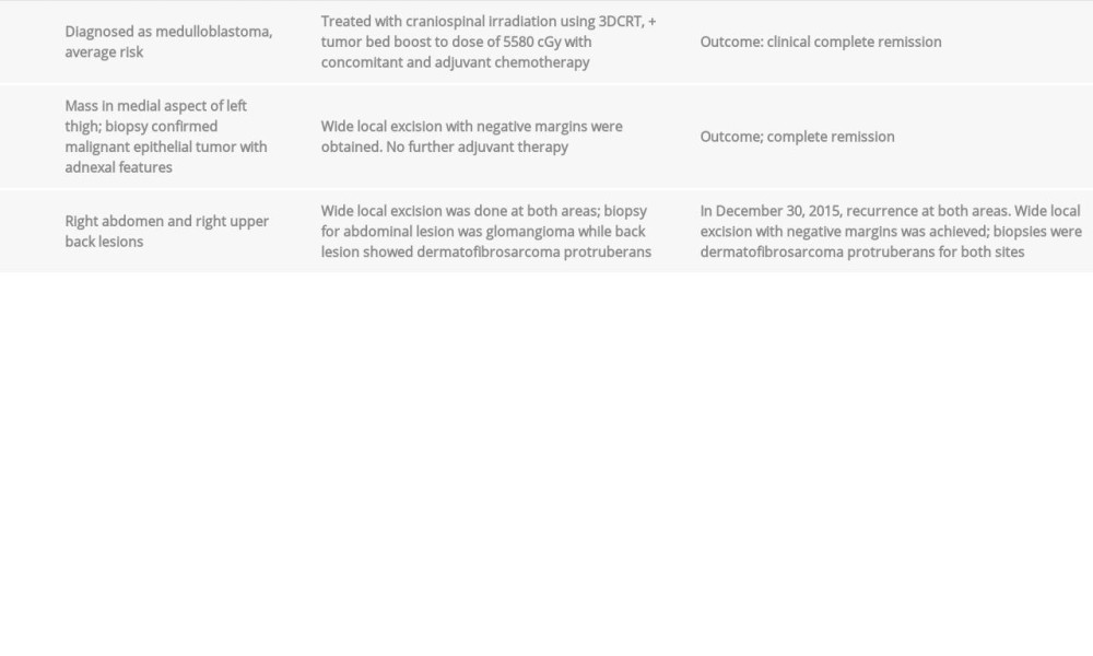
Our case report is of a 19-year-old female who was treated with CSI and chemotherapy for medulloblastoma at the age of 4 years, without any signs of GS, who subsequently developed multiple skin malignancies in previous irradiated areas (Table 1).
Case Report
In 2001, a 4-year-old female underwent craniotomy, gross total resection (GTR) followed by CSI with the doses of 2340 cGy followed by tumor bed boost to complete 5580 cGy using 3-dimensional conformal radiotherapy (3DCRT), with concomitant and adjuvant chemotherapy for average-risk medulloblastoma. Since then, she had been under periodic follow-up checks, and her condition was stable with complete remission on imaging. No comorbidities were found, and her family history was unremarkable.
In February 2015, at the age of 19 years, she complained about a diffuse swelling at the medial aspect of her left thigh. On physical examination, a hard, mobile growth that measured 4×4 cm with indurated margins on the medial left thigh was identified. The rest of her examination was unremarkable. Magnetic resonance imaging (MRI) with contrast of lower limbs demonstrated a mixed solid and cystic subcutaneous nodule involving the left inguinal region, measuring 4.7×4.1×5 cm diameter in antero-posterior, transverse and cranio-caudal directions, without any frank invasion into underlying muscles (Figure 1). Looking retrospectively to treatment planning, this area received point dose of 56 cGy. The patient subsequently underwent surgical excision, with histopathology confirming the malignant epithelial tumor with adnexal features with negative margins (Figure 2). Active surveillance was opted in her case and she was advised to follow-up in the dermatology oncology clinic.
Six months later, in September 2015, she presented in the clinic with complaints of right upper abdominal swelling, which was painless, firm, and with well-defined margins measured approximately 2×2 cm. During examination, another protuberant mass at her right upper back of size 3×3 cm was noticed, which was multi-nodular, non-tender, and ill defined. Excisional biopsy of her abdominal lesion revealed a benign glomangioma, while the excisional biopsy of back mass confirmed dermatofibrosarcoma protruberans (DFSP) (Figure 3). Point doses correspondent to these areas with 209 cGy at the site of abdominal mass, and 1573 cGy at the site of the lesion in the back. The patient was advised for follow-ups.
Two months later in December 2015, she presented with recurrent masses at the right upper abdomen and upper back. A repeat excisional biopsy of the right abdominal mass demonstrated Kaposiform hemangioendothelioma (Figure 4), which was re-excised with negative margins. Recurrent DFSP of the back was confirmed on surgical excision. The chronology of events and point doses are given in Figure 5. Although clinical suspicion for GS was high, no particular genetic testing was performed for this patient.
At the time of publication, she was on regular follow-ups without any evidence of recurrence at any previous tumor sites.
Discussion
Radiation induced vascular proliferative lesions (capillary telangiectasia, cavernous angiomas, or cavernomas) are described with rising incidence in medulloblastoma long-term survivors [7]. We described a case of young female treated with CSI and chemotherapy for medulloblastoma at the age of 4 years who developed metachronous malignant epithelial adnexal tumor of the skin, glomangioma of the skin, DFSP, and Kaposiform hemangioendothelioma of the skin within the previous irradiated areas. To our knowledge, this kind of clinical presentation has never been described before in the literature.
Radiation induced skin tumors are known to develop with a variable latency period with an aggressive biology and multi-focality [8]. However, histopathological examination of our patient’s tumors did not suggest any evidence of high grade or a diffuse pattern. All of these tumors were successfully treated with surgical resection, without any further adjuvant radiotherapy.
A retrospective look at our case, following clinicopathological features, did not correlate these tumors with possible underlying GS: a) classic-Wnt variant medulloblastoma was observed in our patient. GS is known to be associated with desmoplastic/nodular medulloblastoma-SHH origin [9]. b) No clinical or radiological signs of GS at the time of medulloblastoma diagnosis [5], and all the patient’s skin lesions were within previous irradiated areas; although no thorough genetic testing was conducted to exclude underlying GS.
Conclusions
In conclusion, the tumorigenic effects of the CSI for medulloblastoma are well known in long-term survivors. Metachronous of development of 4 histopathologically different skin tumors has not previously been reported. The present case warrants a systematic follow-up with dermatological inspection in long-term survivors of medulloblastoma who have received CSI.
Figures
References:
1.. Christopherson KM, Rotondo RL, Bradley JA, Late toxicity following craniospinal radiation for early stage medulloblastoma: Acta Oncol, 2014; 53(4); 471-80
2.. Hope AJ, Mansur DB, Tu PH, Metachronous secondary atypical meningioma and anaplastic astrocytoma after postoperative craniospinal irradiation for medulloblastoma: Childs Nerv Syst, 2006; 22(9); 1201-7
3.. Stavrou T, Bromley CM, Nicholson HS, Prognostic factors and secondary malignancies in childhood medulloblastoma: J Pediatr Hematol Oncol, 2001; 23(7); 431-36
4.. Salloum R, Chen Y, Yasui Y, Late morbidity and mortality among medulloblastoma survivors diagnosed across three decades: A report from the childhood cancer survivor study: J Clin Oncol, 2019; 37(9); 731-40
5.. Lo Muzio L, Nevoid basal cell carcinoma syndrome (Gorlin syndrome): Orphanet J Rare Dis, 2008; 3; 32
6.. Jiang T, Wang J, Wang Y, Li C, Development of mediastinal lymphoma after radiotherapy for concurrent medulloblastoma and PNET in a patient with Gorlin syndrome: World J Surg Oncol, 2016; 14(1); 215
7.. Jain R, Robertson PL, Gandhi D, Radiation-induced cavernomas of the brain: Am J Neuroradiol, 2005; 2013; 1158-62
8.. Lew SM, Morgan JN, Psaty E, Cumulative incidence of radiation-induced cavernomas in long-term survivors of medulloblastoma: Neurosurg, 2006; 2013(2 Suppl.); 103-7
9.. Mangum R, Varga E, Boué DR, SHH desmoplastic/nodular medulloblastoma and Gorlin syndrome in the setting of Down syndrome: Case report, molecular profiling, and review of the literature: Childs Nerv Syst, 2016; 32(12); 2439-46
Figures
In Press
Case report 
Am J Case Rep In Press; DOI: 10.12659/AJCR.953173
Case report 
Am J Case Rep In Press; DOI: 10.12659/AJCR.953192
Case report 
Am J Case Rep In Press; DOI: 10.12659/AJCR.952818
Case report
Am J Case Rep In Press; DOI: 10.12659/AJCR.953608
Most Viewed Current Articles
07 Dec 2021 : Case report
22,364,578
DOI :10.12659/AJCR.934347
Am J Case Rep 2021; 22:e934347
06 Dec 2021 : Case report  174,245
DOI :10.12659/AJCR.934406
Am J Case Rep 2021; 22:e934406
21 Jun 2024 : Case report
119,744
DOI :10.12659/AJCR.944371
Am J Case Rep 2024; 25:e944371
07 Mar 2024 : Case report
64,648
DOI :10.12659/AJCR.943133
Am J Case Rep 2024; 25:e943133