29 November 2020: Articles 
A 37-Year-Old Saudi Man with a Calcified Intraventricular Astrocytoma
Unusual clinical course, Challenging differential diagnosis
Maryam Abdullah Al Saleem1EF*, Noor Said Alsafwani1E, Tarek Mohammad ElSharkawy1E, Sari Saleh Al-Suhibani2E, Wisam Essawi3EDOI: 10.12659/AJCR.928224
Am J Case Rep 2020; 21:e928224


Abstract
BACKGROUND: Pilocytic astrocytoma is a low-grade glioma that is common in children. Pilocytic astrocytoma is a slow-growing neoplasm that may calcify and occurs throughout the central nervous system, but it has a preference to be located infratentorial in children.
CASE REPORT: Herein, we report an unusual intraventricular location of pilocytic astrocytoma with extensive calcification in a 37-year-old Saudi man who mainly presented with a headache. Although pilocytic astrocytoma can arise throughout the central nervous system, it very rarely arises from the ventricles, especially the lateral ventricle.
CONCLUSIONS: The majority of intraventricular tumors arise within the third and fourth ventricles. The unusual intraventricular location and the unexpected age group are the main difficulties in diagnosing an adult with intraventricular pilocytic astrocytoma. Intraventricular pilocytic astrocytoma can be missed radiologically and misled pathologically; therefore, it should be considered within the differential diagnosis of intraventricular tumors. To the best of our knowledge, this is the first case to be reported in Saudi Arabia.
Keywords: Astrocytoma, Calcinosis, Cerebral Ventricle Neoplasms, Child, Diagnosis, Differential, Magnetic Resonance Imaging, Saudi Arabia
Background
Pilocytic astrocytoma (PA) is the most common glioma in children and accounts for 6% of all gliomas, with a slight predominance in males [1]. Radiologically, PA is usually a cystic tumor with mural nodule formation. Histologically, it is a low-grade glioma corresponding to WHO grade 1 with the characteristic bipolar cells and Rosenthal fibers. Sometimes it tends to form microcysts and/or eosinophilic granular bodies [2]. Therefore, the term “pilocytic” is used, with the first part, “pilo”, meaning “hair-like”, referring to the morphology of the bipolar cells, and the second part, “cytic”, referring to cystic nature of the tumor. Rarely, PA exhibits calcification as a degenerative change. PA occurs throughout the central nervous system, but it tends to have an infratentorial location in children [2]. Only a few cases of intraventricular tumors have been reported in the literature (about 10%) [3]. The majority of intraventricular tumors arise within the third and fourth ventricles. Here, we report a case of calcified pilocytic astrocytoma in the lateral ventricle in an adult. To the best of our knowledge, this is the first case to be reported in Saudi Arabia.
Case Report
A 37-year-old Saudi man, not known to have any other medical illnesses, presented to King Fahad Hospital of Imam Abdulrahman Bin Faisal University, Khobar with a left-sided headache and blurred vision for 2 months. The headache was severe, lasting for 10 min and recurring every 90 min. There was no loss of consciousness, numbness, weakness, nausea, vomiting, gait disturbance, or change in speech.
On physical examination, the patient was conscious, alert, and oriented to time, place, and person. The results of cranial nerves assessment and motor and sensory examinations were normal, so the patient was referred to the Radiology Department for further investigation. Magnetic resonance imaging (MRI) revealed an ill-defined lobulated heterogeneous large intraventricular lesion centered within the posterior horn of the right lateral ventricle, which was heterogenous in T2 and T1, with evidence of blooming seen in gradient echo sequences (GRE) (Figures 1–5). The lesion measured 4×3.6×2.5 cm in maximum diameter, with isolated dilatation of the posterior horn of right lateral ventricle and extension to the right parietal lobe (Figure 3). The periventricular white matter along the right posterior horn of the right lateral ventricle displayed hyperintense T2 signal with blooming, which shows pathological enhancement representing extension of disease (Figure 4). The radiological impression at the time of examination was suspicious for choroid plexus neoplasm. Then, no further radiological investigations were done and the (MRI) findings was sufficient to schedule the patient for diagnostic and therapeutic surgical resection.
A fresh right intraventricular tumor consisting of multiple fragments measuring in aggregate 0.2×0.2×0.2 cm was removed for intra-operative frozen section. Microscopy revealed a mild-to-moderate cellular lesion and indistinct cellular borders in a background of fibrillary eosinophilic stroma with extensive calcification. The nuclei were bland oval-to-round with occasional bipolar cells. Therefore the impression was low-grade glioma.
Additional right intraventricular tumor tissue was excised and placed in 10% buffered formalin for permanent sections consisting of multiple fragments measuring in aggregate 2×1.5×1 cm.
Microscopically, hematoxylin and eosin (H&E)-stained sections showed a neoplastic moderately cellular glial lesion, as well as areas of hypocellular edematous glial tissue at the periphery. Higher magnification revealed bipolar piloid cells arranged in a diffuse pattern within a fibrillary background. Moreover, an extraordinary calcification formed by psammoma bodies (Figure 6A), in addition to microcysts formation (Figure 6B), vascular proliferation (Figure 6C), and eosinophilic granular bodies (EGBs), were noted.
The immunohistochemical studies showed diffuse positivity of glial fibrillary acidic protein (GFAP) (Figure 6D) and S100 protein. Synaptophysin, neuron-specific enolase (NSE), and epithelial membrane antigen (EMA) were all negative. A low mitotic index using Ki67 (less than 1%), along with the case findings above, suggested low-grade intraventricular tumor compatible with PA. Since the diagnosis was confirmed histologically, and because there is to date no targeted therapy approved for PA, no further molecular analysis was recommended or performed.
Discussion
Tumors originating from the lateral ventricles account for less than 1% of all brain tumors [4]. Among these, only 10% are PA, which predominantly affects children and adolescents [3]. The unusual intraventricular location and the unexpected age group are the main difficulties in diagnosing an adult with intraventricular PA.
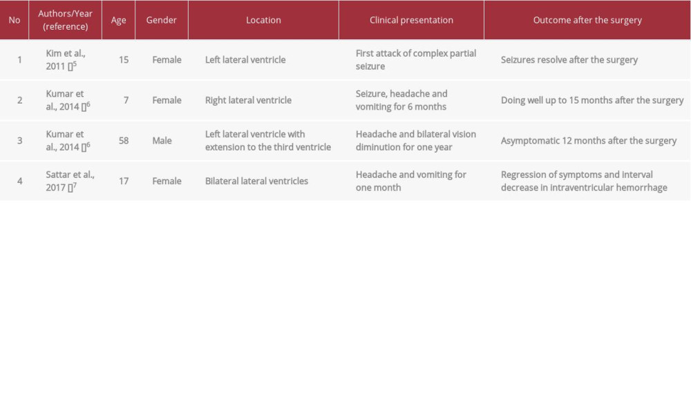
Kim et al. described the first case of calcified intraventricular PA originating from the left lateral ventricle, which presented with seizure in a 15-year-old girl [5]. Kumar et al. reported another 2 cases. The first was a 7-year-old girl who presented as seizure with underlining right lateral ventricular calcified PA [6]. The second was a 58-year-old man with a headache and bilateral vision diminution caused by calcified PA that originated from the left lateral ventricle with extension to the third ventricle [6]. Sattar et al. also reported a 17-year-old girl with bilateral lateral ventricular calcified PA, presenting with headache and vomiting [7]. The Table 1 summarizes the cases of calcified intraventricular pilocytic astrocytoma reported in the literature. Extensive calcification within an intraventricular tumor may obscure the diagnosis of PA. Hence, it requires special concern and awareness.
The pathophysiology of calcification remains unclear, but typically appears in benign or slow-growing tumors [5]. A small proportion of gliomas (9.3% to 19%) are known to harbor calcifications, such as oligodendrogliomas, low-grade astrocytomas, and ependymomas [8–10]. Other nonglial brain tumors may also calcify, including craniopharyngiomas, meningiomas, and pineal gland tumors [11]. Intraventricular tumors that may be calcified are ependymomas, choroid plexus tumors, central neurocytomas, and some metastatic tumors [5].
Ventricular tumors cause mechanical obstruction of CSF (cerebrospinal fluid), thereby increasing intracranial pressure, manifested as headache, blurred vision nausea, and vomiting. Progression to obstructing hydrocephalus with subsequent focal neurological deficits and/or seizures is a potential complication.
The main differential diagnoses of intraventricular tumors are choroid plexus tumors, central neurocytomas, subependymal giant cell astrocytoma, tanycytic ependymoma, and low-grade gliomas, which include PA, diffuse astrocytoma, and oligodendroglioma. Choroid plexus tumors originate from the choroid plexus within the ventricles, mostly the lateral ventricle, and consist of cuboidal-to-columnar cells arranged on true papillary architecture. Even though central neurocytoma arises within the ventricles with preference to the lateral ventricle, it morphologically consists of monomorphic rounded cells that lacks glial fibrillary acidic protein (GFAP) expression. Tanycytic ependymoma (TE) is typically central intramedullary tumors within the thoracic/cervicothoracic region, but still can occur in the ventricles. TE lacks rosette formation, and only focal perivascular pseudorosettes are present, which is why TE remains a challenging entity with spindle bipolar cells arranged in a fascicular pattern with bland nuclei, similar to PA.
In contrast to PA, diffuse astrocytoma shows nuclear atypia and increases mitosis. Lack of giant pyramidal-like cells with a ganglionic appearance and the lack of synaptophysin and neuron-specific enolase (NSE) immunohistochemical expression render subependymal giant cell astrocytoma less likely. Although oligodendroglioma is usually located within the cortex, there are rare cases reported to involve the ventricles. Diagnosis of oligodendroglioma depends only on the detection of IDH-mutation and 1p/19q codeletion. While PA is sometimes easily diagnosed by H&E staining, molecular study is often not required. More than 70% of PA harbor tandem duplication of 7q34, which results in a fusion gene KM 1549-BRAF that activates the MAPK pathway. However, there are other ways to activate the MAPK pathway, depending on the anatomical location. For instance, KM 1549-BRAF fusion is more common in infratentorial PA, while BRAF V600E mutation and NTRK-family fusions are more common in supratentorial PA [2]. It is unknown if intraventricular PA has a specific mutation to activate the MAPK pathway, due to the limited number of reported cases of this entity. Surgery is the primary treatment for PA, and BRAF-targeted therapy is still under study and in trails [12].
Conclusions
Although PA can arise throughout the central nervous system, it is rarely located intraventricularly. Intraventricular PA can be missed radiologically and can mislead pathology, so it should be considered in the differential diagnosis of intraventricular tumors.
Figures
References:
1.. Perry A, Brat D: Practical surgical neuropathology, 2018; 125, Philadelphia, Elsevier
2.. Louis D, Ohgaki H, Wiestler O, Cavenee W: WHO classification of tumours of the central nervous system, 2016; 80-89, International Agency for Research on Cancer
3.. Youmans J, Winn H: Youmans neurological surgery, 2011; 68-1534, Philadelphia, PA, Saunders/Elsevier
4.. Spencer D, Collins W, Sass K: Surgical management of lateral ventricular tumors, 1988; 583-607, Philadelphia, Saunders
5.. Kim Y, Shin H, Suh Y, Pilocytic astrocytoma with extensive psammomatous calcification in the lateral ventricle: A case report: Childs Nerv Syst, 2011; 28(4); 649-52
6.. Kumar R, Garg K, Kakkar A, Sharma M, Giant calcified intraventricular pilocytic astrocytoma: A rare entity: Neurol India, 2014; 62(1); 71
7.. Sattar S, Akhunzada N, Javed G, Pilocytic astrocytoma: A rare presentation as intraventricular tumor: Surg Neurol Int, 2017; 8(1); 116
8.. Friedman JH, Chiucchini I, Tucci JR, Idiopathic hypoparathyroidism with extensive brain calcification and persistent neurologic dysfunction: Neurology, 1987; 37(2); 307-9
9.. Gilbertson E, Good C, Roentgenographic signs of tumors of the brain: Am J Roentgenol Radium Ther Nucl Med, 1956; 76; 226-47
10.. Kalan C, Burrows E, Calcification in intracranial gliomata: Br J Radiol, 1962; 35(417); 589-602
11.. Makariou E, Patsalides A, Intracranial calcifications: Appl Radiol, 2009; 38; 48-60
12.. Selt F, Hohloch J, Hielscher T, Establishment and application of a novel patient-derived KIAA1549: BRAF-driven pediatric pilocytic astrocytoma model for preclinical drug testing: Oncotarget, 2016; 8(7); 11460-79
Figures
In Press
Case report 
Am J Case Rep In Press; DOI: 10.12659/AJCR.949976
Case report 
Am J Case Rep In Press; DOI: 10.12659/AJCR.950290
Case report 
Am J Case Rep In Press; DOI: 10.12659/AJCR.950607
Case report
Am J Case Rep In Press; DOI: 10.12659/AJCR.950985
Most Viewed Current Articles
07 Dec 2021 : Case report  17,691,734
DOI :10.12659/AJCR.934347
Am J Case Rep 2021; 22:e934347
06 Dec 2021 : Case report  164,491
DOI :10.12659/AJCR.934406
Am J Case Rep 2021; 22:e934406
21 Jun 2024 : Case report
113,090
DOI :10.12659/AJCR.944371
Am J Case Rep 2024; 25:e944371
07 Mar 2024 : Case report
59,175
DOI :10.12659/AJCR.943133
Am J Case Rep 2024; 25:e943133