25 November 2024: Articles 
Multi-Modality Imaging in Caseous Calcification of the Mitral Annulus: Case Series and Clinical Implications
Challenging differential diagnosis, Rare disease
Emil DadańskiDOI: 10.12659/AJCR.944821
Am J Case Rep 2024; 25:e944821


Abstract
BACKGROUND: Echogenic masses in the mitral annulus are often incidental findings during routine echocardiography examinations. Most represent the wide spectrum of underlying mitral annular calcifications and usually do not require further diagnostic workup. However, a rare variant called “caseous calcification of the mitral annulus” (CCMA) can be challenging to diagnose and often requires an extensive workup with the use of multiple imaging studies in order to accurately confirm the pathology.
CASE REPORT: The aim of this case series was to illustrate the range of challenges associated with CCMA in various clinical scenarios to highlight the importance of multi-modality imaging in the differential diagnosis. Patient 1 was a 78-year-old asymptomatic woman with a round echo-dense structure incidentally found during a routine echocardiographic examination. Patient 2 was a 79-year-old woman with a suspected mitral abscess. Patient 3 was a 73-year-old woman with an incidentaloma (lung mass), with a concomitant lesion of the mitral annulus. All 3 patients required a unique combination of confirmatory imaging studies to ultimately confirm their diagnosis.
CONCLUSIONS: The incidental detection of CCMA-like changes requires multi-modality imaging to differentiate from abscesses and proliferative changes in the mitral annulus. Transthoracic echocardiography, transesophageal echocardiography, computed tomography, and cardiac magnetic resonance imaging play a key role in this diagnostic workup. Using a combination of these imaging modalities can enhance diagnostic accuracy and strongly influence the subsequent treatment and management strategies.
Keywords: Echo-Planar Imaging, Tomography Scanners, X-Ray Computed, Heart, Heart Diseases, Heart Valve Diseases, Humans, Female, Aged, Calcinosis, Mitral Valve, Multimodal Imaging, Echocardiography, Transesophageal, Tomography, X-Ray Computed, Echocardiography, Diagnosis, Differential, Incidental Findings, Magnetic Resonance Imaging
Introduction
Mitral annular calcification is a frequently encountered echocardiographic observation, especially in older populations. A less common variant is caseous calcification of the mitral annulus (CCMA), which has a prevalence of 0.6% in patients exhibiting mitral annular calcification on echocardiograms. The differential diagnosis of CCMA can be challenging. Clinical cardiologists need to recognize the complexities of this diagnosis and be aware of the potential necessity of using multiple imaging studies to confirm it. This condition can be camouflaged initially, as it shares many identifying markers with more severe conditions, such as an underlying infection, abscess of the mitral valve, or a potential tumor. Generally, caseous calcification is non-malignant, with a good prognosis, and requires only annual follow-up visits, unlike the more serious conditions mentioned above.
An incorrect and hasty diagnosis can lead to the omission of a potentially life-threatening disease or a qualification for a potentially unnecessary and invasive procedure. The aim of this case series was to provide clinical insight into CCMA and highlight a patient-specific approach to selecting the proper imaging studies for accurate diagnosis.
Case Reports
CASE 1: CCMA AS THE INCIDENTAL FINDING:
A 78-year-old female patient with a history of hypertension and paroxysmal atrial fibrillation was referred to the Cardiology Outpatient Clinic for a wellness assessment. At the time of the visit, the patient was in a generally good condition, without symptoms of angina and heart failure. She was not limited when performing mild exertion and was able to walk up 2 flights of stairs without symptoms. During the physical examination, a mild systolic murmur at Erb’s point was audible (Levine scale 2/6). The patient’s blood pressure was well controlled, with an office blood pressure of 132/71 mmHg. An ECG showed a normal sinus rhythm at 68 beats/min and features of left atrial enlargement.
Routine transthoracic echocardiography (TTE) showed preserved ejection fraction, mild diastolic dysfunction, left atrium enlargement, and calcification localized on the posterior mitral annulus. The mass was a round, echo-dense structure with a diameter of 1.8 cm and central areas of echolucency. It was bulging into the left atrium but did not significantly impair mitral inflow (Figure 1). The initial workup ruled out possible underlying neoplasms or abscesses (Table 1). Based on the localization of the mass and its characteristics, a diagnosis of CCMA was established. Considering the entire clinical picture, there was no need to modify antihypertensive, anti-arrhythmic, and anticoagulation therapy. The patient was advised to report for an annual echocardiographic assessment, to monitor the lesion. In the annual follow-up, both the echocardiography and the patient’s condition remained stable, with no significant changes observed.
CASE 2: CCMA IMPAIRING MITRAL VALVE INFLOW AND DIFFERENTIATED WITH AN ABSCESS:
A 79-year-old female patient with underlying obesity, hypertension, type 2 diabetes mellitus, and hypercholesterolemia presented with a paroxysmal complete heart block, which required an implantation of a permanent pacemaker. After implantation, the pocket site became infected, requiring the subsequent removal of the device, intravenous antibiotics, and a reimplantation on the right side of the chest. Over the next year, the patient was admitted multiple times for recurrent urinary tract infections. During one of these hospitalizations, in the Internal Medicine Department, the patient required a cardiology consultation for an exacerbation of heart failure, with increased episodic dyspnea and fatigue. A TTE was performed revealing a 5×3-cm mass protruding into the left atrium, resulting in dysfunction of the posterior leaflet and a drop in the mitral inflow (Figure 2).
A chest computed tomography (CT) scan was performed, which suggested a chronic abscess of mitral annulus. Further workup was performed, with inconclusive findings after transesophageal echocardiography (TEE) and coronary angiography (Figure 3). Considering the complexity of the case, a cardiological consilium was performed, consisting of interventional and clinical cardiologists as well as cardiosurgeons. This heart team recognized that, despite inconclusive findings, there was a high clinical suspicion to warrant exploratory surgery.
Intraoperatively, after a partial resection of the posterior leaflet of the mitral valve, a 5×3×2-cm abscess within the mitral valve annulus that impeded the flow of blood into the left ventricle was discovered. Serous exudates measuring 15 to 20 g were removed, and the leaflet was sutured to the mitral annulus to conclude the procedure. The content was sent for further bacteriological identification (the result was unavailable at the time of this manuscript’s writing), and the patient quickly returned to a good general condition. She was ultimately discharged from the Cardiothoracic Surgery Department and, at the time of this report, remained under the care of cardiologists in an outpatient setting.
CASE 3: CLINICAL TRIBUTE TO MULTIMODALITY ASSESSMENT:
A 73-year-old female patient with well-controlled hypertension was admitted to the Emergency Department for shortness of breath and stabbing chest pain. Initial diagnostics ruled out any possible acute coronary syndrome, and exploratory high-resolution CT scanning of the chest revealed a subpleural mass measuring 10×16×9 mm, as well as a calcification measuring 36×35×20 mm around the border of the left atrium and ventricle near the mitral valve (Figure 4).
After consulting with a pulmonologist, a positron emission tomography (PET) scan using 18-fluoro-deoxyglucose (18F-FDG) to evaluate the lung mass was performed. The study confirmed the presence of a spiculated metabolically active lung tumor (max standardized uptake value [SUV] 4.5) and moderately increased FDG metabolism (max SUV 5.2) around a prominent calcification (30×21 mm) in the posterior part of the left ventricle, involving the mitral valve and a small part of the left atrium (Figure 5). SUV max reference ranges were at 3.8 when the neighboring liver was assessed. Further, a histopathological diagnosis was conducted by sampling nearby lymph nodes and bronchial scrapings, which determined the lesion to be benign in nature.
TTE further revealed an oval-shaped structure in the posterior mitral annulus measuring 20 mm in diameter (Figure 6). Afterward, TEE was performed, and a cardiac mass measuring 20×21×27 mm, surrounded by a capsule producing an acoustic shadow from the atrial side, was visualized (Figure 7).
A mitral ring tumor was initially suspected but was ultimately ruled out after cardiac magnetic resonance imaging was performed, revealing a mass measuring 20×28 mm beneath the posterior leaflet of the mitral valve in the basal segment of the posterior wall, with tissue characteristic of CCMA: low signal intensity in cine steady-state free precession and T1 sequences, hyperintense rim in short TI inversion recovery sequence, no contrast enhancement in first pass perfusion, and peripheral late gadolinium enhancement (Figure 8). No other pathologies within the myocardium were detected.
Based on the available imaging studies and diagnostic work-up, the most likely diagnosis was determined to be CCMA with a benign lung tumor; both of which required only annual follow-up but no aggressive treatment management, considering the benign course of the independent diseases. Over the next 3 years, the nodular lesion in the lung completely regressed, and no CCMA complications were observed.
Discussion
Mitral annulus calcification is a chronic degenerative process. Thickening of the mitral valve leaflets becomes more prevalent after the age of 50 to 60 years [1]. There is generally also fibrous accumulation and calcifications involving the posterior wall of the left ventricle, posterior leaflet of the mitral valve, and posterior portion of the mitral annulus [2–4]. In most cases, deposits of calcium slowly begin to accrue over time on the surface of the valve, but they generally maintain their shape, to have very limited effects of the hemodynamic properties of valvular flow. In 0.6% of all mitral annular calcification cases [5], there is an atypical progression of mitral annular calcification [6], which is a starting point for the formation of CCMA.
The exact pathomorphological mechanism of CCMA development is currently unknown. Considering its close pathological relation with mitral annular calcification, age appears to be one of the most important risk factors for CCMA, as seen in the cases we chose to highlight for this case series. CCMA is also more prevalent in women and patients with concomitant kidney diseases [6].
CCMA is most often asymptomatic and is usually discovered incidentally after simple imaging studies, as depicted throughout our case series. Most asymptomatic pathologies require only careful observation (case 1, case 3). Rare but potential complications of CCMA include a rupture of the calcified shell, with embolic material formation, heart block due to an impingement on the conduction system, increased frequency of infective endocarditis, or dysfunction of the mitral valve.
Despite the benign course of the disease, it is important to remember that CCMA can often mimic more severe pathologies, such as abscesses or tumors; therefore, an urgent but thorough diagnostic workup is paramount to ensure that the patients are adequately treated from this initial consultation [4,5]. Characteristic features of these diseases useful in the differential diagnosis are presented in Table 1 [7,8]. A comparison of echocardiographic images of mitral annular calcification, myxomas, and abscesses is shown in Figure 9. If the mass is localized to a specific leaflet, and there are particular radiological features, such as echolucency, a TTE is generally sufficient for confirming the diagnosis of CCMA, as described in case 1. However, in cases in which there is a high clinical suspicion, particularly those characterized by restricted acoustic viewing or suboptimal TTE image quality, individuals can require a more advanced TEE for a more precise assessment of the mass and improved visualization of posteriorly situated structures. In the most complicated cases, in which the risk of malignancy or infection is substantial, multiple imaging studies must be considered. Characteristic features in different imaging modalities are shown in Table 2 [9].
Case 2 demonstrates how abscess formation in the mitral annulus in patients with a history of recurrent infections and more severe infections of the pacing pocket after implantation are possible. Case 3 illustrates a clinical investigation of a mass in the mitral annulus complicated by the coexistence of a lung tumor, raising concerns of malignancy.
These cases were intentionally selected to help illustrate how complex this diagnosis to rule in CCMA can be and how various techniques and diagnostic modalities can be utilized, considering the unique characteristics of each case. This is a cumbersome but essential part of the diagnostic workup to ensure that more severe conditions are not erroneously excluded. Because of these underlying nuances that make each case of CCMA unique, it is impossible to propose standardized guidelines or to suggest a criterion standard approach, due to the need to tailor-make the workup on a case-by-case basis.
As highlighted by case 2, there can be cases in which imaging studies provide an ambiguous conclusion, and this is where clinical experience and reasoning help supplement limitations to these modalities. Many medical systems worldwide are starting to incorporate a multi-disciplinary consilium of specialists in order to have various perspectives to help expedite the diagnostic process without making premature conclusions.
Despite the customizable diagnostic workup that may ultimately be needed, as authors of this case series, we advocate for the prioritization of excluding life-threatening possibilities, such as abscesses and neoplasms that can often mimic typical CCMA cases, in order to make sure that adequate treatment can begin as quickly as possible, if needed. Once these possibilities are excluded, a more intentional but targeted approach can be used to confirm the presence of CCMA, considering the benign nature of this condition in most cases.
We advocate for more in-depth investigations in the future to determine if there are diagnostic modalities that have greater specificity and sensitivity, to help ascertain the diagnosis of CCMA using evidence-based medicine. Perhaps with further analysis, a more generalized approach to the diagnosis of CCMA would be possible in the future.
Conclusions
Multiple imaging modalities are often but not always necessary to make the diagnosis of CCAM. The use of TTE, TEE, CT, cardiac magnetic resonance imaging, and PET scanning can help differentiate CCMA from more significant pathologies. The incorporation of multi-disciplinary teams can improve the diagnostic process, which ultimately improves the therapeutic decisions dealing with incidental opacities found on the mitral valve.
Figures


References:
1.. Sahasakul Y, Edwards WD, Naessens JM, Tajik AJ, Age-related changes in aortic and mitral valve thickness: Implications for two-dimensional echocardiography based on an autopsy study of 200 normal human hearts.: Am J Cardiol, 1988; 62(7); 424-30
2.. Agrawal T, Nagueh SF, Changes in cardiac structure and function with aging: J Cardiovasc Aging, 2022; 2(1); 13
3.. Massera D, Kizer JR, Dweck MR, Mechanisms of mitral annular calcification: Trends Cardiovasc Med, 2020; 30(5); 289-95
4.. Nakanishi K, Daimon M, Aging and myocardial strain: J Med Ultrason (2001), 2022; 49(1); 53-60
5.. Elgendy IY, Conti CR, Caseous calcification of the mitral annulus: A review.: Clin Cardiol, 2013; 36(10); E27-31
6.. Mallisho M, Hwang I, Alsafwah SF, Liquefaction necrosis of mitral annulus calcification.: J Clin Ultrasound, 2014; 42(6); 382-83
7.. Islam AKMM, Cardiac myxomas: A narrative review.: World J Cardiol., 2022; 14(4); 206-19
8.. Senel M, Schlensak C, Gawaz MP, Advantages of multimodal imaging detecting the rare case of fungal abscess.: JACC Case Rep, 2022; 6; 101694
9.. Srivatsa SS, Taylor MD, Hor K, Liquefaction necrosis of mitral annular calcification (LNMAC): Review of pathology, prevalence, imaging and management: proposed diagnostic imaging criteria with detailed multi-modality and MRI image characterization: Int J Cardiovasc Imaging, 2012; 28(5); 1161-71
Figures
Tables
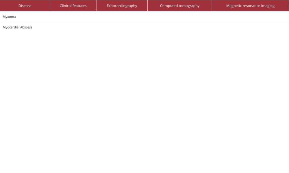 Table 1.. Characteristic clinical and imaging features of myxoma and myocardial abscess.
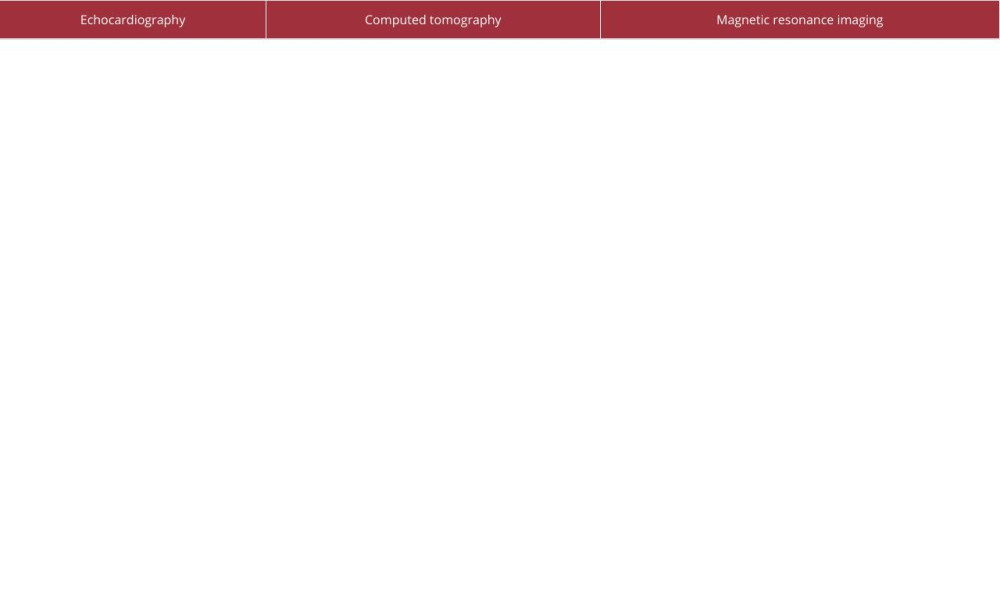 Table 2.. Characteristic features of caseous calcification of the mitral annulus in particular medical imaging techniques.Table 1.. Characteristic clinical and imaging features of myxoma and myocardial abscess.Table 2.. Characteristic features of caseous calcification of the mitral annulus in particular medical imaging techniques.
 In Press
Case report 
Am J Case Rep In Press; DOI: 10.12659/AJCR.953173
Case report 
Am J Case Rep In Press; DOI: 10.12659/AJCR.953192
Case report 
Am J Case Rep In Press; DOI: 10.12659/AJCR.952818
Case report
Am J Case Rep In Press; DOI: 10.12659/AJCR.953608
Most Viewed Current Articles
07 Dec 2021 : Case report
22,364,578
DOI :10.12659/AJCR.934347
Am J Case Rep 2021; 22:e934347
06 Dec 2021 : Case report  174,245
DOI :10.12659/AJCR.934406
Am J Case Rep 2021; 22:e934406
21 Jun 2024 : Case report
119,744
DOI :10.12659/AJCR.944371
Am J Case Rep 2024; 25:e944371
07 Mar 2024 : Case report
64,648
DOI :10.12659/AJCR.943133
Am J Case Rep 2024; 25:e943133