15 June 2025: Articles 
Successful Conservative Treatment of Tracheobronchial Injury as a Complication After Emergency Tracheal Intubation in an Infant: A Case Report
Diagnostic / therapeutic accidents, Management of emergency care, Clinical situation which can not be reproduced for ethical reasons, Rare coexistence of disease or pathology
Kazuki Kikuchi ABCDEF 1, Keisuke SuzukiDOI: 10.12659/AJCR.947693
Am J Case Rep 2025; 26:e947693


Abstract
BACKGROUND: Tracheal intubation is a routine but potentially hazardous procedure, with complications ranging from mild mucosal injury to severe tracheobronchial trauma. Tracheobronchial injuries are particularly critical, with high morbidity and mortality rates, necessitating prompt diagnosis and appropriate management. However, there is no clear consensus on optimal treatment, especially in pediatric patients.
CASE REPORT: We present a case of a previously healthy 11-month-old girl who developed a tracheobronchial injury following endotracheal intubation for status epilepticus. Computed tomography (CT) revealed a 6×4 mm tracheobronchial injury, left pneumothorax, and mediastinal emphysema. Given the minor nature of the injury, stable respiratory status, and the expectation of early extubation, conservative management was selected. The patient was treated with nasal intubation, antibacterial therapy, and close monitoring. She was successfully extubated on the second day and discharged on the tenth day without complications.
CONCLUSIONS: This case highlights the importance of recognizing tracheobronchial injury as a potential intubation-related complication, especially in infants with fragile airway anatomy. Key factors contributing to the injury included deep tube insertion, inappropriate tube size, and elevated cuff pressure. Our experience suggests that conservative management can be a viable option in select cases of pediatric tracheobronchial injury when symptoms are minimal and early extubation is feasible. Additionally, meticulous intubation techniques, proper tube selection, and experienced personnel are critical in preventing such complications. Further studies are needed to establish standardized guidelines for managing tracheobronchial injuries in infants and young children.
Keywords: Case Reports, conservative treatment, Infant, Intubation, Trachea, Humans, Female, Intubation, Intratracheal, Bronchi, Tomography, X-Ray Computed, Pneumothorax, Mediastinal Emphysema, Status epilepticus
Introduction
Tracheal intubation is a routine procedure. However, it carries various risks of complications, including sore throat, laryngitis, glottal edema, mucosal ulceration, laryngeal and tracheal stenosis, tracheal wall necrosis, fistula, aspiration, esophageal intubation, bronchial intubation, atelectasis, and tracheal injury [1–3]. Among these, tracheobronchial injury is a particularly serious complication, with a mortality rate of up to 22%. However, there is no clear consensus on the optimal management for this complication [1]. The most common site of tracheobronchial injury is the membranous wall of the trachea, which is the posterior portion of the trachea that lacks support from cartilaginous tissue [1]. Symptoms of tracheobronchial injury include subcutaneous emphysema, respiratory failure, pneumothorax, and hemoptysis. While some injuries are recognized immediately after intubation or extubation, others may not manifest until several days after the procedure, often delaying diagnosis [4]. Delayed diagnosis may contribute to mediastinitis development and symptom worsening. Therefore, early diagnosis and appropriate treatment selection are crucial for managing this condition.
The present report describes the successful conservative management of a tracheobronchial injury in an infant, highlighting critical considerations for early diagnosis and treatment planning, particularly in infants.
Case Report
The patient was a previously healthy 11-month-old girl who weighed 8 kg. The patient experienced a fever of 38°C one evening, followed by symmetrical clonic convulsions that appeared in the extremities the next day, prompting a request for emergency medical assistance. Upon arrival at the emergency department after experiencing seizures for 1 hour, the patient continued to have ongoing clonic convulsions, with a Glasgow Coma Scale score of 3 (E1V1M1), body temperature of 37.8°C, blood pressure of 104/84 mmHg, and pulse rate of 190 beats/min. The patient’s weak spontaneous breathing necessitated mechanical ventilation via a Jackson-Rees system. The seizure activity rapidly terminated when midazolam was administered 5 minutes after presentation. However, the patient’s respiratory depression persisted, requiring continued assisted ventilation.
Blood gas analysis (Table 1) at the time of admission revealed marked acidosis and hypercapnia, prompting the decision to proceed with intubation and mechanical ventilation. The patient was well-ventilated via an endotracheal tube with a cuff and an internal diameter of 3.5 mm. However, she required reintubation as the tube was accidentally pulled out. During the reintubation, clonic convulsions reoccurred 20 minutes after presentation, and midazolam was re-administered. The patient was intubated with the endotracheal tube inserted to a depth of 15 cm from the right corner of the mouth. Auscultation revealed decreased breath sounds in the left lung field, accompanied by poor thoracic movement. Right, single-lung intubation was diagnosed based on the chest radiographic findings (Figure 1). The endotracheal tube depth was adjusted to 12 cm from the right mouth angle, and the patient was admitted to the intensive care unit with good ventilation. A computed tomography (CT) scan performed at admission revealed infiltrative shadows indicative of aspiration, left pneumothorax, mediastinal emphysema, and tracheobronchial injury (Figure 2). Measurement on the CT scan indicated a tracheobronchial injury of 6×4 mm in size. Although the possibility of barotrauma was considered as a differential diagnosis, tracheal damage was observed at the carina where the intubation tube would have reached; therefore, the primary condition was diagnosed as tracheobronchial injury. Therefore, nasal intubation was performed 3 hours after presentation, and the tube depth was adjusted to ensure stable fixation. Considering that the CT scan showed only minor tracheobronchial injury, the patient’s seizures had been controlled, and she had begun to breathe spontaneously, conservative treatment was preferred.
As the nasal discharge was positive for influenza A antigen, peramivir hydrate (80 mg) was intravenously administered. Additionally, to prevent exacerbation of the leak due to elevated pressure in the trachea, we planned for early extubation. The patient was accordingly administered antibacterial therapy to treat the pneumonia. A continuous infusion of 0.04 mg/h midazolam was administered to maintain sedation, and triclofos sodium was additionally administered in case of restlessness. The patient was extubated and received oxygen via a high-flow nasal cannula on the second day of admission and an oxygen mask on the third day. A CT scan performed on the sixth day revealed resolution of the pneumonia, pneumothorax, and mediastinal emphysema (Figure 3). The patient was discharged from the hospital on the tenth day.
Discussion
The present case report describes the case of an infant with a tracheobronchial injury who effectively responded to conservative treatment. The injury in our patient likely resulted from multiple factors, including deep intubation, inappropriate tube size, and high cuff pressure. These issues highlight the importance of proper preparation and technique, particularly when treating pediatric patients.
The estimated incidence of tracheal injury during intubation is 1/20 000 [5]. Although this complication does occur at a certain rate, reports of tracheobronchial injuries in healthy infants are extremely rare. This is likely due to the limited need for intubation in healthy infants. Conservative treatment of bronchial injuries in neonates and infants has been reported previously, including in patients requiring mechanical ventilation for respiratory disease and tracheostomy for airway management because of congenital anomalies [6]. In comparison with those patients, our patient had no preexisting respiratory disease and the injury was likely iatrogenic, highlighting the existence of different etiologies and treatment approaches. Tracheobronchial injury can be attributed to both mechanical and anatomical factors. Mechanical factors include multiple attempts before successful intubation, inexperienced personnel, protrusion of the stylet from the endotracheal tube, cuff overinflation, improper tube tip position, failure to deflate the cuff when repositioning the tube, improper tube size, severe coughing, and patient movement during intubation. Anatomical factors include congenital tracheal abnormalities, the fragility of the tracheobronchial area, inflammatory conditions such as chronic obstructive pulmonary disease, neoplastic lesions near the trachea, chronic steroid use, advanced age, and female sex [1,7,8]. In our patient, the tracheobronchial injury occurred during intubation due to several contributing factors. First, the medical staff had limited experience in intubating infants. Second, the initially inserted endotracheal tube was inadvertently pulled out while the cuff inflated, leading to an elevated cuff pressure immediately post-intubation. Third, the endotracheal tube was inserted up to a depth of 15 cm, which exceeded the recommended tube insertion length of 10.5–11.0 cm. Finally, the size of the tube used (internal diameter: 3.5 mm) exceeded the recommended size (internal diameter: 3.0 mm). The deep insertion of the tube could have damaged the tracheobronchial area near the endotracheal tube tip, and pressure injury could have occurred as the patient was ventilated via a Jackson-Rees system. Given that the injury was located just above the carina and the airway was anatomically narrow owing to the patient’s age, surgical repair was considered technically challenging and a potentially high-risk procedure. These anatomical and technical limitations, combined with the expectation of early extubation and stable vital signs, prompted us to opt for conservative management. The patient was a well-developed but anatomically fragile infant [6]. The adhesive strength of the tube fixation tape decreased due to the patient’s uncontrolled seizures, saliva, and fever-induced sweating, and we were concerned about skin damage from the fixation tape. Therefore, we believe that using a device for tube fixation would have been more effective than using tape [9]. In pediatric cases, intubation is less common than in adults, and not all tube sizes are readily available in every medical setting. Additionally, given variations in tube size, the amount of air injected into the cuff and insertion depth differ for each patient. In infants, the tracheal length is short, and the position of the tube is considerably affected by factors such as tube deflection in the oral cavity, neck position, and body movements. As the tube position can easily shift, careful technique, adequate preparation, and confirmation of proper tube placement are essential [10]. Moreover, in children, the endotracheal tube is less likely to shift with changes in head posture with nasotracheal intubation than with oral intubation [10]. Therefore, this should be considered when inserting the tracheal tube, especially when achievement of the recommended depth is difficult or when body movement is a concern. It is also worth noting that nasal intubation is less likely to induce the gag reflex than oral intubation, and the tape used to secure the intubation tube is less likely to be affected in patients with a lot of saliva. On the other hand, nasal intubation may cause nosebleeds and is contraindicated in patients with suspected skull base fractures. Cuff pressures exceeding 30 cmH2O may lead to tracheal injury due to ischemic necrosis of the tracheal mucosa [8]. Several reports suggest that even experienced emergency physicians and anesthesiologists may not be able to accurately adjust the cuff pressure solely through palpation alone, emphasizing the need for a cuff manometer when inflating the cuff [11,12].
Although no consensus exists on the management of tracheobronchial injuries, patients with small injuries (<20 mm), those with minimal and nonprogressive symptoms, those with no air leak during spontaneous breathing, and those in whom extubation within 24 h is expected, are often treated conservatively [1,6,13,14]. However, size-based criteria for injury assessment, such as the 20-mm threshold often used in adults, may not directly apply to infants. Instead, clinical features such as air leaks, symptom progression, and readiness for extubation should guide clinical management. Conservative management includes cuff placement distal to the injury site, regular endotracheal suctioning, chest drain placement if necessary, and appropriate empiric antibacterial therapy. If the lesion is near the tracheal bifurcation, a thin cuffed tube can, reportedly, be placed in each main bronchus [13]. Surgical treatment is recommended when mediastinitis or esophageal injury is present, as the complications of mediastinitis are associated with increased mortality, and esophageal injury is a risk factor for the development of mediastinitis [15]. In our patient, the endotracheal tube cuff could not be placed distal to the injury site, as the injured area was directly above the carina of the trachea. Moreover, placing a tube in each main bronchus was not feasible because the patient had a narrow airway, which was still developing. However, her seizures had been controlled, and spontaneous breathing had recovered at the time of intensive care unit admission. Given that extubation within 24 h was expected, we implemented conservative treatment, intending to remove the tube as soon as possible. Notably, the patient demonstrated good progress. This case is notable because it demonstrates that even in a small infant with a complex medical presentation, conservative treatment, without surgical intervention, can be successful. This case report adds valuable evidence for clinicians managing similar cases of high-risk, pediatric airway injuries. Although surgical treatment may be considered for tracheobronchial injury, conservative management should be the first choice if the injury appears minor on CT or if early extubation is expected.
Finally, this case underscores the need for skilled personnel during pediatric intubation. Given the technical challenges, procedures in infants should ideally be performed by or under the supervision of experienced practitioners. This is especially important in emergency settings, where suboptimal intubation techniques can lead to severe complications.
Conclusions
Tracheobronchial injuries in infants are rare and challenging to manage. The present case demonstrates that under specific conditions – such as small injury size, minimal symptoms, and stable respiratory status – conservative treatment may be effective. However, the unique anatomical characteristics of infants necessitate careful monitoring and skilled clinical judgment. Further cases should be reported to establish infant-specific guidelines for managing tracheobronchial injuries.
Figures
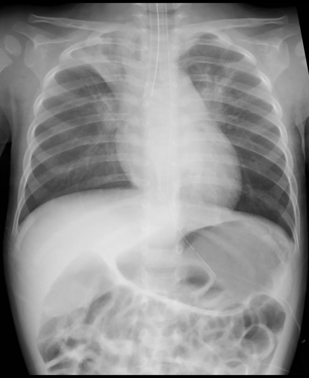 Figure 1. At the time of arrival, a chest radiograph revealed right single-lung intubation.
 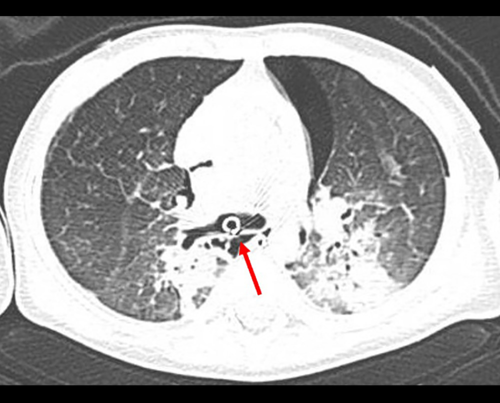 Figure 2. At admission, computed tomography revealed infiltrative shadows indicative of aspiration, left pneumothorax, mediastinal emphysema, and tracheobronchial injury (arrow).
 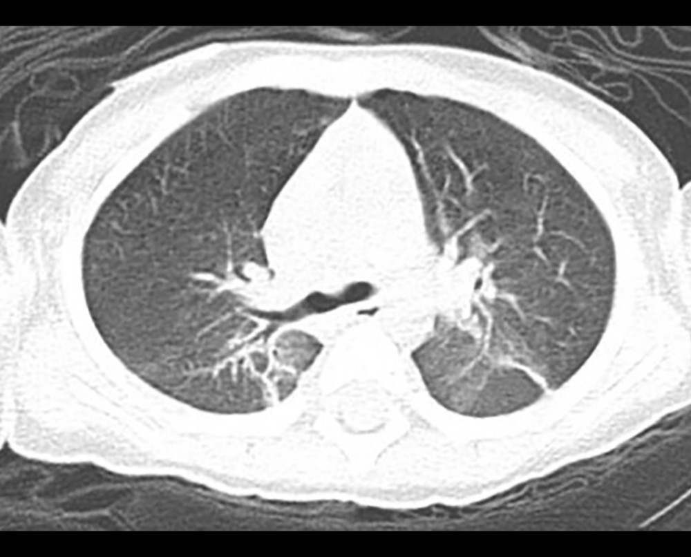 Figure 3. On the sixth day, computed tomography revealed the resolution of the pneumothorax and mediastinal emphysema.
References
1. Miñambres E, Burón J, Ballesteros MA, Tracheal rupture after endotracheal intubation: A literature systematic review: Eur J Cardiothorac Surg, 2009; 35; 1056-62
2. Jaber S, Rollé A, Godet T, Effect of the use of an endotracheal tube and stylet versus an endotracheal tube alone on first-attempt intubation success: A multicentre, randomised clinical trial in 999 patients: Intensive Care Med, 2021; 47; 653-64
3. Russotto V, Myatra SN, Laffey JG, Intubation practices and adverse peri-intubation events in critically ill patients from 29 countries: JAMA, 2021; 325; 1164-72 [published erratum appears in JAMA. 2021;325:2507]
4. Jougon J, Ballester M, Choukroun E, Conservative treatment for postintubation tracheobronchial rupture: Ann Thorac Surg, 2000; 69; 216-20
5. Orta DA, Cousar JE, Yergin BM, Olsen GN, Tracheal laceration with massive subcutaneous emphysema: A rare complication of endotracheal intubation: Thorax, 1979; 34; 665-69
6. Cunningham LC, Jatana KR, Grischkan JM, Conservative management of iatrogenic membranous tracheal wall injury: A discussion of 2 successful pediatric cases: JAMA Otolaryngol Head Neck Surg, 2013; 139; 405-10
7. Marty-Ané CH, Picard E, Jonquet O, Mary H, Membranous tracheal rupture after endotracheal intubation: Ann Thorac Surg, 1995; 60; 1367-71
8. Sudhoff TH, Seidl RO, Estel B, Coordes A, Association of oversized tracheal tubes and cuff overinsufflation with postintubation tracheal ruptures: Clin Exp Otorhinolaryngol, 2015; 8; 409-15
9. Buckley JC, Brown AP, Shin JS, Rogers KM, Hoftman NN: Anesth Analg, 2016; 122; 1439-43
10. Olufolabi AJ, Charlton GA, Spargo PM, Effect of head posture on tracheal tube position in children: Anaesthesia, 2004; 59; 1069-72
11. Michlig SA, Anaesthetic staff cannot identify extremely high tracheal tube cuff pressures by palpation of the pilot balloon: Br J Anaesth England, 2013; 111; 300-1
12. Hoffman RJ, Parwani V, Hahn IH, Experienced emergency medicine physicians cannot safely inflate or estimate endotracheal tube cuff pressure using standard techniques: Am J Emerg Med, 2006; 24; 139-43
13. Conti M, Pougeoise M, Wurtz A, Management of postintubation tracheobronchial ruptures: Chest, 2006; 130; 412-18
14. Conti M, Porte H, Wurtz A, Management of tracheobronchial ruptures in critically ill patients: Minerva Anestesiol, 2007; 73; 7-8
15. Cardillo G, Carbone L, Carleo F, Tracheal lacerations after endotracheal intubation: a proposed morphological classification to guide non-surgical treatment: Eur J Cardiothorac Surg, 2010; 37; 581-87
Figures
Figure 1. At the time of arrival, a chest radiograph revealed right single-lung intubation.Figure 2. At admission, computed tomography revealed infiltrative shadows indicative of aspiration, left pneumothorax, mediastinal emphysema, and tracheobronchial injury (arrow).Figure 3. On the sixth day, computed tomography revealed the resolution of the pneumothorax and mediastinal emphysema. In Press
Case report 
Am J Case Rep In Press; DOI: 10.12659/AJCR.949566
Case report 
Am J Case Rep In Press; DOI: 10.12659/AJCR.952005
Case report 
Am J Case Rep In Press; DOI: 10.12659/AJCR.952443
Case report
Am J Case Rep In Press; DOI: 10.12659/AJCR.953173
Most Viewed Current Articles
07 Dec 2021 : Case report
22,282,456
DOI :10.12659/AJCR.934347
Am J Case Rep 2021; 22:e934347
06 Dec 2021 : Case report  174,072
DOI :10.12659/AJCR.934406
Am J Case Rep 2021; 22:e934406
21 Jun 2024 : Case report
119,650
DOI :10.12659/AJCR.944371
Am J Case Rep 2024; 25:e944371
07 Mar 2024 : Case report
64,568
DOI :10.12659/AJCR.943133
Am J Case Rep 2024; 25:e943133