02 July 2025: Articles 
A 69-Year-Old Woman with an Occipital Giant Proliferating Trichilemmal (Pilar) Cyst Treated with Surgical Resection
Rare disease
Mahammad Davudov A 1*, Sahib Bilalzade F 1, Oktay Mehdiyev B 1, Javid Huseynov E 1, Veli Nasirov E 2, Roya Basser F 1DOI: 10.12659/AJCR.947743
Am J Case Rep 2025; 26:e947743


Abstract
BACKGROUND: Proliferating trichilemmal cysts, also referred to as pilar cysts, are benign adnexal tumors in the scalp diagnosed most commonly in middle-aged women. The cysts can be large and require careful surgical resection, as malignant variants can exist. Although uncommon, it is rare for these tumors to reach large sizes. This report describes a 69-year-old woman with an occipital giant proliferating trichilemmal (pilar) cyst treated with surgical resection and flap reconstruction.
CASE REPORT: A 69-year-old female patient presented with a slowly growing mass in the occipital region of the head, which had been present for 11 years. The mass exhibited areas of hyperkeratosis and an unpleasant odor. The tumor was first excised with electrocautery to minimize bleeding during removal. We initially attempted to repair the defect using a local rotation skin flap. Although this approach provided some coverage, it quickly became clear that the available tissue would not be enough. As a result, we decided to supplement the repair with a supraclavicular artery island flap to ensure adequate closure.
CONCLUSIONS: This case highlights how crucial it is to combine thorough clinical evaluation, imaging, and histopathological analysis when dealing with proliferative pilar tumors. In this instance, we successfully removed a large tumor located in the occipital region of a 69-year-old woman. The diagnosis was later confirmed histologically as a benign proliferative pilar tumor. Regular follow-up is essential to ensure the absence of recurrence and to address potential future complications.
Keywords: Female, Occipital Bone, Rare Diseases, Surgery, Plastic, Humans, Aged, Epidermal Cyst, Scalp, Surgical Flaps, Skin Neoplasms, Hair Diseases
Introduction
Adnexal tumors are a heterogeneous group of skin neoplasms originating from pilosebaceous units and sweat glands. Among these, proliferating trichilemmal cysts (PTCs) are rare benign lesions derived from the outer root sheath of the hair follicle, most commonly found on the scalp of older women. Clinically, they typically present as slow-growing, firm, and superficial nodules. Histologically, these cysts have a lobular structure, produce a type of keratin typical of hair follicles (trichilemmal keratinization), and show increased growth of skin cells, and they lack the usual granular layer seen in normal skin. Although generally benign, rare cases of malignant transformation have been reported. Early diagnosis and complete surgical excision remain the mainstays of treatment to prevent local complications and recurrence [1].
Case Report
HISTOLOGICAL EXAMINATION:
Histopathological analysis revealed trichilemmal keratinization characteristic of pilar cysts, with irregularly contoured lobular-cystic morphology and calcified areas. A foreign body-type reaction to keratin was observed sporadically (Figure 7). Focal invasion into the deep dermis and subcutaneous tissue was noted. Low-grade nuclear atypia and focal mitosis were observed (Figures 7, 8). The tumor, measuring 15×12×7.5 cm, had intact surgical margins. No perineural or lymphovascular invasion was observed. High-grade atypia and atypical mitosis were absent, and no necrosis was detected. According to the classification criteria proposed by Ye et al, the tumor was classified as Group 2. The irregular borders and invasion into the deep dermis and subcutaneous adipose tissue supported this classification [1].
Discussion
This case report contributes to the current understanding of proliferating trichilemmal cysts by highlighting the clinical and surgical challenges posed by giant lesions, particularly those involving the occipital region. Unlike typical pilar cysts, which are often small and asymptomatic, this case demonstrates how neglected lesions can attain significant size, necessitating advanced reconstructive techniques. The chronic growth history, absence of overt malignancy, and successful use of local and regional flaps offer valuable insights into diagnostic differentiation and surgical planning for similar tumors. Moreover, the report emphasizes the necessity of a multidisciplinary approach, integrating pathology, imaging, and reconstructive surgery for optimal outcomes in extensive adnexal neoplasms.
Proliferating pilar tumors (PPTs) are most commonly seen in postmenopausal women, particularly those over the age of 60 [2,3]. This sex- and age-related pattern may be influenced by hormonal and biological changes that occur after menopause, which are thought to play a role in the development of these tumors. Interestingly, PPTs are rarely seen in bald individuals, likely because these tumors originate from hair follicle structures, which are absent in bald areas. Since baldness is far more common in men than in women, this may partly explain why women tend to develop PPTs more often. These trends highlight how factors like age and hair follicle density can help us better understand who is more at risk. [4].
When PPTs exhibit prolonged growth, necrosis, and calcification within the tumor can occur due to impaired blood supply [5]. As these tumors increase in size, the risk of malignant transformation also rises, necessitating lymph node dissection in such cases. In our case, the enlarged lymph node was evaluated as reactive [6].
In a study by Je Nappi involving 76 patients, PPTs were histopathologically classified into 3 grades. Based on histopathological findings, our case was classified as Grade 2, which have a 15% risk of recurrence [1].
While Alam et al emphasized the diagnostic challenge in differentiating PTC from squamous cell carcinoma due to ulceration and rapid growth, our case exhibited no such clinical ambiguity, as the lesion lacked ulcerative features and resembled neither an epidermoid cyst nor a malignant cutaneous tumor. The lesion in their report was excised without the need for advanced reconstructive techniques. In contrast, our case required a 2-stage closure involving a local rotation flap and a supraclavicular artery island flap, highlighting the importance of reconstructive planning in large tumors [7].
Although these tumors are known for their recurrence rates, close and extended follow-up remains crucial to catch any signs of regrowth or progression early. When recurrence does occur, it can be clinically challenging, sometimes suggesting a more aggressive course. For that reason, regular monitoring plays a key role, not just in spotting recurrence, but also in allowing timely intervention when needed. This kind of careful follow-up helps manage complications more effectively and supports better overall outcomes for the patient [8].
In this case, CT imaging proved particularly useful in assessing the tumor’s characteristics. The scan did not reveal any signs of bone invasion or resorption, which supported the initial impression of a benign lesion. These radiological features suggested aggressive tumor behavior, as malignant tumors often infiltrate and destroy surrounding bone structures, in contrast to benign lesions, which typically exhibit more localized growth patterns without significant bone destruction [2]. In significant defects, partial necrosis of the local rotation flap highlights the need for alternative reconstruction methods [9]. The necrotic area presents a considerable risk of infection, which can complicate the healing process. To reduce this risk and promote optimal wound healing, topical 1% gentamicin was applied following surgical debridement. This approach helps control infection and supports tissue repair, facilitating the progression of secondary wound healing [10]. Following removal of the rotation flap, a defect was created in the neck, which was reconstructed using a supraclavicular fascio-cutaneous flap [11].
Conclusions
Proliferating trichilemmal cysts are a rare subset of benign scalp tumors with the potential for extensive growth and, in some cases, malignant transformation. This case, involving a 69-year-old woman with a giant occipital lesion, illustrates the importance of early recognition, thorough radiologic and histopathologic assessment, and a tailored surgical approach to achieve complete resection and functional reconstruction. The use of combined flap techniques in a vascular-rich area demonstrates the complexity of surgical management in advanced cases. Regular follow-up remains essential to monitor for recurrence or late-onset complications.
Figures
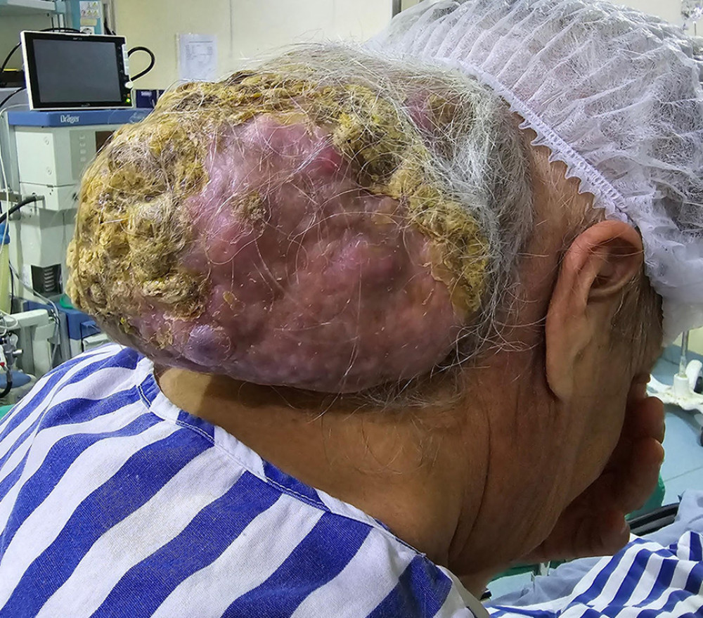 Figure 1. Giant tumor in the occipital region showing areas of hyperkeratosis, erythema, and cystic regions. The mass had been slowly growing for 11 years.
 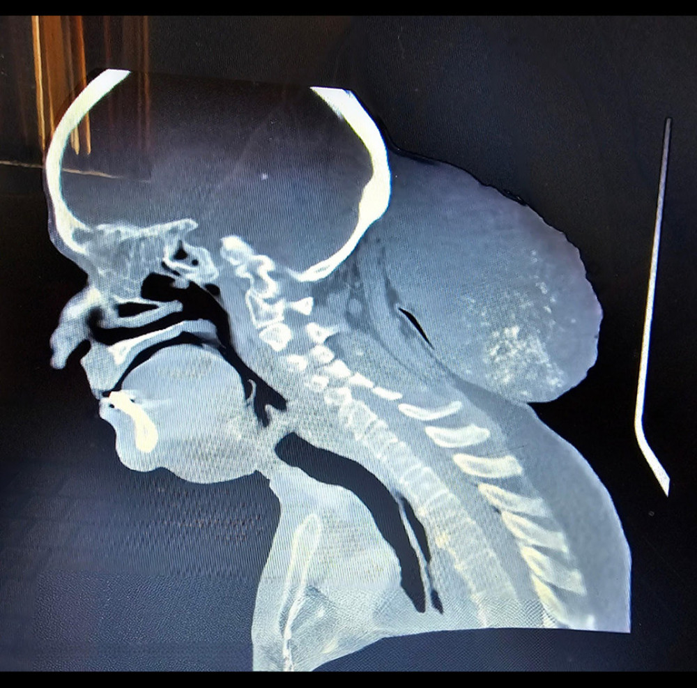 Figure 2. The mass, measuring approximately 15×14×11 cm, was located in the occipital area. Imaging techniques revealed no evidence of bone infiltration or resorption, although areas of calcification were observed within the mass.
 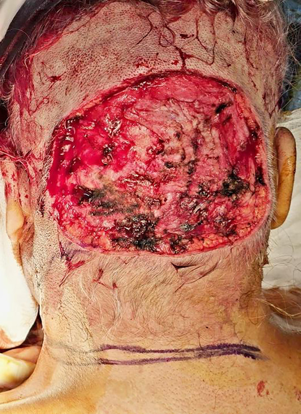 Figure 3. After the mass was surgically excised, a significant defect area was left in the occipital region.
  Figure 4. The defect was initially managed using a local rotational skin flap. However, a supraclavicular artery island flap was utilized for complete closure due to inadequate tissue availability.
 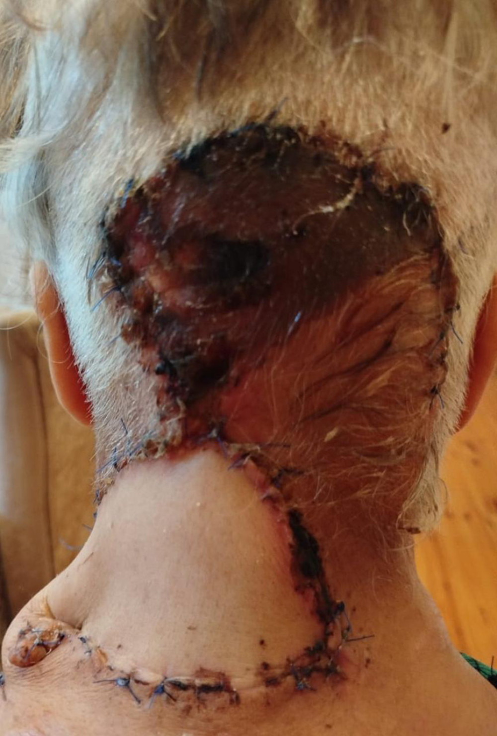 Figure 5. Two weeks postoperatively, necrosis of the damaged tissue and a clear demarcation line were distinctly visible.
 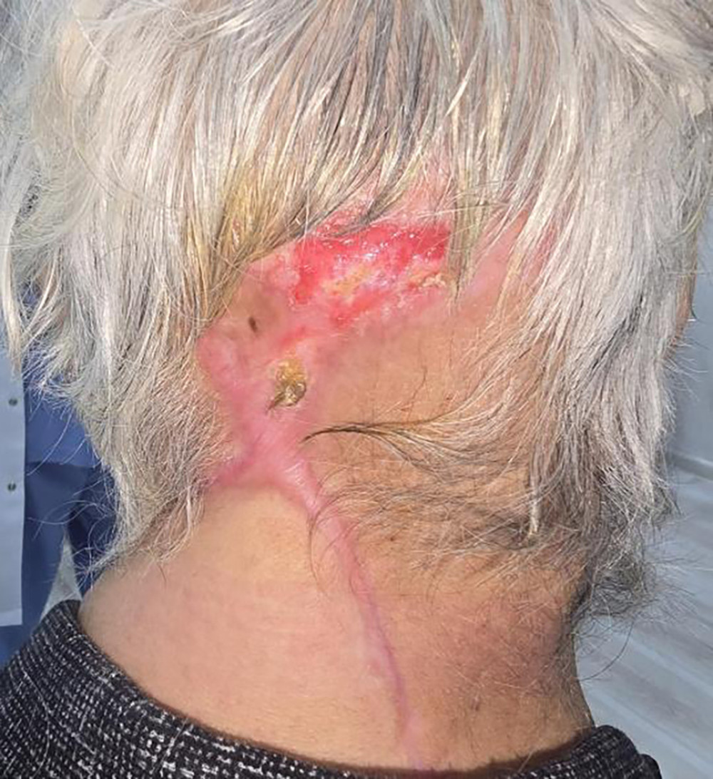 Figure 6. Six months after the surgery, the necrotic tissue was surgically debrided, and the wound was left to heal by secondary intention.
 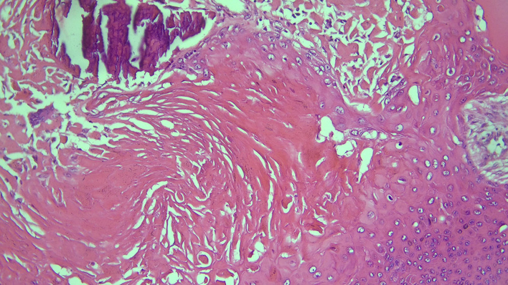 Figure 7. Trichilemmal keratinization, foreign body-type reaction, and calcification (H&E: ×200).
 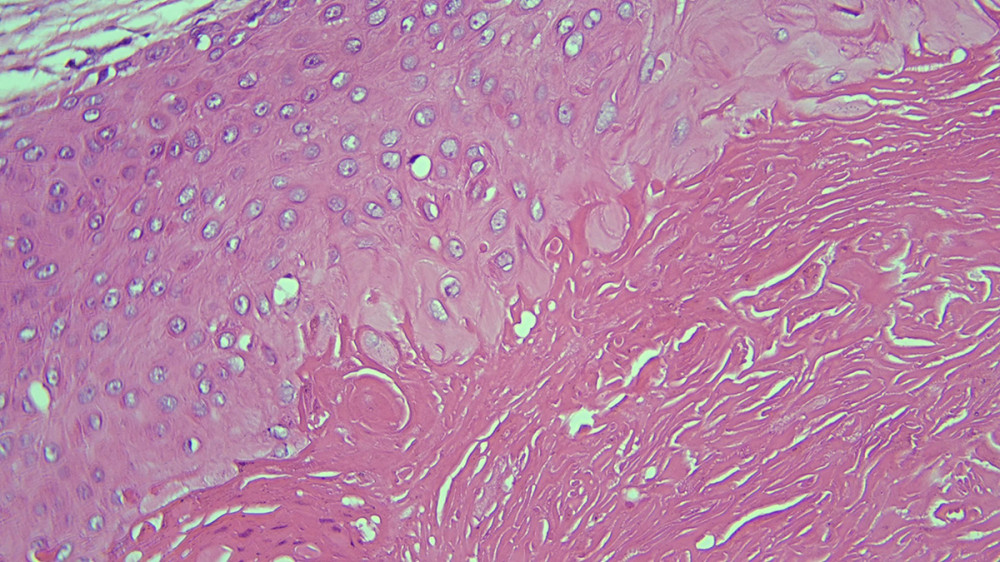 Figure 8. Slight atypia, eosinophilic compact keratin showing lamination in squamous epithelium without granular layer (H&E: ×400).
 References
1. Ye J, Nappi O, Swanson PE, Patterson JW, Wick MR, Proliferating pilar tumors: A clinicopathologic study of 76 cases with a proposal for definition of benign and malignant variants: Am J Clin Pathol, 2004; 122(4); 566-74
2. Noorbakhsh SI, Harris BR, Adelanwa AO, Thomay AA, Large proliferating trichilemmal tumor (PTT) of the occipital scalp: ACS Case Rev Surg, 2024; 4(6); 91-95
3. Alarcón Pérez CE, Gómez Ángulo D, Olmos Pérez M, Management of 3 proliferating pilar tumors: Definition, differential diagnosis, and treatment options: Actas Dermosifiliogr (Engl Ed), 2019; 110(10); 850-54
4. Sharma R, Verma P, Yadav P, Sharma S, Proliferating trichilemmal tumor of scalp: Benign or malignant, a dilemma: J Cutan Aesthet Surg, 2012; 5(3); 213-15
5. Narra R, Kamaraju SK, Putcha A, Trichilemmal cysts of scalp: Imaging findings: Indian J Case Reports, 2018; 4(6); 429-30
6. Kim UG, Kook DB, Kim TH, Kim CH, Trichilemmal carcinoma from proliferating trichilemmal cyst on the posterior neck: Arch Craniofac Surg, 2017; 18(1); 50-53
7. Alam K, Gupta K, Maheshwari V, A large proliferating trichilemmal cyst masquerading as squamous cell carcinoma: Indian J Dermatol, 2015; 60(1); 104
8. Morgado B, Agostini P, Rivero A, Silva N, Extensive and ulcerated malignant proliferating trichilemmal (pilar) tumor arising from multiple large degenerated trichilemmal cysts: BMJ Case Rep, 2016; 2016; bcr2015209785
9. Al-Shanawani B, Abdelhamid MM, Al-Shomer FM, Giant proliferating trichilemmal tumor: Arch Plast Surg, 2013; 40(4); 461-63
10. Junker JPE, Lee CCY, Samaan S, Topical delivery of ultrahigh concentrations of gentamicin reduces bacterial levels in infected porcine wounds: Plast Reconstr Surg, 2015; 135(1); 151-59
11. Kokot N, Mazhar K, Reder LS, Use of the supraclavicular artery island flap for reconstruction of cervicofacial defects: Otolaryngol Head Neck Surg, 2014; 150(2); 222-28
Figures
Figure 1. Giant tumor in the occipital region showing areas of hyperkeratosis, erythema, and cystic regions. The mass had been slowly growing for 11 years.Figure 2. The mass, measuring approximately 15×14×11 cm, was located in the occipital area. Imaging techniques revealed no evidence of bone infiltration or resorption, although areas of calcification were observed within the mass.Figure 3. After the mass was surgically excised, a significant defect area was left in the occipital region.Figure 4. The defect was initially managed using a local rotational skin flap. However, a supraclavicular artery island flap was utilized for complete closure due to inadequate tissue availability.Figure 5. Two weeks postoperatively, necrosis of the damaged tissue and a clear demarcation line were distinctly visible.Figure 6. Six months after the surgery, the necrotic tissue was surgically debrided, and the wound was left to heal by secondary intention.Figure 7. Trichilemmal keratinization, foreign body-type reaction, and calcification (H&E: ×200).Figure 8. Slight atypia, eosinophilic compact keratin showing lamination in squamous epithelium without granular layer (H&E: ×400). In Press
Case report 
Am J Case Rep In Press; DOI: 10.12659/AJCR.953173
Case report 
Am J Case Rep In Press; DOI: 10.12659/AJCR.953192
Case report 
Am J Case Rep In Press; DOI: 10.12659/AJCR.952818
Case report
Am J Case Rep In Press; DOI: 10.12659/AJCR.953608
Most Viewed Current Articles
07 Dec 2021 : Case report
22,364,578
DOI :10.12659/AJCR.934347
Am J Case Rep 2021; 22:e934347
06 Dec 2021 : Case report  174,245
DOI :10.12659/AJCR.934406
Am J Case Rep 2021; 22:e934406
21 Jun 2024 : Case report
119,744
DOI :10.12659/AJCR.944371
Am J Case Rep 2024; 25:e944371
07 Mar 2024 : Case report
64,648
DOI :10.12659/AJCR.943133
Am J Case Rep 2024; 25:e943133