31 July 2025: Articles 
Evidence of Vertical Dengue Transmission: A Case of Fever in a 4-Day-Old Neonate
Challenging differential diagnosis, Diagnostic / therapeutic accidents, Management of emergency care, Unexpected drug reaction, Congenital defects / diseases, Clinical situation which can not be reproduced for ethical reasons
Bugis Mardina Lubis BDEF 1,2*, Rycha Dwi Syafutri E 1, Halisa Tiara Ariani Matondang E 1, Dewi Sari BE 2DOI: 10.12659/AJCR.948417
Am J Case Rep 2025; 26:e948417


Abstract
BACKGROUND: Dengue is a virus transmitted by Aedes mosquitoes, with rare cases of vertical transmission occurring through infected mothers. Acute infection can be identified by molecular viral testing or by detecting dengue non-structural protein 1 (NS1) and elevated serum IgM. This report describes the case of a 4-day-old male neonate with fever, rash, and jaundice due to vertical transmission of maternal dengue virus infection, confirmed by NS1 detection.
CASE REPORT: We report a case of a 4-day-old male neonate admitted with high fever, rash, and jaundice. Initial laboratory investigations showed thrombocytopenia, elevated liver enzymes, and signs of systemic inflammation. The mother and infant both tested positive for dengue NS1 antigen, confirming vertical transmission. The neonate received supportive management in the neonatal intensive care unit (NICU), including intravenous fluids and platelet transfusions. He gradually recovered without complications and was discharged in stable condition.
CONCLUSIONS: This case underscores the importance of considering congenital dengue in neonates born to mothers with peripartum dengue infection. NS1 antigen testing can aid early diagnosis, even before seroconversion occurs. Prompt supportive care, especially monitoring and treating thrombocytopenia, is essential for a favorable outcome. Increased awareness and standardized management guidelines are needed to improve neonatal care in similar cases.
Keywords: Antigens, Viral, Dengue, infections, Thrombocytopenia, Vertical Dimension, Humans, Infant, Newborn, Male, Fever, Infectious Disease Transmission, Vertical
Introduction
Dengue hemorrhagic fever (DHF) has become increasingly prevalent due to urbanization, environmental changes, and evolving epidemiology [1]. It is caused by a single-stranded RNA viruses belonging to the
Clinical manifestations of congenital dengue vary and can include fever, thrombocytopenia, hepatomegaly, rash, and respiratory distress. Often, symptoms are mild but can escalate to severe forms resembling DHF in adults. Dengue with warning signs is characterized by clinical features such as abdominal pain, persistent vomiting, clinical fluid accumulation such as ascites or pleural effusion, mucosal bleeding, lethargy, hepatomegaly (liver enlargement >2 cm), increase in hematocrit, and thrombocytopenia [2]. The lack of maternal protective antibodies and direct viral transmission are significant contributors to neonatal dengue [4–8].
Laboratory findings can include thrombocytopenia, leukopenia, and elevated levels of aspartate aminotransferase (AST). Confirmatory diagnosis can be established through detection of viral antigens using enzyme-linked immunosorbent assay (ELISA), polymerase chain reaction (PCR) or by isolating the virus from body fluids. Another important diagnostic examination is the detection of NS1, a highly conserved glycoprotein essential for dengue virus viability. The RNA genome of the dengue virus is processed by cellular and viral proteases to generate 3 structural proteins and 7 non-structural proteins: NS1, NS2a, NS2b, NS3, NS4a, NS4b, and NS5. NS1 is typically associated with intracellular organelles during cell infection. During the acute phase of infection, NS1 can also be found circulating in the bloodstream. [2,9]
Management of dengue is guided by the clinical phase of illness. For patients without warning signs, symptomatic treatment with acetaminophen and adequate oral fluids intake is recommended. In contrast, patient exhibiting warning signs may require medical observation and carefully monitored intravenous fluid therapy [2].
Symptomatic dengue infection during pregnancy has been associated with an increased risk of preterm birth and low birth weight, although the overall incidence of vertical transmission remains low [9]. Notably, maternal dengue infection during the third trimester has been linked to adverse perinatal outcomes. The risk of vertical transmission is highest when maternal infection occurs near the time of delivery [10].
Despite its growing relevance, congenital dengue remains under-reported, with limited case studies in the literature. This report describes the case of a 4-day-old male neonate with fever, rash, and jaundice due to vertical transmission of maternal dengue virus infection, confirmed by NS1 detection.
Case Report
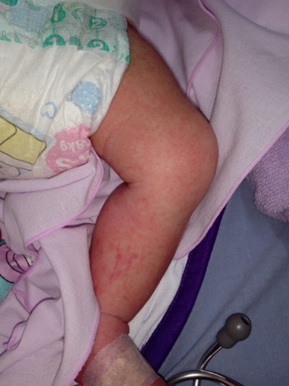
A 4-day-old male neonate was admitted to the Neonatal Intensive Care Unit (NICU) with a 3-day history of fever, jaundice, and a convalescent maculopapular rash on the extremities (Figure 1). He was born via cesarean section due to cephalopelvic disproportion at 39 week’s gestation, weighing 4380 g, measuring 57 cm in length, APGAR scores of 8 and 9 at 1 and 5 minutes, respectively, and he cried immediately after birth. He received the Bacille Calmette-Guérin (BCG) vaccine immediately after birth and was breastfed.
The mother, a 29-year-old multiparous woman, experienced a febrile illness 1 day before delivery. Her fever persisted for 3 days postpartum, prompting testing for dengue NS1 antigen rapid test (SD Bioline Dengue NS1 Ag Test, Abbott Diagnostics), which returned positive. Dengue IgM and IgG serologies were not tested in the mother. No complications were reported during her pregnancy, and routine antenatal care was unremarkable.
On examination, the neonate was febrile (38.5°C) with generalized jaundice and mild hepatomegaly. Laboratory investigations revealed significant thrombocytopenia, with a platelet count of 15 000/mm3 (normal range: 150 000–450 000/mm3) on the third hospital day. Hemoglobin was mildly decreased at 12.4 g/dL (normal range for neonates: 14–22 g/dL), indicating mild anemia. Liver function tests showed elevated transaminases, with AST at 180 IU/L and alanine transaminase (ALT) at 68 IU/L (normal range for neonates: AST <70 IU/L; ALT <45 IU/L). The NS1 antigen test result was positive. Dengue IgM antibodies were tested in the neonate using ELISA on day 4 of life, and the titers were negative. Blood cultures and sepsis screening were negative. Chest X-ray and abdominal ultrasonography were unremarkable.
Initial management included intravenous fluids, broad-spectrum antibiotic therapy with cefotaxime, and topical gentamicin cream for the rash. On hospital day 5, a 50-mL thrombocyte concentrate transfusion was performed due to persistent thrombocytopenia and the presence of petechial rash. Post-transfusion platelet counts showed significant improvement, increasing from 15 000/mm3 on day 3 to 37 000/mm3 on day 4, and stabilizing at 47 000/mm3 on day 7.
The fever resolved by day 6, and the rash showed marked regression. Liver enzymes remained elevated but showed a declining trend. No bleeding episodes or other complications were observed during hospitalization. The neonate was discharged in stable condition on day 8, with advice for follow-up in the outpatient department for monitoring of liver function and platelet counts.
Discussion
This case highlights the diagnostic and clinical challenges of congenital dengue, a rare but important manifestation of vertical transmission of dengue virus. It emphasizes the importance of considering dengue in the differential diagnosis of febrile neonates, particularly in endemic areas, and underscores the potential severity of complications such as thrombocytopenia and hepatitis in affected neonates, even when maternal infection was unrecognized at delivery.
Dengue virus, transmitted via Aedes mosquitoes, poses a significant public health challenge in tropical and subtropical regions, affecting up to 50% of the global population. Congenital dengue occurs through vertical transmission, typically when mothers contract the infection during late pregnancy [11]. The incubation period of the dengue virus is 3–10 days. The etiological virus has a half-life of 40 days in neonates. The onset of fever is typically occur 1–11 days after birth and persisting for 1–5 days [11]. In our case, fever in the neonate developed on the second day of life and persisted for 6 days, which is slightly longer than the usual clinical course.
A maculopapular rash is observed in 50–80% of dengue patients and may be the first visible sign of illness. The initial rash is due to capillary dilation and often appears as facial flushing within the first 1–2 days of fever. A secondary rash, appearing 3–7 days after fever onset, consists of flat or raised red spots and can resemble measles [12,13]. We found similar dermatologic features in our patient.
Occasionally, separate rashes combine and appear as extensive areas of redness with tiny bleeding spots and circular patches of unaffected skin, creating a distinctive pattern resembling “white islands in a sea of red”. The cutaneous rash typically does not cause any symptoms, with itching only reported in 16–27% of all cases. Bleeding episodes are rare in dengue; they can manifest as epistaxis, gingival bleeding, heavy menstruation, petechiae and purpura, or gastrointestinal tract hemorrhage [14]. The few published case reports on congenital dengue suggest that most of the symptoms in newborns are generally mild, such as fever and rash, but can involve more severe features, such as thrombocytopenia, respiratory distress, and hepatitis [1,5,15]. In our neonatal patient, thrombocytopenia was detected on the third hospital day, and we transfused platelets on the fifth day as there was significant drop in the count. After thorough monitoring, the platelet count had increased and became stable.
The absence of standardized guidelines for the diagnosis and managing congenital dengue continues to pose clinical challenges. Limited data and variability in clinical presentations make standardized management difficult. This case underscores the need for high clinical suspicion and early laboratory testing, particularly NS1 antigen detection and serial platelet monitoring, which can improve diagnostic accuracy during the acute phase. NS1 antigen is detectable within the first few days of illness, while IgM antibodies typically become detectable by day 4 of fever onset. Diagnosis is confirmed through seroconversion or rising IgM titers in paired acute and convalescent samples [6,16].
In our case, NS1 antigen was positive while dengue IgM and IgG antibodies were negative on day 3 of the illness, supporting the observation that IgM may be less sensitive in early congenital infections. This finding agrees with a prospective study of 2531 mother–infant pairs, in which 2.5% of mothers had positive dengue IgM, yet only 1.6% of the neonates had serologic evidence of vertical transmission, and neither maternal nor neonatal samples were positive for viral RNA by PCR [5]. These results highlight potential limitations of serologic testing and the need for improved diagnostic strategies in neonates.
Our observations are consistent with a 2023 review by Chong et al [18], which emphasizes that perinatal dengue poses diagnostic and therapeutic challenges in Southeast Asia due to variable clinical presentations and a lack of region-specific management guidelines. They note that neonatal dengue is often under-recognized and may mimic sepsis, potentially delaying appropriate care. Our case reinforces their findings, demonstrating that prompt recognition and supportive management, including fluid therapy and platelet transfusion, can lead to favorable outcomes [17].
Prevention remains the most effective strategy to reduce the burden of congenital dengue. Public health efforts focusing on vector control, community education, and early identification of dengue in pregnant women are essential. Pregnant women in endemic areas should be closely monitored for signs of infection, as timely diagnosis and management can mitigate risks to the fetus and neonate [18].
Conclusions
This case illustrates a rare but clinically significant instance of vertically transmitted dengue infection in a neonate, presenting with fever, rash, thrombocytopenia, and elevated liver enzymes. It highlights the importance of recognizing dengue virus infection in neonates, particularly in endemic regions. Early diagnosis through NS1 antigen testing and timely supportive management, including platelet transfusion and fluid therapy, are essential to improve neonatal outcomes. Preventive strategies, such as vector control and increased awareness among pregnant women, remain vital. Further research is warranted to enhance understanding and establish evidence-based guidelines for the diagnosis and management of congenital dengue.
References
1. Karim SA, Climate change and its impact on the increase in dengue hemorrhagic fever cases in North Maluku Province (Case study in Tidore Islands City): Ilomata Int J Soc Sci, 2024; 5(1); 176-89
2. Schaefer TJ, Panda PK, Wolford RW, Dengue fever. [Updated 2024 Mar 6]: StatPearls [Internet], 2025, Treasure Island (FL), StatPearls Publishing Available from: https://www.ncbi.nlm.nih.gov/books/NBK430732/
3. Ranjan R, Kumar K, Nagar N, Congenital dengue infection: Are we missing the diagnosis?: Pediatr Infect Dis, 2016; 8(4); 120-23
4. Thomas J, Thomas P, George CR, Neonatal dengue: Int J Contemp Pediatr, 2017; 4; 2234
5. Gupta S, Choudhury V, Gupta NP, Congenital dengue in neonate: Clin Case Rep, 2021; 9(2); 704-6
6. Yin X, Zhong X, Pan S, Vertical transmission of dengue infection: The first putative case reported in China: Rev Inst Med Trop Sao Paulo, 2016; 58; 90
7. Brar R, Sikka P, Suri V, Maternal and fetal outcomes of dengue fever in pregnancy: A large prospective and descriptive observational study: Arch Gynecol Obstet, 2021; 304(1); 91-100
8. World Health Organization (WHO): Dengue and severe dengue: Fact sheet Available from: https://www.who.int/news-room/fact-sheets/detail/dengue-and-severe-dengue
9. Brady OJ, Gething PW, Bhatt S, Refining the global spatial limits of dengue virus transmission by evidence-based consensus: PLoS Negl Trop Dis, 2012; 6(8); e1760
10. Vélez Jaramillo Y, Reveiz Montes MA, Galván-Barrios JP, Picón-Jaimes YA, Maternal and foetal outcomes in women with gestational dengue: A systematic review: Le Infezioni in Medicina, 2025; 33(1); 15-28
11. Chan M, Johansson MA, The incubation periods of dengue viruses: PLoS One, 2012; 7(11); e50972
12. Alcon S, Talarmin A, Debruyne M, Enzyme-linked immunosorbent assay specific to dengue virus type 1 nonstructural protein NS1 reveals circulation of the antigen in the blood during the acute phase of disease: J Clin Microbiol, 2002; 40(2); 376-81
13. Hasan S, Jamdar SF, Alalowi M, Al Ageel Al Beaiji SM, Dengue virus: A global human threat – review of literature: J Int Soc Prev Community Dent, 2016; 6(1); 1-6
14. World Health Organization (WHO): Dengue: Guidelines for diagnosis, treatment, prevention and control, 2009; 10-12, Geneva, World Health Organization Part 1.1.6: Dengue Case Classification
15. Kariyawasam S, Senanayake H, Dengue infections during pregnancy: Case series from a tertiary care hospital in Sri Lanka: J Infect Dev Ctries, 2010; 4(11); 767-75
16. Duarte AO, Oliveira JV, Carvalho TCX, Maternal and congenital infections arising from Zika, dengue and Chikungunya arboviruses in Salvador, Brazil: Trans R Soc Trop Med Hyg, 2019; 114; 222-25
17. Nguyen TM, Huan VT, Reda A, Clinical features and outcomes of neonatal dengue at the Children’s Hospital 1, Ho Chi Minh, Vietnam: J Clin Virol, 2021; 138; 104758
18. Chong V, Tan JZL, Arasoo VJT, Dengue in pregnancy: A Southeast Asian perspective: Trop Med Infect Dis, 2023; 8(2); 86
In Press
Case report 
Am J Case Rep In Press; DOI: 10.12659/AJCR.949566
Case report 
Am J Case Rep In Press; DOI: 10.12659/AJCR.952005
Case report 
Am J Case Rep In Press; DOI: 10.12659/AJCR.952443
Case report
Am J Case Rep In Press; DOI: 10.12659/AJCR.953173
Most Viewed Current Articles
07 Dec 2021 : Case report
22,282,456
DOI :10.12659/AJCR.934347
Am J Case Rep 2021; 22:e934347
06 Dec 2021 : Case report  174,072
DOI :10.12659/AJCR.934406
Am J Case Rep 2021; 22:e934406
21 Jun 2024 : Case report
119,650
DOI :10.12659/AJCR.944371
Am J Case Rep 2024; 25:e944371
07 Mar 2024 : Case report
64,568
DOI :10.12659/AJCR.943133
Am J Case Rep 2024; 25:e943133