


29 August 2026: Articles 
Coronary-Subclavian Steal Syndrome in a Post-Coronary Artery Bypass Grafting Patient: A Reversible Cause of Myocardial Ischemia
Unusual clinical course, Challenging differential diagnosis, Unusual or unexpected effect of treatment
Abdallah Rayyan EF 1*, Daniel Bishev EF 2, Nasir Nawaz EF 3
DOI: 10.12659/AJCR.951303
Am J Case Rep 2026; 27:e951303


Abstract
BACKGROUND: Coronary-subclavian steal syndrome (CSSS) is a rare but clinically significant cause of myocardial ischemia in patients with prior coronary artery bypass grafting (CABG), particularly when the left internal mammary artery (LIMA) is used as a conduit. Proximal subclavian artery stenosis can lead to reversal of LIMA graft flow, resulting in compromised myocardial perfusion that can mimic progression of native coronary disease.
CASE REPORT: A 63-year-old man with a history of multi-vessel coronary artery disease, prior PCI, and CABG with a LIMA-to-LAD graft presented with progressive exertional chest pain and left upper-extremity paresthesia. Physical examination revealed a diminished left radial pulse and inability to obtain blood pressure in the affected arm, raising suspicion for a proximal inflow lesion. CTA subsequently identified high-grade stenosis of the proximal left subclavian artery, and coronary angiography confirmed underfilling of the LIMA-LAD graft consistent with CSSS physiology. After evaluating medical therapy, redo surgical revascularization, and endovascular intervention, percutaneous stent placement was selected due to its lower procedural morbidity and favorable reported outcomes. A balloon-expandable stent was successfully deployed, restoring antegrade flow with immediate improvement in graft perfusion.
CONCLUSIONS: This case emphasizes the importance of recognizing coronary-subclavian steal syndrome in post-CABG patients presenting with recurrent angina and upper-extremity vascular findings. Targeted vascular imaging and bedside examination facilitated the diagnosis, and endovascular subclavian revascularization proved safe and effective in restoring graft perfusion. A practical diagnostic flowchart is also presented to support clinical evaluation and management in similar cases.
Keywords: Cardiovascular Diseases, Coronary Artery Disease, Myocardial Infarction, Subclavian Steal Syndrome
Introduction
Coronary-subclavian steal syndrome (CSSS) is an uncommon but clinically significant complication occurring in approximately 0.5% to 6% of patients after coronary artery bypass grafting (CABG) using the left internal mammary artery (LIMA) [1,2]. However, the true incidence is likely underdiagnosed because symptoms are often misattributed to progression of native coronary artery disease rather than proximal subclavian obstruction [2]. The underlying mechanism involves reversal of blood flow within the LIMA graft due to hemodynamically significant subclavian artery stenosis, which ultimately compromises myocardial perfusion and precipitates recurrent ischemia [3,4].
Delayed recognition of CSSS can result in recurrent angina, graft dysfunction, and potentially avoidable myocardial infarction or graft failure [3]. Clinical features such as inter-arm blood pressure discrepancy, diminished pulse amplitude, and upper-extremity ischemic symptoms can serve as early diagnostic clues and should prompt targeted vascular imaging in post-CABG patients [4].
According to the 2024 ESC Guidelines for the management of elevated blood pressure and hypertension, bilateral arm blood pressure measurement is recommended at the initial clinical evaluation to identify significant inter-arm systolic differences that can indicate underlying arterial stenosis [5]. Although routine CTA screening of the subclavian artery is not specifically recommended, imaging can be useful in selected high-risk patients with extensive atherosclerotic burden to support vascular risk stratification and management decisions.
This condition carries important clinical implications given its potential to mimic progressive coronary artery disease and its association with adverse cardiac outcomes if left untreated. Endovascular stenting has emerged as an effective therapeutic strategy when anatomically feasible, with a growing literature supporting its role in restoring antegrade LIMA flow [6,7]. Given the relationship between peripheral arterial disease and subclavian stenosis, routine vascular assessment—particularly evaluation of subclavian inflow—may warrant consideration in high-risk patients scheduled for CABG using a LIMA graft. Future studies should explore whether early detection and prophylactic optimization of subclavian patency can reduce the incidence of CSSS and improve long-term graft outcomes.
In this report, we present a case of CSSS characterized by recurrent angina and upper-extremity ischemic symptoms in a post-CABG patient, highlighting the diagnostic importance of peripheral vascular examination and the successful use of endovascular revascularization to restore graft perfusion.
Case Report
A 63-year-old White man presented to his cardiologist’s office with a 1-month history of progressively worsening, crushing substernal exertional chest pain associated with left upper-extremity paresthesia. He had a past medical history of coronary artery disease (CAD) with prior percutaneous coronary intervention (PCI) to his native left circumflex artery and subsequent 2-vessel CABG in 2013. CABG was done with a LIMA graft to the left anterior descending (LAD) artery and saphenous vein graft (SVG) to the first diagonal branch. He also had a known occluded co-dominant right coronary artery (RCA) with right-to-right collaterals, and normal left ventricular systolic function. Other history was significant for peripheral artery disease status post aorto bi-iliac angioplasty and stenting, bilateral carotid artery stenosis, hypertension, and hyperlipidemia.
During the initial evaluation, his left radial pulse was not palpable and a true left brachial blood pressure could not be obtained. A marked inter-arm systolic pressure discrepancy was documented, with a right upper-extremity blood pressure of 148 mm Hg and a left upper-extremity blood pressure of 94 mm Hg; blood pressure measurement on the left arm was initially difficult to obtain due to severely diminished arterial flow. The patient also reported left arm pain and numbness consistent with upper-extremity ischemia, and neurological examination demonstrated mildly reduced motor strength in the left upper extremity (4/5). Electrocardiography revealed normal sinus rhythm with non-specific ST-segment changes in the inferolateral leads. Given his history of CAD and recurrent angina, the initial differential diagnosis included progression of native coronary artery disease, graft occlusion, microvascular angina, and coronary-subclavian steal physiology due to proximal subclavian artery stenosis. The concurrent presence of anginal symptoms and upper-extremity ischemic complaints, along with pulse asymmetry, shifted diagnostic suspicion toward an inflow obstruction rather than isolated coronary disease progression.
CTA of the chest and abdomen demonstrated a high-grade stenosis of the proximal left subclavian artery with associated narrowing at the origin of the left vertebral artery. Due to the progressive nature of symptoms and the imaging-confirmed inflow obstruction, coronary and bypass graft angiography with left subclavian artery assessment was performed.
He was taken to the cardiac catheterization lab for coronary and bypass graft angiography, as well as angiography of the left upper extremity. He was found to have a patent left main stem, a large anatomically co-dominant circumflex artery with a patent proximal stent, and only mild irregularities of the obtuse marginal branches and posterior descending artery. The left anterior descending artery was occluded proximally with patent LIMA-LAD, which seemed underfilled due to proximal subclavian artery stenosis. The saphenous vein graft to the major diagonal branch was patent. Angiography of the left subclavian was done next, which demonstrated a 99% hazy eccentric left subclavian stenosis proximal to the internal mammary artery takeoff with slow flow beyond and poor retrograde filling from a diffusely diseased vertebral artery. Sequential angiographic images demonstrated the pre-intervention subclavian stenosis, positioning of the stent, restoration of vessel lumen, and improved antegrade flow through the LIMA graft following revascularization (Figure 1A–1D).
Management options considered included medical therapy alone, redo surgical bypass, or endovascular revascularization. Medical therapy would not address the fixed mechanical obstruction, and redo surgery was considered higher risk due to prior sternotomy and the focal nature of the lesion. Based on current evidence supporting the safety and efficacy of percutaneous treatment in this context [6,7], endovascular stenting was selected as the preferred strategy.
Angioplasty and stenting of the subclavian stenosis was done with a 7.0 × 37 mm express balloon-expandable bare metal stent deployed at 12 atmospheres. He was placed on dual antiplatelet therapy with aspirin 81 mg and clopidogrel 75 mg daily and continued on high-intensity statin. He was monitored in the cardiovascular intensive care unit for 24 hours without any complications and was discharged home the next day. At follow-up, both anginal symptoms and left upper-extremity perfusion had improved, supporting successful restoration of graft inflow physiology.
Discussion
CSSS remains an underrecognized cause of recurrent angina in post-CABG patients despite increased awareness. Subclavian artery stenosis proximal to the LIMA graft compromises coronary perfusion, resulting in myocardial ischemia that may be misinterpreted as progression of native coronary disease. Early recognition is essential to prevent graft compromise and recurrent ischemic symptoms. Bedside examination findings, particularly marked inter-arm systolic blood pressure discrepancy and asymmetric pulse strength, can provide critical early diagnostic clues. In this patient, these clinical findings raised immediate suspicion for inflow obstruction and guided further targeted vascular imaging, underscoring their diagnostic importance.
Multiple therapeutic approaches have been described for CSSS, including conservative medical management, surgical revascularization, and endovascular therapy. Medical therapy alone does not resolve the fixed anatomical obstruction of subclavian inflow and therefore does not prevent ongoing ischemia in symptomatic patients. Redo surgical bypass, while effective in restoring graft patency, carries higher morbidity in reoperative thoracic surgery, particularly in patients with diffuse atherosclerotic disease or prior sternotomy [8].
Endovascular stenting has emerged as an effective strategy due to its minimally invasive profile, rapid recovery, and strong clinical outcomes. More than 90% of interventions achieve technical success with sustained symptom relief in over 80% of treated patients [6]. Chatterjee et al demonstrated significantly higher restenosis rates with angioplasty alone compared to stent-supported angioplasty (22% vs 9%), reinforcing stent placement as the preferred strategy for durable patency [7]. In a large single-center study spanning over 2 decades, Bradaric et al reported a 97.7% technical success rate for endovascular therapy, including 100% success in stenotic lesions and 86% in total occlusions, with very low periprocedural complication rates (stroke 0.8%, TIA 1.5%, access complications 3.8%) and only 12% restenosis over a mean follow-up of 28 months [8]. These data support endovascular intervention as a safe and effective first-line therapy in anatomically suitable cases, with significantly lower morbidity compared to redo surgical bypass.
Open surgical intervention, including carotid–subclavian bypass or subclavian artery transposition, remains an important therapeutic option in selected patients. Surgery is generally considered following failed or recurrent stent occlusion after endovascular therapy, as described in reports where multiple repeat interventions ultimately required bypass reconstruction [9,10]. In addition, anatomical factors can guide treatment selection, as endovascular outcomes are less favorable in complete occlusions and longer lesions (particularly those exceeding 2 cm), or when it is not possible to cross the lesion endovascularly [9,10]. While endovascular management offers a minimally invasive approach with shorter recovery, surgical revascularization may provide more durable long-term patency in low-risk patients, supporting its consideration when long-term graft and vessel durability is a clinical priority [9,10].
What distinguishes this case is the dual presentation of recurrent angina and upper-extremity ischemic symptoms, along with easily detectable bedside findings such as pulse asymmetry and inability to obtain blood pressure in the affected limb. These clinical indicators prompted targeted subclavian artery imaging rather than an assumption of native coronary progression or graft failure, demonstrating the diagnostic value of incorporating peripheral vascular assessment into the evaluation of post-CABG patients. Additionally, the coexistence of symptomatic upper-extremity ischemia and compromised graft flow is infrequently reported in the literature, making this case unique in highlighting the combined systemic and coronary manifestations of subclavian inflow obstruction. Furthermore, this report provides a structured bedside-to-imaging pathway supported by a flowchart, which has not been previously incorporated into similar case descriptions.
For practical clinical application, awareness of a structured diagnostic and management approach is essential. CSSS should be considered in post-CABG patients with recurrent angina, especially when a LIMA graft is present. Simple bedside findings such as inter-arm blood pressure discrepancy or diminished radial pulse can immediately direct suspicion toward a proximal inflow lesion rather than isolated coronary disease progression. In cases where ischemic symptoms persist without a clear coronary lesion, dedicated imaging of the subclavian artery using CTA or angiography is warranted. When a focal stenosis is confirmed, endovascular stenting should be prioritized due to its high procedural success, low complication rates, and favorable long-term outcomes compared with angioplasty alone or redo surgery. To support clinical decision-making, we summarize this diagnostic and interventional pathway in a simplified flowchart (Figure 2), which may serve as a practical reference in similar cases.
This case reinforces that early incorporation of peripheral vascular examination and targeted imaging into the evaluation of post-CABG angina can facilitate timely recognition of CSSS, guide appropriate endovascular intervention, and potentially improve graft longevity and clinical outcomes.
Conclusions
In conclusion, this case highlights the importance of maintaining a high index of clinical suspicion for coronary-subclavian steal syndrome in post-CABG patients presenting with recurrent angina, particularly when physical examination reveals pulse asymmetry or inter-arm blood pressure discrepancy. Prompt recognition through targeted vascular imaging and timely endovascular subclavian artery revascularization, which should be prioritized when anatomically feasible, can restore graft perfusion and prevent misinterpretation of symptoms as graft failure or native coronary progression. By combining bedside vascular assessment with evidence-based decision-making, this case supports a practical diagnostic and therapeutic pathway—illustrated in the flowchart—to assist clinical evaluation and management in similar scenarios.
Figures
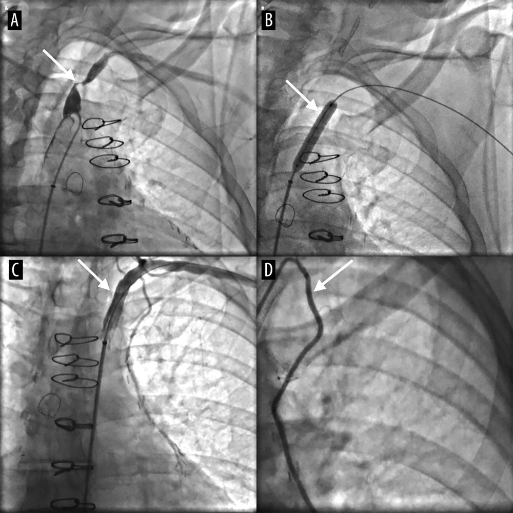 Figure 1. Sequential angiographic images demonstrating the interventional management of coronary-subclavian steal syndrome. (A) Severe proximal left subclavian artery stenosis prior to intervention. (B) Deployment of a balloon-expandable stent across the lesion. (C) Restoration of vessel lumen following stent placement. (D) Improved antegrade flow through the LIMA-to-LAD graft following revascularization.
 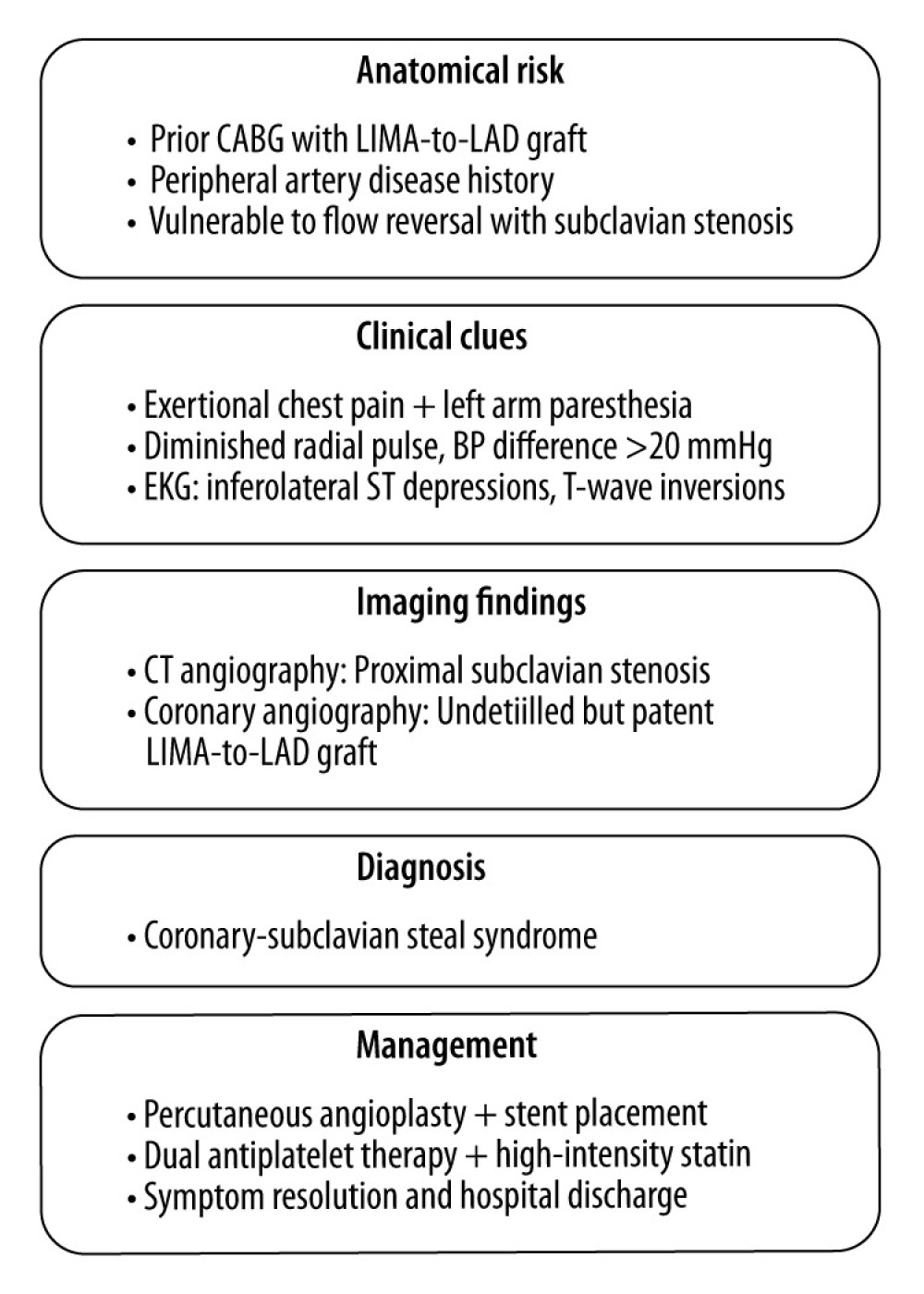 Figure 2. Flowchart summarizing the evaluation and management of coronary-subclavian steal syndrome (CSSS) in a post-CABG patient.
 References
1. Cua B, Mamdani N, Halpin D, Review of coronary subclavian steal syndrome: J Cardiol, 2017; 70(5); 432-37
2. Müller AM, Bertram J, Bradaric C, Frequency of subclavian artery stenosis in patients with mammarian artery coronary bypass and suspected coronary artery disease progression: Clin Res Cardiol, 2023; 112(9); 1204-11
3. Bicknell CD, Subramanian A, Wolfe JHN, Coronary subclavian steal syndrome: Eur J Vasc Endovasc Surg, 2004; 27(2); 220-21
4. Saha T, Naqvi SY, Ayah OA, Subclavian artery disease: Diagnosis and therapy: Am J Med, 2017; 130(4); 409-16
5. McEvoy JW, McCarthy CP, Bruno RM, 2024 ESC Guidelines for the management of elevated blood pressure and hypertension: Eur Heart J, 2024; 45(38); 3912-4018
6. Ahmed AT, Mohammed K, Chehab M, Comparing percutaneous transluminal angioplasty and stent placement for treatment of subclavian arterial occlusive disease: A systematic review and meta-analysis: Cardiovasc Intervent Radiol, 2016; 39(5); 652-67
7. Chatterjee S, Nerella N, Chakravarty S, Angioplasty alone versus angioplasty and stenting for subclavian artery stenosis—A systematic review and meta-analysis: Am J Ther, 2013; 20(5); 520-23
8. Bradaric C, Kuhs K, Groha P, Endovascular therapy for steno-occlusive subclavian and innominate artery disease: Circ J, 2015; 79(3); 537-43
9. Caesar-Peterson S, Bishop MA, Subclavian artery stenosis: StatPearls June 21, 2023, Treasure Island (FL), StatPearls Publishing
10. Salman R, Hornsby J, Wright LJ, Treatment of subclavian artery stenosis: A case series: Int J Surg Case Rep, 2016; 19; 69-74
Figures
Figure 1. Sequential angiographic images demonstrating the interventional management of coronary-subclavian steal syndrome. (A) Severe proximal left subclavian artery stenosis prior to intervention. (B) Deployment of a balloon-expandable stent across the lesion. (C) Restoration of vessel lumen following stent placement. (D) Improved antegrade flow through the LIMA-to-LAD graft following revascularization.Figure 2. Flowchart summarizing the evaluation and management of coronary-subclavian steal syndrome (CSSS) in a post-CABG patient. In Press
Case report 
Am J Case Rep In Press; DOI: 10.12659/AJCR.954074
Case report
Am J Case Rep In Press; DOI: 10.12659/AJCR.953592
Case report 
Am J Case Rep In Press; DOI: 10.12659/AJCR.954225
Case report 
Am J Case Rep In Press; DOI: 10.12659/AJCR.952870
Most Viewed Current Articles
07 Dec 2021 : Case report
28,712,481
DOI :10.12659/AJCR.934347
Am J Case Rep 2021; 22:e934347
06 Dec 2021 : Case report  180,580
DOI :10.12659/AJCR.934406
Am J Case Rep 2021; 22:e934406
21 Jun 2024 : Case report
124,319
DOI :10.12659/AJCR.944371
Am J Case Rep 2024; 25:e944371
08 May 2026 : Case report
95,142
DOI :10.12659/AJCR.952158
Am J Case Rep 2026; 27:e952158