


11 September 2026: Articles 
Appendico-Sigmoid Fistula as a Rare Complication of Chronic or Recurrent Appendicitis: A Case Report
Unusual clinical course, Challenging differential diagnosis, Diagnostic / therapeutic accidents, Management of emergency care, Rare disease, Clinical situation which can not be reproduced for ethical reasons, Rare coexistence of disease or pathology
Shashi Kush AEF 1*, Suvendu Sekhar Jena AE 1, Amitabh Yadav AE 1, Samiran Nundy AE 1
DOI: 10.12659/AJCR.952691
Am J Case Rep 2026; 27:e952691


Abstract
BACKGROUND: Appendico-colic fistula is a rare condition characterized by an abnormal communication between the appendix and colon, most commonly involving the sigmoid colon (ie, appendico-sigmoid fistula) because of their close anatomical proximity. It is usually associated with chronic or recurrent appendicitis, diverticular disease, inflammatory bowel disease, or malignancy. Preoperative diagnosis is challenging due to nonspecific clinical and radiologic findings.
CASE REPORT: A 46-year-old woman presented with a 3-month history of lower abdominal pain, which had worsened over the preceding 15 days and was associated with vomiting. Initial contrast-enhanced computed tomography (CECT) suggested sigmoid diverticular perforation with a localized collection. Because of discordant radiologic findings, repeat CECT with rectal contrast was performed, revealing inflammatory changes involving the appendix and sigmoid colon, along with rectal contrast entering the appendiceal lumen; these observations were suggestive of an appendico-sigmoid fistula. Exploratory laparotomy demonstrated a fistulous communication between the tip of the appendix and the sigmoid colon with surrounding inflammatory adhesions. The patient underwent sigmoid colectomy with appendectomy, colorectal anastomosis, and diverting loop ileostomy. Histopathology demonstrated inflammatory changes without evidence of diverticula or malignancy. The postoperative course was uneventful, and ileostomy reversal was performed after 2 months.
CONCLUSIONS: Appendico-colic fistula is a rare complication of chronic or recurrent appendicitis that can mimic sigmoid diverticular perforation. Careful interpretation of imaging, particularly CECT with rectal contrast, is important for diagnosis. Surgical management should be individualized according to the underlying etiology and intraoperative findings.
Keywords: appendicitis, Case Reports, complications, Fistula, Gastroenterology, Sigmoid Colon
Introduction
Appendico-colic fistula, an abnormal communication between the appendix and the colon, is a rare entity—fewer than 100 cases have been reported [1]. This type of fistula is commonly associated with benign conditions such as complicated or recurrent appendicitis, diverticular perforation, and inflammatory bowel disease; appendiceal and sigmoid malignancies have also been reported [2]. Preoperative diagnosis is challenging due to nonspecific symptoms and complex radiologic findings. However, contrast-enhanced computed tomography (CECT) of the abdomen with rectal contrast may provide an early clue by demonstrating a fistulous communication between the appendix and sigmoid colon (ie, appendico-sigmoid fistula). This report describes an appendico-colic fistula secondary to chronic or recurrent appendicitis in a 46-year-old woman, which was managed using a 2-stage surgical approach.
Case Report
A 46-year-old woman with no known comorbidities presented to the emergency department with a 15-day history of severe lower abdominal pain, associated with nonbilious vomiting for 1 to 2 days. She also reported a 3-month history of dull, aching lower abdominal pain localized to the hypogastric region without migration. She had received conservative treatment from her primary physician with analgesics and broad-spectrum antibiotics. There was no history of fever, diarrhea, dysuria, hematemesis, melena, or weight loss.
The patient had previously undergone contrast-enhanced computed tomography (CECT) of the abdomen at another hospital, which suggested sigmoid diverticular perforation with a localized collection. She was initially treated with oral antibiotics; however, because of persistent symptoms, she presented to our center.
On examination, the patient was afebrile, hemodynamically stable, and in good general condition. Abdominal examination revealed minimal tenderness in the hypogastric region without rebound tenderness, guarding, or tenderness at McBurney’s point. Obturator, psoas, and Rovsing signs were negative. Laboratory investigations, including complete blood count (hemoglobin, 10.3 g/dL; total leukocyte count, 5440/mm3; platelets, 467 000/mm3), kidney function tests (blood urea nitrogen, 5.0 mg/dL; creatinine, 0.5 mg/dL; sodium/potassium, 137/3.80 mEq/L), and liver function tests (total/direct bilirubin, 0.18/0.08 mg/dL; albumin, 2.80 g/dL; alanine aminotransferase/aspartate aminotransferase, 16/10 IU/L; alkaline phosphatase, 57 IU/L), showed results within normal limits. Upon discussion with the radiology team, discordance was noted in the previous imaging findings. Therefore, repeat CECT of the abdomen with oral and rectal contrast was performed, revealing a loculated fluid collection measuring approximately 3 × 3 × 4 cm near the tip of the appendix with associated inflammatory changes involving both the appendix and sigmoid colon. A thin streak of rectal contrast was seen entering the appendiceal lumen, raising suspicion for an appendico-sigmoid fistula. Additionally, the distal ileum was adherent to the collection site, with secondary small bowel dilation (Figure 1). Colonoscopy was deferred because the patient was in an active inflammatory phase with associated small bowel dilation and localized pelvic inflammatory changes, increasing the risk of procedural complications and limiting its diagnostic utility in the acute setting.
After informed consent and preoperative clearance had been obtained, the patient underwent exploratory laparotomy. The initially planned laparoscopic approach was deferred because of abdominal distension caused by dilated small bowel loops. Intraoperatively, seropurulent fluid (approximately 25–30 mL) was identified in the pelvis. Careful adhesiolysis revealed a fistulous communication between the tip of the appendix and the distal sigmoid colon, whereas the appendiceal base appeared healthy. The terminal ileal loops were dilated and densely adherent to the fistula site; they were carefully dissected free (Figure 2).
A sigmoid colectomy with resection of approximately 25 cm of sigmoid colon was performed due to the preoperative radiologic suspicion of diverticular perforation and intraoperative uncertainty regarding the extent of sigmoid involvement and underlying pathology. This was followed by end-to-end hand-sewn colorectal anastomosis and appendectomy. A diverting loop ileostomy was created because of the unprepared, edematous bowel.
Histopathologic examination demonstrated granulation tissue and inflammatory changes in both the sigmoid colon and appendix, without evidence of diverticula or malignancy. Postoperative recovery was uneventful. Ileostomy reversal was performed after 2 months, and the patient remained well at the 1-year follow-up.
Discussion
Appendico-colic fistulas are extremely rare, such that fewer than 100 cases have been documented [1]. The earliest reported case was described by Cherigie et al in 1953 [2]. The most common cause of fistula formation is chronic or recurrent appendicitis managed conservatively and associated with a localized abscess or delayed presentation to a healthcare facility [3]. Such fistulas usually are associated with a long pelvic appendix lying in close proximity to the sigmoid colon. Other reported causes include diverticular perforation, Crohn’s disease, inflammatory bowel disease, appendiceal malignancy, and sigmoid malignancy [4].
Appendicitis can have a varied clinical presentation. Typical symptoms, such as migratory right lower quadrant pain, fever, and vomiting, usually suggest acute-phase presentation; malignancy often presents with subtle symptoms, leading to delayed diagnosis. Inflammatory bowel diseases such as Crohn’s disease may manifest as abdominal pain, altered bowel habits, features of subacute intestinal obstruction, and imaging findings suggestive of multiple small bowel strictures, bowel wall thickening, and lymphadenopathy. Sigmoid diverticular perforation or sigmoid malignancy may involve large bowel obstruction or bleeding per rectum. Investigations such as sigmoidoscopy and CECT may reveal a sigmoid mass in cases of malignancy or findings suggestive of diverticular perforation, including a localized collection and pelvic inflammation [3]. Patients with appendico-sigmoid fistulas caused by diverticular disease usually present at an older age with a chronic history of symptoms related to previous episodes of diverticulitis. A barium enema with air contrast is considered the best investigation because it demonstrates free flow of contrast from the sigmoid colon into the appendix via the fistulous communication. Colonoscopy should be performed to exclude malignancy and evaluate the morphology of the fistulous communication [5]. Accurate preoperative diagnosis often requires multiple diagnostic modalities because management substantially differs between benign and malignant causes. Plain abdominal radiographs have limited diagnostic value. CECT of the abdomen with rectal contrast is considered among the most sensitive investigations because it can demonstrate opacification of the fistulous tract and appendiceal lumen. The addition of colonoscopy further improves diagnostic accuracy by excluding malignancy and inflammatory bowel disease [6]. However, CECT interpretation is subject to interobserver variation, as demonstrated in the present case: initial images suggested localized diverticular perforation, whereas repeat imaging identified a fistulous communication between the appendix and sigmoid colon, resulting in modified management. Findings suggestive of an appendico-colic fistula on CECT with rectal contrast include rightward displacement of the sigmoid colon, ill-defined fat planes between the appendix and sigmoid colon, and passage of rectal contrast into the appendiceal lumen before opacification of the proximal colon. Colonoscopy may identify associated malignant pathology or benign conditions (eg, ulcerative colitis and diverticular disease). Magnetic resonance imaging is also useful because of its superior soft tissue resolution, particularly for differentiating between inflammatory and malignant causes. Fluorodeoxyglucose positron emission tomography (FDG-PET) can help to differentiate benign from malignant etiologies [2]. However, FDG uptake may also be increased in inflammatory lesions, although differences in maximum standardized uptake values (SUVmax) may occasionally help distinguish the 2 entities. SUVmax does not always reflect the degree of malignancy. Thus, several methods have been proposed to improve FDG-PET sensitivity and specificity. Experimental approaches using amino acid-tagged molecules might improve FDG-PET specificity, although no human studies have been published [2]. Intraoperative frozen section examination can also help differentiate benign from malignant pathology.
Management should be individualized according to the etiology, intraoperative findings, and surgeon expertise. In benign cases, conservative management with intravenous antibiotics may be attempted; however, spontaneous fistula closure is uncommon, and conservative treatment is associated with a high risk of complications [7]. Surgical treatment can be performed as a 1-stage procedure with resection of the diseased bowel segment and appendectomy or as a 2-stage procedure with the addition of a diverting ileostomy when the bowel is edematous or unprepared. Minimally invasive approaches may provide faster recovery and improved outcomes, although they require expertise and accurate preoperative delineation of the anatomy [8]. In cases of suspected malignancy, radical surgery with appropriate lymphadenectomy is recommended [9].
Conclusions
Although appendico-colic fistula is a rare condition, it remains difficult to diagnose preoperatively. Management should be tailored according to the underlying etiology. Benign causes can usually be managed with limited surgery (eg, appendectomy with limited sigmoid colon resection or primary repair of the colonic fistula) and may also be approached via minimally invasive techniques. In contrast, malignant etiologies require radical surgical resection with lymphadenectomy, followed by adjuvant chemotherapy when indicated. This case report has highlighted diagnostic challenges associated with appendico-colic fistula and reviewed the available diagnostic modalities and management options.
Figures
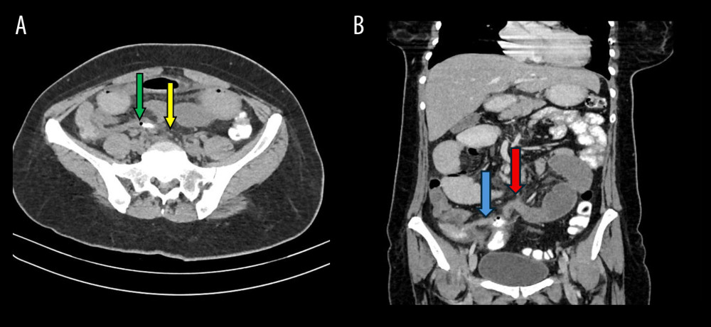 Figure 1. (A) Axial computed tomography image showing a fistulous communication between the appendix and sigmoid colon, with a thin streak of rectal contrast entering the appendiceal lumen (green arrow) and the adjacent sigmoid colon (yellow arrow). (B) Coronal computed tomography image showing an inflamed appendix containing an air bubble (blue arrow) in close proximity to the sigmoid colon, with dilated small bowel loops (red arrow).
 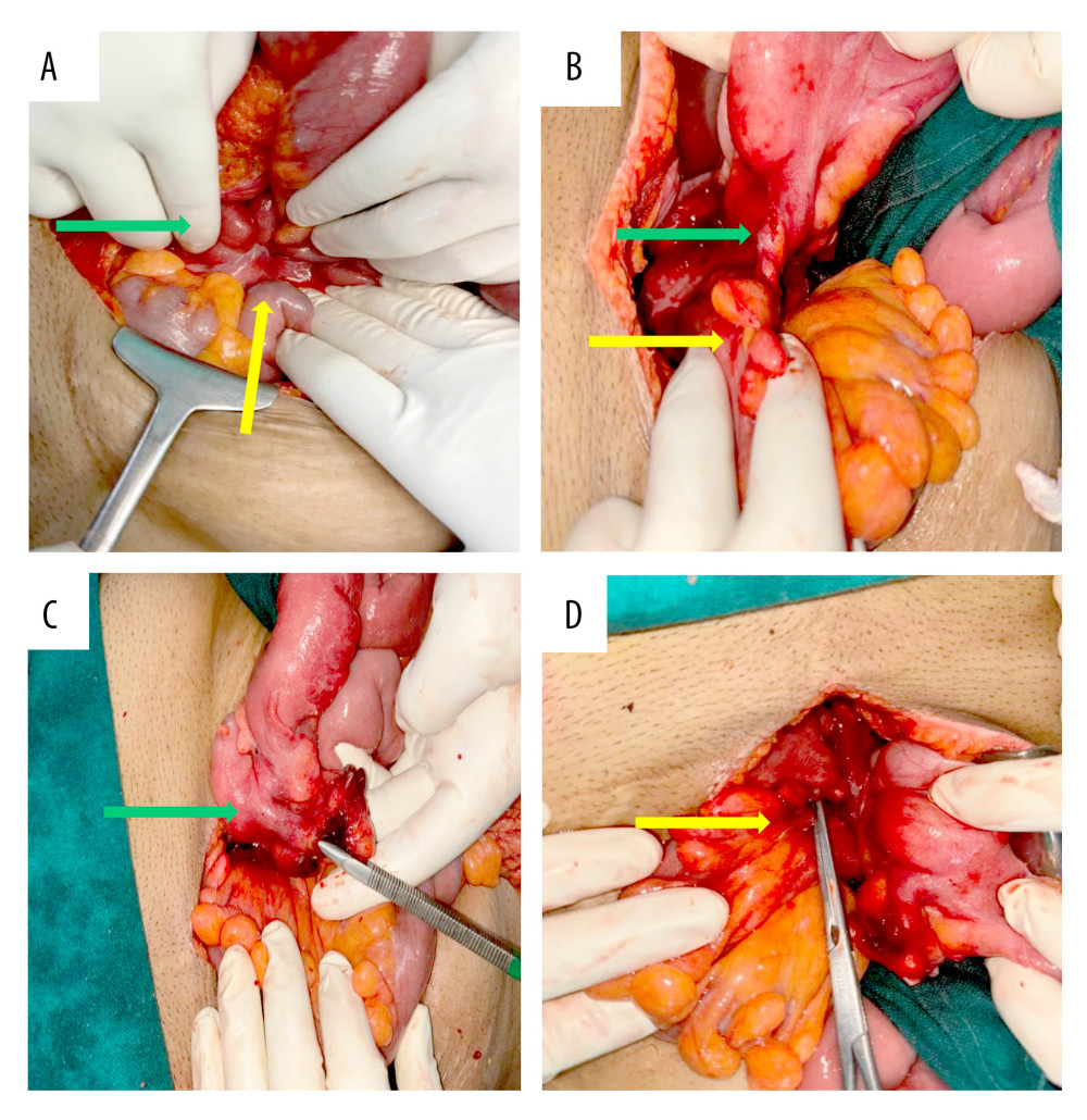 Figure 2. (A) Intraoperative image showing the appendix (green arrow) adjacent to the small bowel (yellow arrow) and sigmoid colon (white arrow). (B) Fistulous communication between the tip of the appendix (green arrow) and the sigmoid colon (yellow arrow). (C) After division of the fistulous tract, a metal forceps is visible within the appendiceal lumen (green arrow). (D) After division of the fistulous tract, the opening in the sigmoid colon is visible separately, with a metal forceps inserted into the lumen (yellow arrow).
 References
1. Yan H, Wu YC, Wang X, Appendico-vesicocolonic fistula: A case report and review of literature: World J Clin Cases, 2022; 10(10); 3241-50
2. Nakamoto H, Yokota R, Namba H, A benign appendiceal-colonic fistula, diagnosed and managed laparoscopically: A case report: Am J Case Rep, 2020; 21; e925946
3. Belay SA, Negussie MA, Kassie MT, A rare case of congenital appendiceal duplication complicated by an appendiceal-sigmoid fistula: A case report: Int J Surg Case Rep, 2025; 131; 111429
4. Santangelo M, Lanteri R, D’Angelo M, Appendiceal-sigmoid fistula presenting in a man with ulcerative colitis: A case report: J Med Case Rep, 2010; 4(1); 229
5. Yiangou C, Holme TC, Sigmoido-appendiceal fistula due to diverticulitis: J R Soc Med, 1998; 91(10); 544
6. Morris-Stiff GJ, Islam KA, Appendico-colic fistula complicating appendicitis in cystic fibrosis: BMJ Case Rep, 2010; 2010; bcr0220102714
7. Touati MD, Othmane MR, Belhadj A, A rare case report of an appendico-sigmoid fistula complicating untreated acute appendicitis: Unraveling complexity in diagnosis and treatment: Int J Surg Case Rep, 2024; 116; 109298
8. McDonald SJ, Lee F, Dean N, Fistulae involving the appendix: A systematic review of the literature: ANZ J Surg, 2020; 90(10); 1878-87
9. Hakim S, Amin M, Cappell MS, Limited, local, extracolonic spread of mucinous appendiceal adenocarcinoma after perforation with formation of a malignant appendix-to-sigmoid fistula: Case report and literature review: World J Gastroenterol, 2016; 22(38); 8624-30
Figures
Figure 1. (A) Axial computed tomography image showing a fistulous communication between the appendix and sigmoid colon, with a thin streak of rectal contrast entering the appendiceal lumen (green arrow) and the adjacent sigmoid colon (yellow arrow). (B) Coronal computed tomography image showing an inflamed appendix containing an air bubble (blue arrow) in close proximity to the sigmoid colon, with dilated small bowel loops (red arrow).Figure 2. (A) Intraoperative image showing the appendix (green arrow) adjacent to the small bowel (yellow arrow) and sigmoid colon (white arrow). (B) Fistulous communication between the tip of the appendix (green arrow) and the sigmoid colon (yellow arrow). (C) After division of the fistulous tract, a metal forceps is visible within the appendiceal lumen (green arrow). (D) After division of the fistulous tract, the opening in the sigmoid colon is visible separately, with a metal forceps inserted into the lumen (yellow arrow). In Press
Case report 
Am J Case Rep In Press; DOI: 10.12659/AJCR.953831
Case report 
Am J Case Rep In Press; DOI: 10.12659/AJCR.953658
Case report 
Am J Case Rep In Press; DOI: 10.12659/AJCR.953333
Case report 
Am J Case Rep In Press; DOI: 10.12659/AJCR.953693
Most Viewed Current Articles
07 Dec 2021 : Case report
28,709,279
DOI :10.12659/AJCR.934347
Am J Case Rep 2021; 22:e934347
06 Dec 2021 : Case report  180,392
DOI :10.12659/AJCR.934406
Am J Case Rep 2021; 22:e934406
21 Jun 2024 : Case report  124,124
DOI :10.12659/AJCR.944371
Am J Case Rep 2024; 25:e944371
08 May 2026 : Case report
94,633
DOI :10.12659/AJCR.952158
Am J Case Rep 2026; 27:e952158