


26 September 2026: Articles 
Left Bundle Branch Area Pacing as a Rescue Strategy After Failed Cardiac Resynchronization Therapy in a Patient With Persistent Left Superior Vena Cava, Absent Coronary Sinus Branches, After Bilateral Maze Procedure: A Case Report
Unusual or unexpected effect of treatment, Rare disease, Educational Purpose (only if useful for a systematic review or synthesis)
Lucie Kohoutková ABCDEF 1,2*, Martin Augustynek DG 1, David Šipula DEF 2DOI: 10.12659/AJCR.952879
Am J Case Rep 2026; 27:e952879


Abstract
BACKGROUND: Cardiac resynchronization therapy can fail in the presence of anatomical variations, such as persistent left superior vena cava, which can preclude coronary sinus cannulation and left ventricular lead placement. Conduction system pacing, particularly left bundle branch area pacing, has emerged as a physiological alternative for achieving ventricular resynchronization in such anatomically challenging situations and may provide an effective solution when standard cardiac resynchronization therapy (CRT) is not feasible.
CASE REPORT: We report the case of a 78-year-old man with chronic heart failure (left ventricular ejection fraction 35%) and a history of mitral valve replacement with a bioprosthesis, tricuspid valve repair, and a bilateral Maze procedure. The patient was indicated for cardiac resynchronization therapy pacemaker (CRT-P) implantation due to sick sinus syndrome with tachy-brady arrhythmia and left bundle branch block with a QRS duration of 190 ms. Periprocedural venography revealed a persistent left superior vena cava draining into a markedly dilated coronary sinus without suitable lateral branches, preventing left ventricular lead placement. Following device implantation, the patient experienced progressive heart failure deterioration, with a decline in left ventricular ejection fraction (LVEF) to 25% and an increase in NT-proBNP to 7741 ng/L, subsequently fulfilling criteria for implantable cardioverter-defibrillator implantation. Due to unfavorable venous anatomy, conduction system pacing (CSP) was selected as an alternative strategy. A right-sided implantable cardioverter defibrillator (ICD) system with left bundle branch area pacing (LBBAP) was successfully implanted, resulting in significant QRS narrowing to 120 ms and excellent pacing parameters. At 2-month follow-up, the patient remained clinically stable (NYHA class II) with improvement in LVEF to 30-35%.
CONCLUSIONS: This case illustrates that LBBAP can provide effective ventricular activation and resynchronization when conventional CS-based CRT is not feasible due to venous anomalies. LBBAP is a safe and effective alternative in patients with PLSVC and unfavorable CS anatomy.
Keywords: cardiac resynchronization therapy, Cardiology, Case Reports, Heart Failure, persistent left superior vena cava
Introduction
Conduction system pacing (CSP) is a physiological pacing strategy that activates the ventricles through the native His–Purkinje network [1]. Placement of a coronary sinus (CS) lead for conventional cardiac resynchronization therapy (CRT) can fail in patients with complex venous anatomy, particularly in the presence of persistent left superior vena cava (PLSVC), which frequently drains into a large, funnel-shaped coronary sinus lacking suitable lateral branches.
PLSVC is the most common congenital anomaly of systemic venous return, occurring in approximately 0.3% to 0.5% of the general population and up to 10% of patients with congenital heart disease [2]. In such cases, CSP provides a practical alternative for achieving ventricular resynchronization [3].
Case Report
A 78-year-old man with a history of mitral valve replacement with a bioprosthesis, tricuspid valve repair, and a bilateral Maze procedure (2020) was followed for chronic heart failure with reduced left ventricular ejection fraction (LVEF 35%). On January 27, 2025, he was indicated for implantation of a cardiac resynchronization pacemaker (CRT-P, SJM Allure RF) for sick sinus syndrome with tachy-brady arrhythmia, with an LVEF of 35% and left bundle branch block (LBBB) morphology with a QRS duration of 190 ms. The NT-proBNP level at that time was 4636 ng/L. The timeline of the patient’s clinical course is presented in Figure 1.
During the procedure on January 27, 2025, bilateral venography revealed a persistent left superior vena cava. A biventricular pacemaker (Abbott Allure RF) was implanted with a right atrial lead positioned in the right atrial appendage and a ventricular lead placed in the right ventricular septum. The left ventricular port was capped with the prospect of future surgical implantation of an epicardial bipolar Abbott Myodex lead (model 1084T). Computed tomography was not performed, as it is not routinely indicated in this setting and was not expected to provide additional clinically relevant information.
Following ventricular lead implantation, high pacing thresholds and a persistently wide QRS complex were observed, with subsequent deterioration of left ventricular function and a decline in LVEF to 25% by August 12, 2025. The NT-proBNP level increased to 7741 ng/L. The patient subsequently met the indication criteria for implantable cardioverter defibrillator (ICD) implantation according to current guidelines, specifically the 2023 focused update of the 2021 ESC Guidelines for the diagnosis and treatment of acute and chronic heart failure [4].
During the procedure on September 12, 2025, venography revealed venous drainage into a markedly dilated coronary sinus with a wide main trunk and no identifiable lateral branch (Figure 2). Because no suitable coronary sinus branch could be identified and conventional coronary sinus cannulation was not feasible, conduction system pacing (CSP) was selected as an alternative strategy for left ventricular activation. Using a right subclavian approach, a right atrial lead (Tendril 2088TC, Abbott) was positioned in the right atrial appendage, and a defibrillation lead (Durata 7122Q, Abbott) was actively fixed in the apical septal region of the right ventricle. The defibrillation lead served solely for defibrillation, while ventricular pacing was provided by a bipolar lead (QuickFlex 1258T). The lead was advanced to the basal interventricular septum under fluoroscopic guidance and progressively screwed into the septum while monitoring the paced QRS morphology (Figure 3). During lead advancement, a transition from a left bundle branch block-like morphology to a right bundle branch block-like morphology (R-prime pattern) in lead V1 was observed, consistent with engagement of the left bundle branch area. Non-selective left bundle branch area capture was achieved. Periprocedural electrical parameters were excellent: paced QRS duration 120 ms, V6 RWPT 75 ms, V1–V6 Δ = 31 ms, capture threshold 0.5 V at 0.5 ms, impedance 340 Ω, and R-wave amplitude > 12 mV.
Following successful right-sided ICD implantation with ventricular pacing provided by left bundle branch area pacing, the previously implanted left-sided cardiac resynchronization therapy pacemaker (CRT-P) system was completely extracted using simple traction, as the leads had been in place for less than 1 year (Figure 4). A chest X-ray focused on the implanted leads obtained on postoperative day 1 is shown in Figure 5. An electrocardiogram (ECG) demonstrating left bundle branch area pacing (LBBAP) is shown in Figure 6.
At the 2-month follow-up visit on November 5, 2025, the patient remained clinically stable (NYHA class II), without angina, syncope, or heart failure decompensation. Echocardiography demonstrated improvement of left ventricular ejection fraction to 30–35%. The ICD system with LBBAP (Unify Assura) was functioning in DDDR mode with a lower rate limit of 60 bpm. Device parameters were stable: RA impedance 400 Ω, RV impedance 410 Ω (defibrillation function), LV impedance (CSP lead) 340 Ω, pacing threshold 0.5 V at 0.5 ms, and R-wave sensing > 12 mV. Paced QRS duration was 120 ms. Device diagnostics showed atrial pacing 90%, LBBAP pacing 94%, and atrial tachyarrhythmia/atrial fibrillation burden < 1%.
Discussion
The 2023 HRS/APHRS/LAHRS guideline on cardiac physiologic pacing recognizes conduction system pacing as an acceptable and effective strategy in situations where conventional coronary sinus lead placement is not feasible or fails to achieve adequate resynchronization [5]. It can help minimize the adverse hemodynamic consequences associated with long-term right ventricular pacing, including pacing-induced dyssynchrony, functional deterioration, and pacing-induced cardiomyopathy [6].
To the best of our knowledge, this case represents a unique combination of persistent left superior vena cava, absence of suitable coronary sinus branches, and prior bilateral Maze procedure, successfully managed using right-sided ICD implantation with left bundle branch area pacing.
Conclusions
Persistent left superior vena cava (PLSVC) is encountered in approximately 0.5% of patients undergoing cardiac device implantation and can complicate implantation of cardiac implantable electronic devices due to altered venous drainage and dilation of the coronary sinus. In such situations, conduction system pacing (CSP) is an alternative strategy to conventional biventricular pacing, and its use has been increasing as device therapy moves toward more individualized approaches [7].
Rodriguez-Queralto et al described successful conduction system pacing in a patient with persistent left superior vena cava in whom conventional CRT implantation was not feasible. In that report, a modified implantation approach enabled physiological ventricular activation and clinical improvement [8].
Left bundle branch area pacing (LBBAP) is generally considered technically easier and more stable over time than His bundle pacing, typically providing higher R-wave amplitudes and lower and more durable capture thresholds. By directly engaging the left bundle branch region, LBBAP can effectively restore physiological ventricular activation in patients with left bundle branch block or pacing-induced dyssynchrony [9].
These findings suggest that LBBAP can serve not only as a rescue strategy when coronary sinus lead implantation fails but also as a potential primary pacing strategy in patients with persistent left superior vena cava and unfavorable coronary sinus anatomy.
Figures
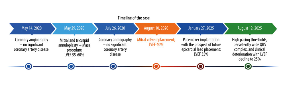 Figure 1. Timeline of the case. Initial failed cardiac resynchronization therapy device implantation was followed by clinical deterioration and subsequent rescue therapy using left bundle branch area pacing, resulting in clinical improvement and partial recovery of left ventricular ejection fraction.
 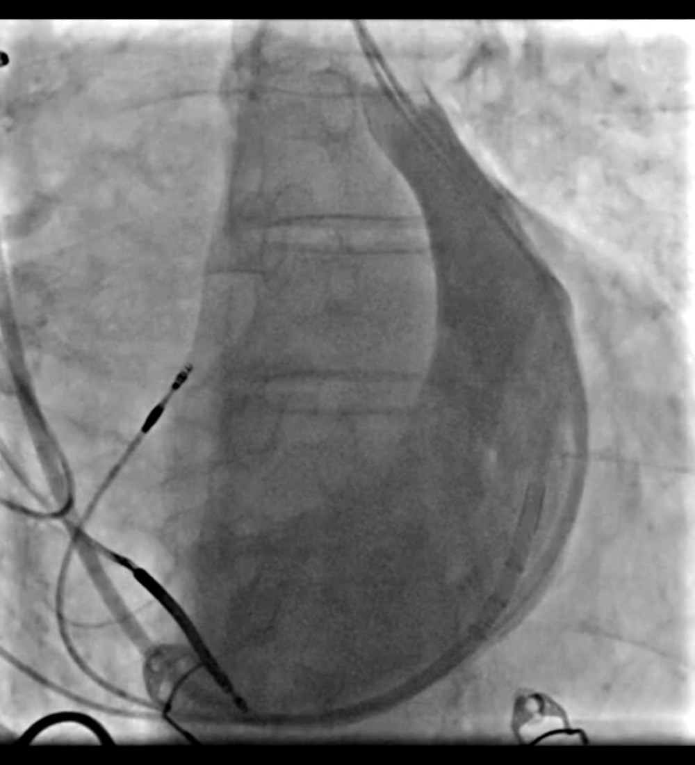 Figure 2. Selective coronary sinus venography (contrast injection into the coronary sinus). The fluoroscopic image shows the coronary sinus opacified after contrast injection through a catheter introduced via right-sided venous access. The contrast outlines the main trunk of the coronary sinus without an adequate lateral or posterolateral branch suitable for left ventricular lead implantation.
 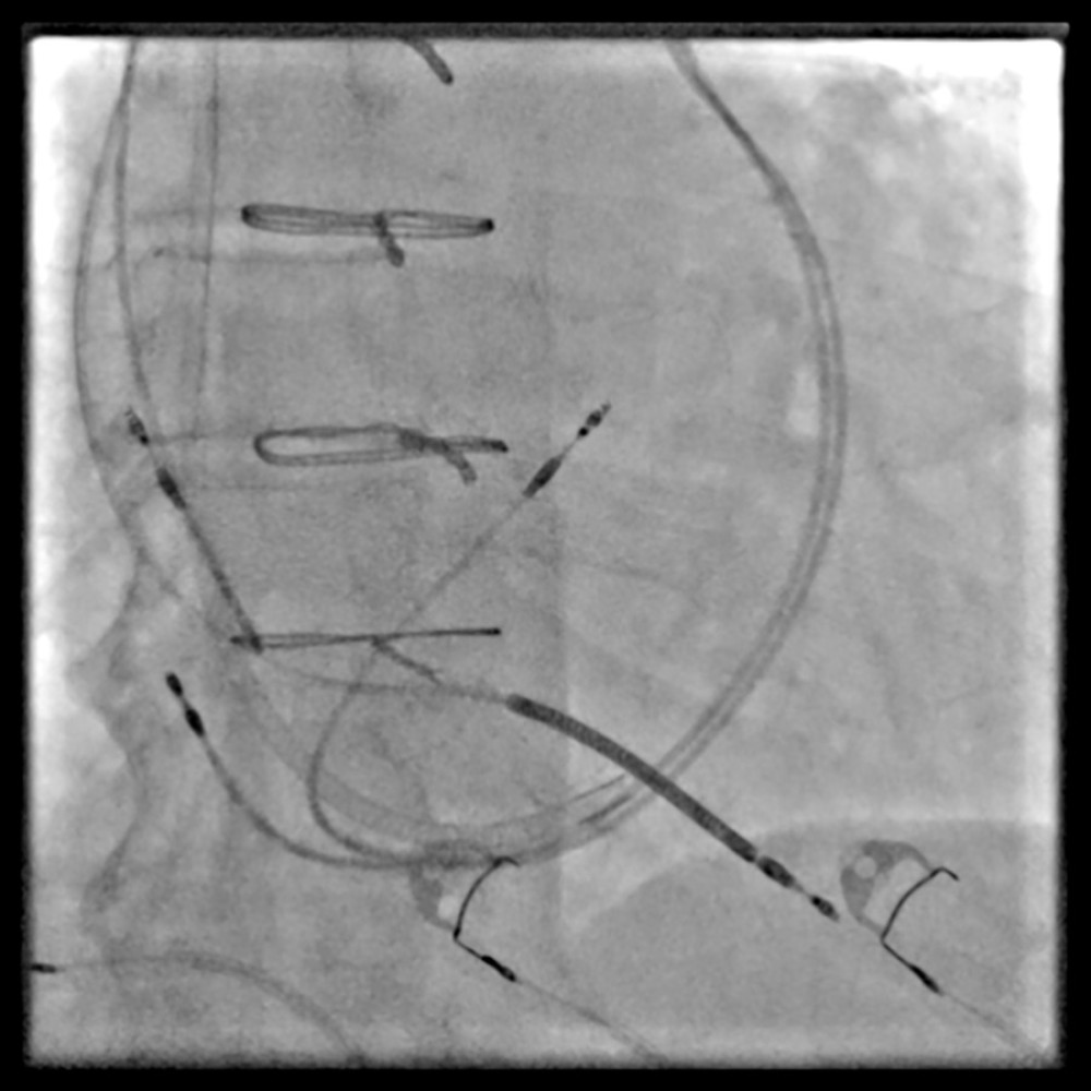 Figure 3. Perioperative X-ray documentation of all implanted leads showing the previously implanted left-sided cardiac resynchronization device leads and the newly implanted right-sided implantable cardiac defibrillator system leads. The image shows the course of the persistent left superior vena cava and the dilated coronary sinus before extraction of the previous cardiac resynchronization therapy pacemaker system, highlighting the anatomical limitation for conventional left ventricle lead placement.
 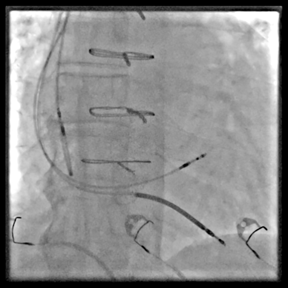 Figure 4. Final perioperative X-ray image with confirmed lead positions. The final fluoroscopic image after implantation of the implantable cardioverter defibrillator system with left bundle branch area pacemaker stimulation demonstrates stable positioning of the left bundle branch area pacemaker lead within the interventricular septum, consistent with effective conduction system capture. The implantable defibrillator lead remains optimally placed in the apical septum, and the right atrial lead shows appropriate fixation. This final radiograph confirms correct depth and orientation of the left bundle branch area pacemaker lead, ensuring reliable physiological pacing and anticipated resynchronization.
 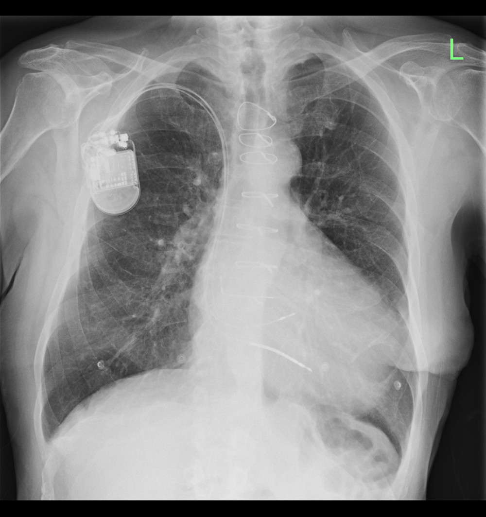 Figure 5. Upright chest X-ray on postoperative day 1. Chest radiograph obtained on postoperative day 1 confirming stable positions of all conduction system pacing-related leads, without evidence of dislodgement. The left bundle branch area pacemaker lead maintains appropriate septal orientation, indicative of sustained conduction system engagement. No pneumothorax or early postoperative complications are present.
 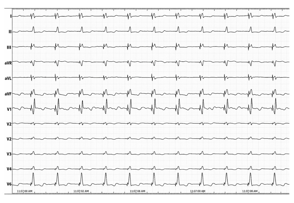 Figure 6. Postoperative electrocardiogram recording. Postoperative electrocardiogram demonstrating non-selective left bundle branch area pacing, characterized by a paced QRS duration of 120 ms.
 References
1. Jastrzębski M, Moskal P, Bednarek A, Electrocardiographic characterization of left bundle branch area pacing: Criteria for left bundle branch capture: JACC Clin Electrophysiol, 2021; 7(5); 605-16
2. Povoski SP, Khabiri H, Skolnick ML, Persistent left superior vena cava: Clinical implications: World J Surg Oncol, 2006; 4; 24
3. Goyal SK, Punnam SR, Verma G, Ruberg FL, Persistent left superior vena cava: A case report and review of literature: Cardiol Res Pract, 2010; 2010; 798304
4. McDonagh TA, Metra M, Adamo M, 2023 focused update of the 2021 ESC Guidelines for the diagnosis and treatment of acute and chronic heart failure: Eur Heart J, 2023; 44(37); 3627-25
5. Chung MK, Patton KK, Lau CP, 2023 HRS/APHRS/LAHRS guideline on cardiac physiologic pacing: Heart Rhythm, 2023; 20(9); e17-e91
6. Vijayaraman P, Ponnusamy SS, Cano Ó, Conduction system pacing: The state of the art: Heart Rhythm, 2021; 18(7); 1080-90
7. Biffi M, Boriani G, Frabetti L, Left superior vena cava persistence in patients undergoing pacemaker or cardioverter-defibrillator implantation: Clinical and procedural implications: Europace, 2001; 3(3); 227-34
8. Rodriguez-Queralto O, Gonzalez-Matos C, Zaraket F, Conduction system pacing in a patient with persistent left superior vena cava and absent right superior vena cava: A case report: J Med Case Rep, 2025; 19; 538
9. Kohoutková L, Augustynek M, Szymeczek H, Personalised treatment strategies in heart failure: A literature review and new proposed algorithm for device therapy selection: Biomed Pap Med Fac Univ Palacky Olomouc Czech Repub, 2025; 169(1); 1-9
Figures
Figure 1. Timeline of the case. Initial failed cardiac resynchronization therapy device implantation was followed by clinical deterioration and subsequent rescue therapy using left bundle branch area pacing, resulting in clinical improvement and partial recovery of left ventricular ejection fraction.Figure 2. Selective coronary sinus venography (contrast injection into the coronary sinus). The fluoroscopic image shows the coronary sinus opacified after contrast injection through a catheter introduced via right-sided venous access. The contrast outlines the main trunk of the coronary sinus without an adequate lateral or posterolateral branch suitable for left ventricular lead implantation.Figure 3. Perioperative X-ray documentation of all implanted leads showing the previously implanted left-sided cardiac resynchronization device leads and the newly implanted right-sided implantable cardiac defibrillator system leads. The image shows the course of the persistent left superior vena cava and the dilated coronary sinus before extraction of the previous cardiac resynchronization therapy pacemaker system, highlighting the anatomical limitation for conventional left ventricle lead placement.Figure 4. Final perioperative X-ray image with confirmed lead positions. The final fluoroscopic image after implantation of the implantable cardioverter defibrillator system with left bundle branch area pacemaker stimulation demonstrates stable positioning of the left bundle branch area pacemaker lead within the interventricular septum, consistent with effective conduction system capture. The implantable defibrillator lead remains optimally placed in the apical septum, and the right atrial lead shows appropriate fixation. This final radiograph confirms correct depth and orientation of the left bundle branch area pacemaker lead, ensuring reliable physiological pacing and anticipated resynchronization.Figure 5. Upright chest X-ray on postoperative day 1. Chest radiograph obtained on postoperative day 1 confirming stable positions of all conduction system pacing-related leads, without evidence of dislodgement. The left bundle branch area pacemaker lead maintains appropriate septal orientation, indicative of sustained conduction system engagement. No pneumothorax or early postoperative complications are present.Figure 6. Postoperative electrocardiogram recording. Postoperative electrocardiogram demonstrating non-selective left bundle branch area pacing, characterized by a paced QRS duration of 120 ms. In Press
Case report 
Am J Case Rep In Press; DOI: 10.12659/AJCR.953543
Case report 
Am J Case Rep In Press; DOI: 10.12659/AJCR.954216
Case report 
Am J Case Rep In Press; DOI: 10.12659/AJCR.954932
Case report 
Am J Case Rep In Press; DOI: 10.12659/AJCR.954146
Most Viewed Current Articles
07 Dec 2021 : Case report
28,712,695
DOI :10.12659/AJCR.934347
Am J Case Rep 2021; 22:e934347
06 Dec 2021 : Case report  180,721
DOI :10.12659/AJCR.934406
Am J Case Rep 2021; 22:e934406
21 Jun 2024 : Case report
124,419
DOI :10.12659/AJCR.944371
Am J Case Rep 2024; 25:e944371
08 May 2026 : Case report
95,462
DOI :10.12659/AJCR.952158
Am J Case Rep 2026; 27:e952158